Review

Role of Hyperspectral Imaging in Forensic Science

1
Department of Computer Science and Engineering, SRM University-AP, Amaravati 522240, Andhra Pradesh, India
2
Centre for Interdisciplinary Research, SRM University-AP, Amaravati 522240, Andhra Pradesh, India
*
Author to whom correspondence should be addressed.
Algorithms 2026, 19(8), 629; https://doi.org/10.3390/a19080629
Submission received: 8 June 2026 / Revised: 7 July 2026 / Accepted: 9 July 2026 / Published: 28 July 2026
(This article belongs to the Section Evolutionary Algorithms and Machine Learning)

Abstract

Hyperspectral imaging (HSI) is a state-of-the-art analytical technique that combines the use of conventional digital imaging and spectroscopy to capture both spatial and spectral information simultaneously in hundreds of narrow, adjacent wavelength bands. In recent decades, the progress in HSI has been rapid, and the technique has been increasingly utilized in forensic sciences, demonstrating its superiority to standard analytical techniques with respect to being non-invasive and contact-free. Although numerous forensic HSI articles have appeared in the literature in recent years, there has yet to emerge a systematic comparison of HSI performance, instrumentation, and cross-domain translational difficulties within forensic science. This review fills this important gap by analyzing the principles, instrumentations, methods of HSI data processing, and potential applications of HSI in forensics in the context of nine important fields: blood stain analysis and estimation of blood age; document authentication; fingerprint detection and enhancement; gunshot residue (GSR) analysis; analysis of trace evidences; detection of biological fluids; postmortem interval (PMI) estimation; determination of bruise age; and multidisciplinary applications. Comparative analysis of over fifty peer-reviewed articles published from 2010 to 2026 in HSI-based forensic sciences is provided herein, with classification accuracies between 81% and 100%. The use of chemometric and machine-learning methods, such as principal component analysis (PCA), support vector machines (SVM), partial least square discriminant analysis (PLS-DA), and Convolutional Neural Networks (CNNs), is carefully analyzed. Some problems concerning standardization, legal acceptance, data sets available, and forensic application are considered alongside future developments of HSI technology.

1. Introduction

Forensic science relies on one simple principle, and that is the scientific and impartial collection, preservation, and analysis of the evidence. The gathering of the physical evidence from the crime scene to the court room requires analytical methods which are accurate, reproducible, and as non-destructive as possible. Traditional forensics face some disadvantages; chemical enhancement techniques lead to destructive and irreversible alteration of the evidence, the inability of traditional imaging techniques to distinguish materials with similar spectra, along with contamination due to extensive sample preparation [1,2].
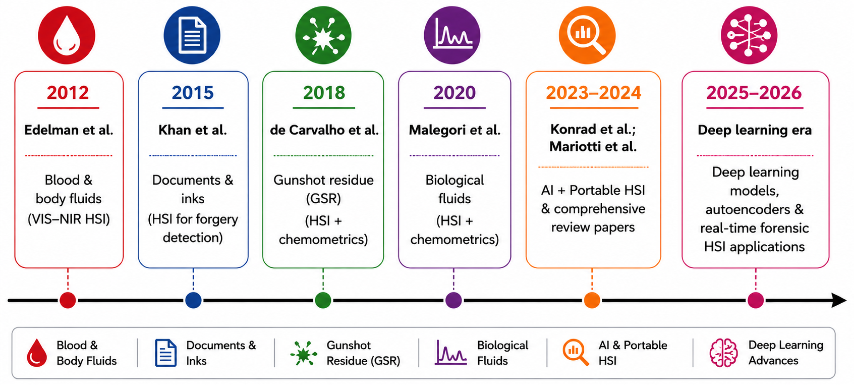
HSI, initially employed for remote sensing and military applications in the 1970s and 1980s [3], has evolved into a fundamental component of forensic sciences. This analytical method utilizes the interplay between electromegnetic radiation and matter, which means that materials reflect, absorb, and emit electromagnetic energy at distinct wavelengths determined by their unique chemical structure. HSI obtains hundreds of narrow-band images to create the three-dimensional dataset known as the hyperspectral data cube (hypercube for short) [4,5]. The importance of this feature from the forensic perspective is very high. Objects that have a visual resemblance to each other, like questioned document ink and hidden traces of blood stains on complex backgrounds, can be discriminated based on their distinctive spectral signatures. Although traditional RGB imaging acquires the data in three spectral bands, HSI provides high-resolution spectral images with a number of bands ranging from 200 nm to 400 nm, which provides an excellent chance to characterize the material [6]. The recent advancements between 2025–2026 have brought about an enormous boost in terms of HSI due to sensor miniaturization, portable and handheld HSI technologies, high-speed data collection, and the use of AI-based spectral analysis. Specifically, the transformer-based deep-learning architectures, graph neural networks, self-supervised learning, and explainable AI have tremendously advanced the process of hyperspectral image classification in terms of understanding the complex relationships between spectra and spatial features with a reduced reliance on large annotated datasets. This advancement in technology has resulted in the expansion of HSI from its usual laboratory applications into other fields including biomedical imaging, agriculture, food quality inspection, environmental monitoring, and forensic science. Therefore, HSI is emerging as a potential technology for the next generation of forensic examinations [7,8,9,10]. Over the last years, there have been observed numerous advances in the employment of HSI in forensics. The first investigations in this area were conducted by Edelman et al. [11] years ago. Since then, a lot of scientists have proven the potentiality of the technique in different areas, including the analysis of blood stains and biological fluids, questioned documents investigation, fingerprints visualization, GSR detection, trace evidence analysis, and PMI estimation [12,13,14]. To demonstrate the achievements of forensics technology throughout the years, a timeline of the key accomplishments is given in Figure 1.
Considering the above-mentioned comprehensive set of applications of forensic sciences where HSI plays a crucial role, one can easily conclude the diversity of this advanced approach for solving various forensics problems. The key advantage of using HSI over many classical methods lies in its ability to conduct an investigation without destruction or invasion. This means not only preservation of evidence integrity but also availability of subsequent testing [19]. Furthermore, due to providing spatial and spectral information, HSI allows detecting and analyzing diverse types of evidence.
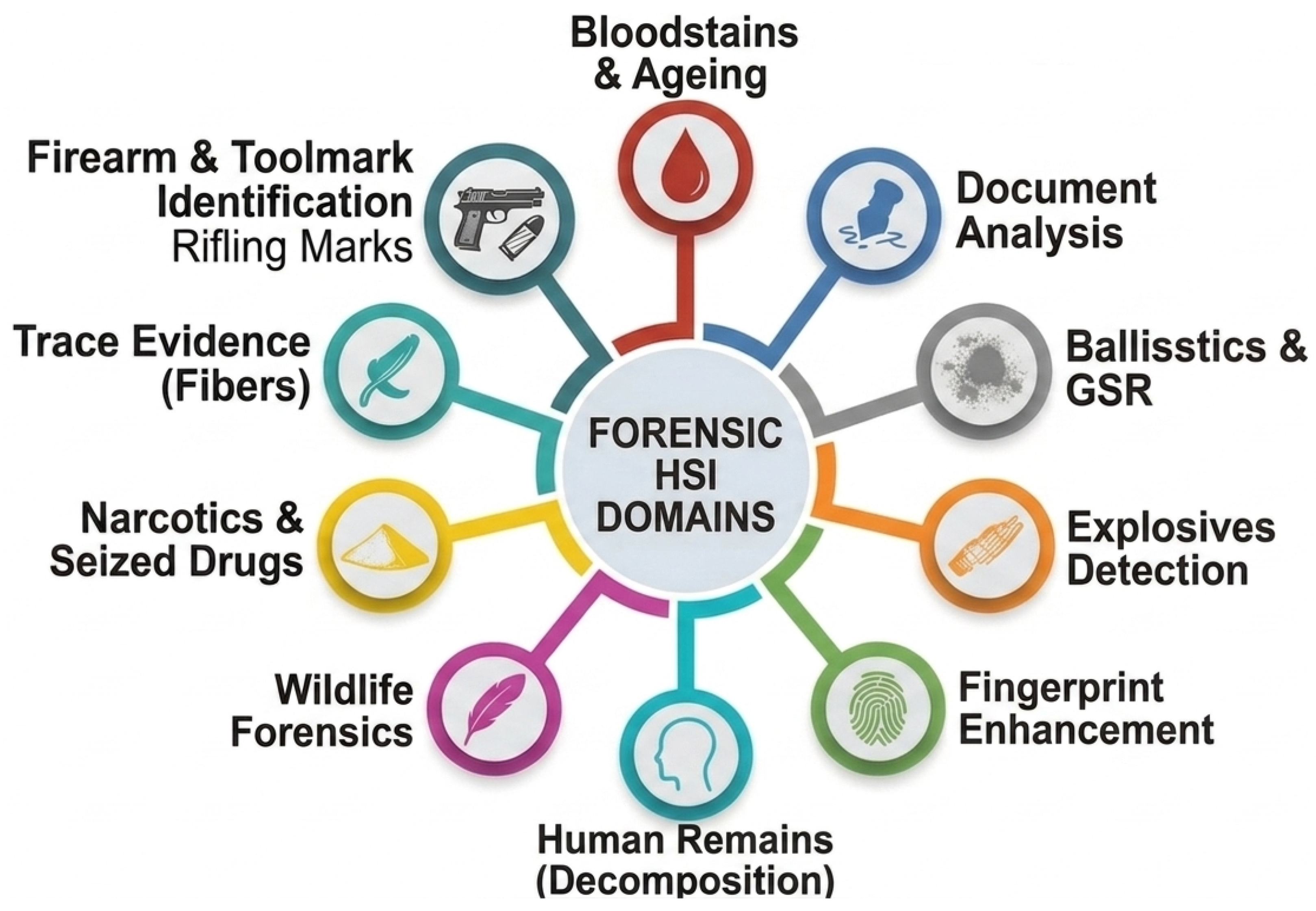
Figure 2 illustrates some examples of important domains where HSI has already been successfully implemented, including blood stain analysis, document forensics, fingerprint identification, gunshot residue analysis, trace evidence examination, biological fluid identification, estimation of postmortem interval, and determination of bruise age.
Even though numerous works have been produced on the topic of HSI-enabled forensic investigations, there is still a lack of thorough and well-structured comparison of various HSI methods used in forensics. The review articles written in this area usually analyze HSI applications for particular fields of forensic investigation without mentioning any comparative analysis of instruments involved, data processing techniques, machine-learning algorithms applied, and problems connected with the transition from one field to another. This paper aims at conducting such a comparative analysis.

1.1. Objectives of This Review

The main objectives of this review are to: (i) present an in-depth understanding of the fundamental aspects, instrumentation, and data processing involved in the implementation of HSI in forensic science; (ii) thoroughly assess the most recent developments in the application of HSI technology to nine forensic areas of importance, such as blood stain, document, fingerprint, GSR, trace, biological fluid, postmortem interval, bruise age estimation, and multidisciplinary forensics; (iii) critically review traditional chemometric methods, machine-learning models, and new deep-learning frameworks used for the forensic HSI analysis; (iv) highlight the existing problems, limitations, and gaps in research concerning the forensic investigation using HSI technology; and (v) outline future research directions in this area, which include explainable artificial intelligence, transformer networks, graph neural networks (GNNs), mobile HSI systems, and forensic imaging protocols.

1.2. Literature Search and Selection Methodology

The review has been carried out based on the Preferred Reporting Items for Systematic Reviews and Meta-Analyses (PRISMA 2026) protocol in order to make sure that there is transparency, replicability, and systematic nature of the literature search process. The literature search was completed using seven major scientific databases including Scopus, Web of Science, PubMed, IEEE Xplore, ScienceDirect, SpringerLink, and Google Scholar. The period of time considered in the search range is from January 2010 to June 2026; however, some landmark papers prior to the date have been considered to give the historical background.
The search protocol consisted of combining Boolean operators along with keywords connected to the topic of the study, which are “HSI”, “Near-Infrared Spectroscopy”, “Forensic Science”, “Crime Scene”, “Bloodstain”, “Fingerprint”, “Document Examination”, “GSR”, “Trace Evidence”, “Biological Fluids”, “Postmortem Interval”, “Bruise”, “Machine Learning”, “Deep Learning”, and “Artificial Intelligence”.
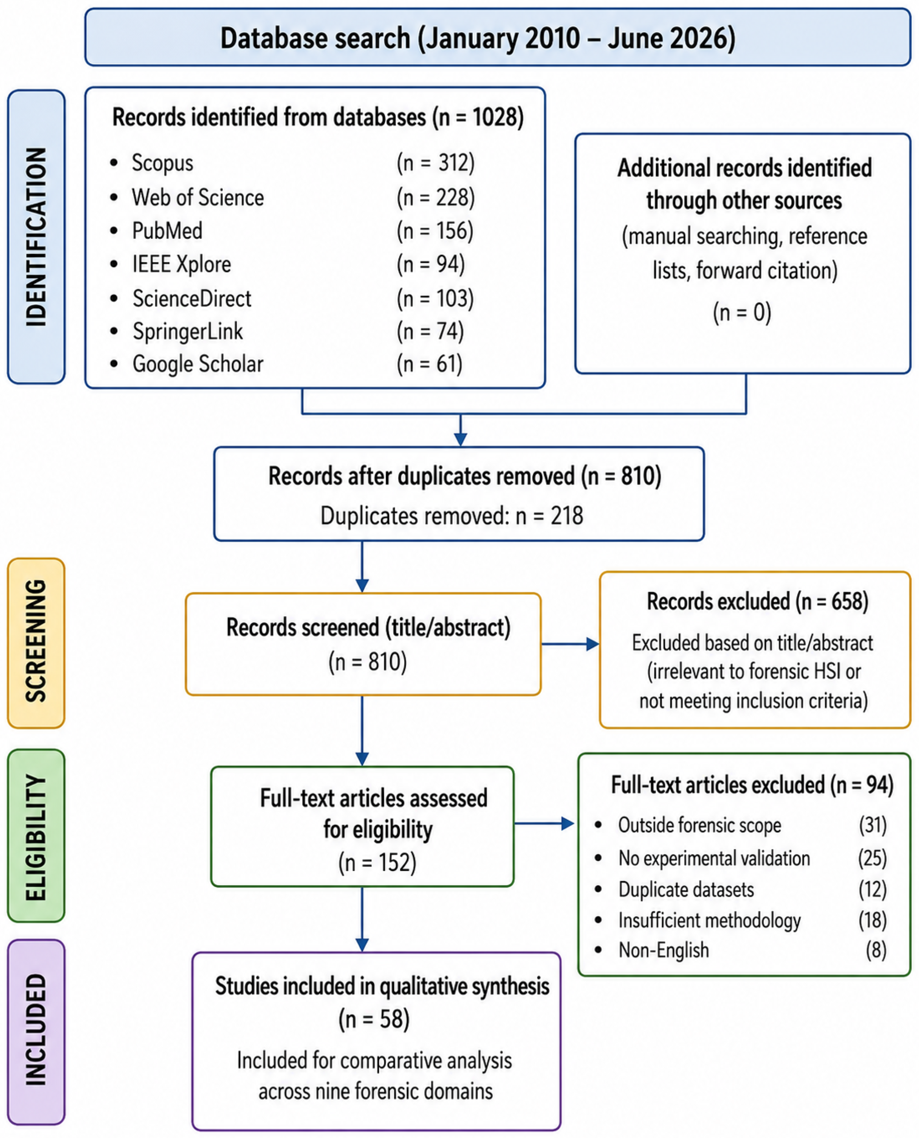
The articles selected for analysis were supposed to meet the following criteria: (i) published in reputable journals or high-quality conferences; (ii) published in English language; (iii) concerning the use of HSI or other spectral imaging technologies for forensic purposes; and (iv) presenting experimental evidence or results from development and review studies. The papers that were duplicate records, irrelevant to the topic under consideration, insufficient in methodology details and validation, using overlapping data sets, and not published in English language were omitted. After eliminating duplicates, all identified papers were screened based on titles and abstracts and further analyzed for their compliance with the defined eligibility criteria. In the case of an eligible article, the relevant information about the application of forensic technology, imaging method, spectral range, imaging system, pre-processing, feature extraction, machine/deep-learning algorithm used, performance measures, used data set, and limitations of the paper was extracted and comparatively analyzed. The entire process of literature identification, screening, eligibility, and selecting process is illustrated in Figure 3. Overall, 1028 documents were retrieved from database searching. Of 1028 retrieved records, 218 duplicates were removed and the title and abstract of 810 articles were screened. Overall, 58 studies fulfilled the inclusion criteria and have been included in this qualitative synthesis.

Quality Evaluation

The quality of each included study was evaluated based on criteria such as clarity of the experimental design, appropriateness of HSI acquisition details, dataset details, preprocessing method employed, validation scheme, evaluation metrics used, reproducibility of the results, and forensic relevance.

2. Principles of HSI

2.1. Fundamental Concepts

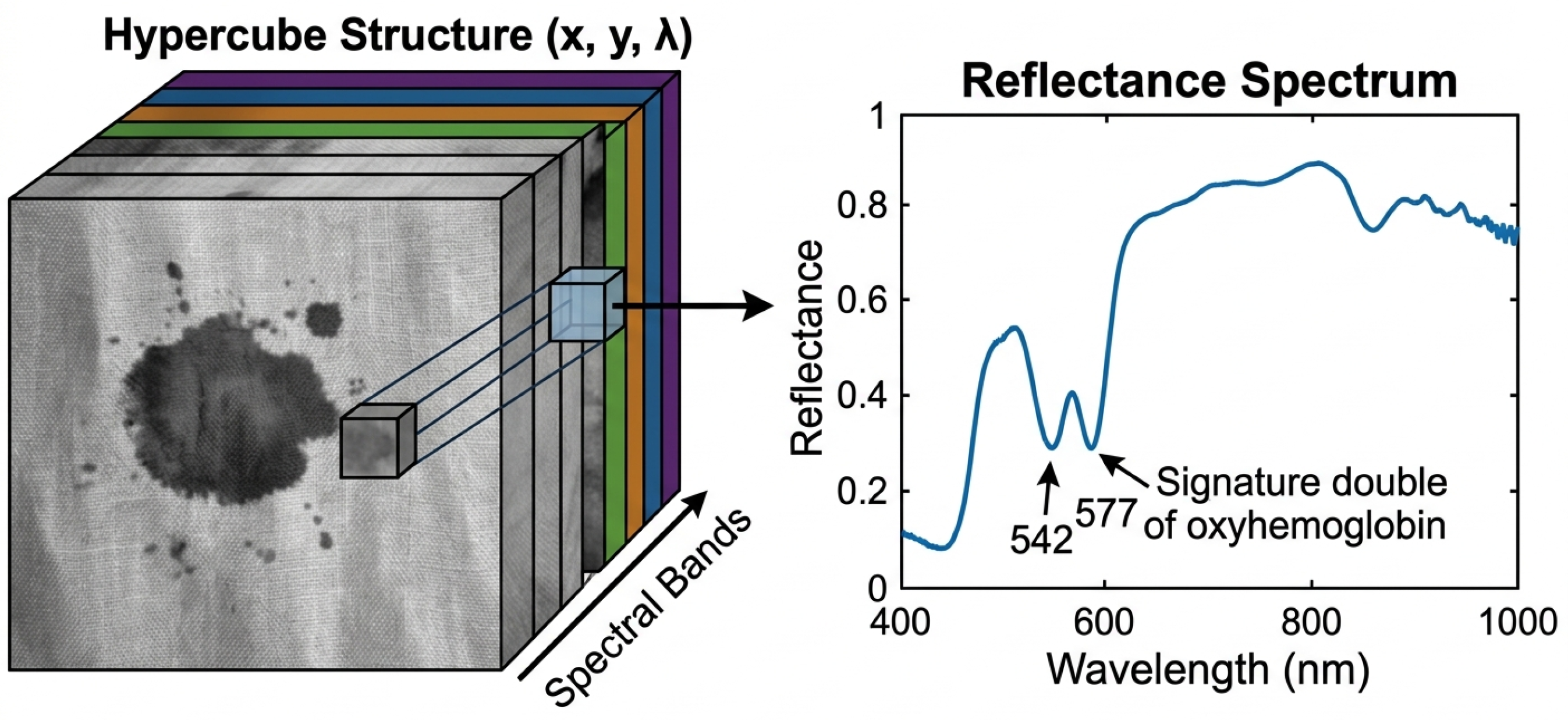
The fundamental basis of HSI relies on the interaction of electromagnetic waves with matter. If light waves of specific wavelengths interact with a specific chemical compound, the probabilities of absorption, reflection, and transmission depend on the molecular structure and electronic configuration of the compound. The dominant process for Ultraviolet (UV) range (200 to 400 nm) is electronic excitation in aromatic compounds and conjugated systems. Chromophoric compounds, such as hemoglobin, melanin, and artificial colorants, have characteristic absorption spectra for visible range (400 to 700 nm). Near-infrared (NIR) range (700 to 2500 nm) comprises multiple overtones and combination bands owing to vibrations of O-H, N-H, and C-H bonds in organic compounds, such as proteins, lipids, and cellulose [20,21]. The most basic element that an HSI system can provide is a three-dimensional image I(x, y, λ), where x and y are spatial coordinates and λ denotes wavelengths. It is known as the hyperspectral data cube, or hypercube, which incorporates both spatial and spectral features at the same time. In other words, every pixel of a particular scene is described by its spectral signature, providing the capability for both spectral discrimination and spatial identification of the material of interest. As shown in Figure 4, the hyperspectral cube is built up from various spectral bands in the direction of wavelengths, and the reflectance spectrum serves as the specific spectral signature of the material [22].

2.2. HSI Acquisition Modalities

HSI systems are differentiated based on the spectral scanning technique used. Whiskbroom (or point-scanning) HSI devices acquire data from only one spatial pixel per image and scan the pixels to form the picture, providing superior spectral resolution, but they need mechanical scanning. The pushbroom (or line-scanning) HSI systems obtain data from one line per snapshot and are popular in forensics for their rapid operation and resilience. Snapshot/staring HSI systems acquire a complete spatial image within a single shot but may lack some spectral resolution [23,24]. The wavelength range in the HSI camera defines what analysis can be performed. Visible-near infrared (VNIR) systems (400–1000 nm) are ideal for detecting hemoglobin derivatives and chromophores. Short-wave infrared (SWIR) systems (1000–2500 nm) offer an abundance of information about organic functional groups. LWIR cameras operate in the mid-infrared thermal emissions band and can perform some very unusual tasks in forensic research [25,26]. Most forensic studies use VNIR cameras for their low cost, high spatial resolution, and standard lab/field environment suitability [27].

2.3. Key Instrumentation Components

The typical components of forensic HSI systems include the light source (tungsten-halogen source for reflectance mode or UV lamp for fluorescent mode activation or laser, collection optics, dispersive device such as diffraction grating or acousto-optic tunable filter (AOTF)), two-dimensional detector array (typically CCD and InGaAs detectors), and computer data acquisition software. Companies producing systems utilized in forensic investigations include Specim (Finland), Headwall Photonics (USA), Resonon (USA), and Cubert (Germany). Mobile and handheld HSI systems for the analysis of crime scenes in situ without transferring specimens to laboratory facilities have been also designed [28]. Systems from the FX line produced by Specim and snapshot imagers represent examples of developments in this direction.

3. Data Processing and Chemometric Methods

3.1. Preprocessing

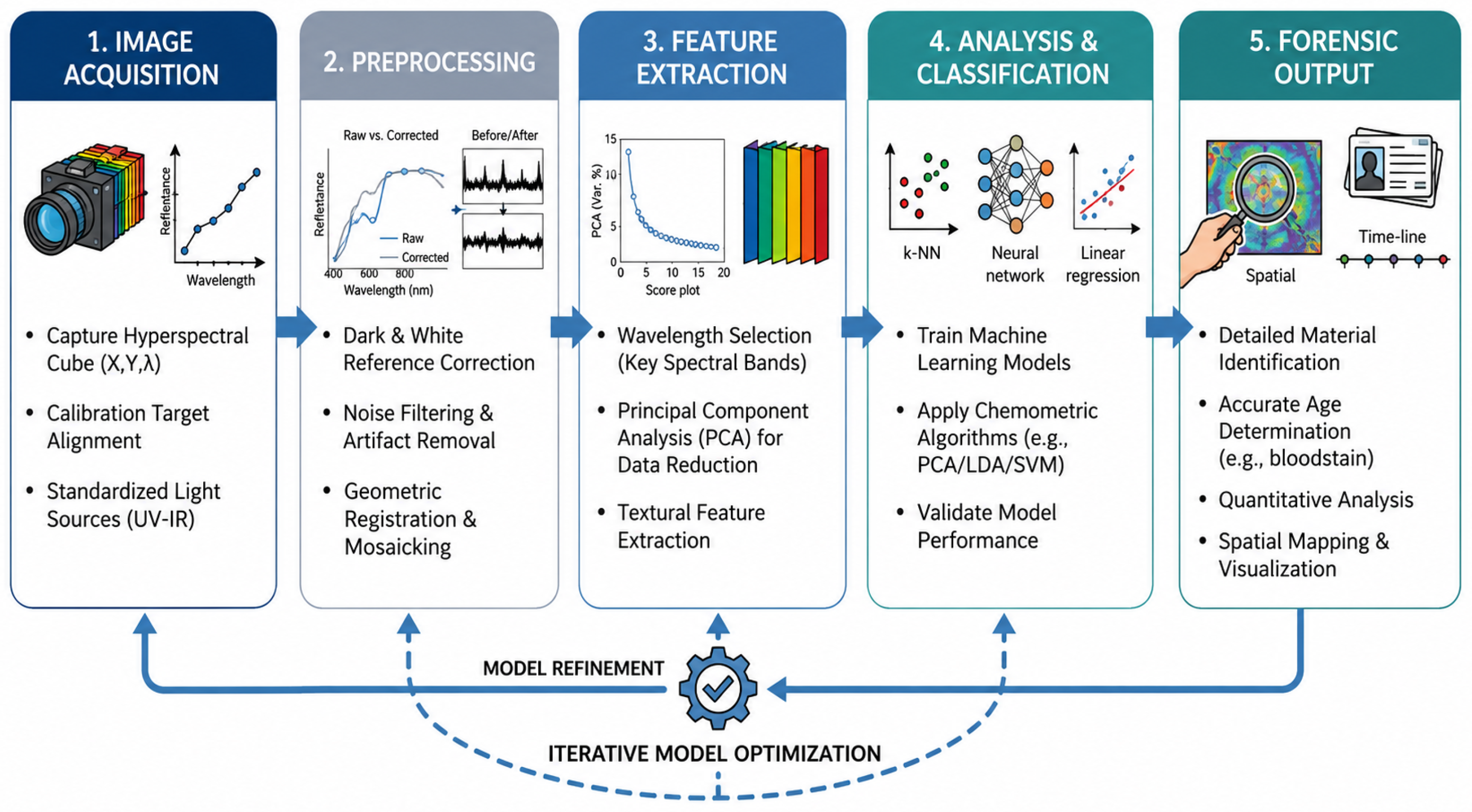
The preprocessing stage plays an important role in hyperspectral data processing since it enhances data quality through the elimination of instrumental distortions, noise reduction, enhancement of spectra, and normalization before applying any other algorithms. Preprocessing is executed between acquisition and extraction phases, as shown in Figure 5.
First of all, dark current subtraction and radiometric calibration of acquired data with white reference enable transforming data into reflectance values. Moreover, data normalization can be conducted to mitigate the effect of different background-related influences and illumination conditions. Also, noise reduction is a common practice for improving data quality.
Different preprocessing methods can be found in the literature, among which there are Standard Normal Variate (SNV) normalization, Multiplicative Scatter Correction (MSC), Savitzky–Golay (SG) filtering, and derivatives (first and second order) [29,30]. These algorithms help to increase spectral discrimination and reduce the impact of scattering and baseline effects on data. Finally, such preprocessing approaches as binning and spatial filtering can be used to enhance images and decrease computations.

3.2. Dimensionality Reduction

Due to the high dimensionality of hyperspectral images (over 200 bands per pixel), there arises a need to perform dimensionality reduction prior to classification. PCA is universally used to project the data into an alternative lower dimensional space characterized by orthogonal axes maximizing variance. MNF transformation is used when there is spectral correlation of noise. ICA technique is employed when independence of sources is of importance [31,32]. These techniques prove useful not only for visualization (false color imagery) but also in preparing data for further classification.

3.3. Classifiers and Machine Learning

Forensic HSI applications heavily rely on classification methods. The traditional chemometric algorithms used for classification purposes include PLS-DA, Linear Discriminant Analysis (LDA), and Soft Independent Modeling of Class Analogy (SIMCA). SVMs have proven themselves as an efficient tool for forensic HSI in particular, owing to their superior generalization ability in relation to high-dimensional spectra [33,34].
In more recent studies, deep-learning models were also employed to solve forensic HSI problems. In this regard, CNN was tested in 1D (spectral), 2D (spatial), and 3D (spectral–spatial) variants for such applications as blood stain classification, ink differentiation, and drugs identification. As reported by Książek et al., the use of 2D CNN model resulted in an overall classification accuracy of 98–100% under controlled settings [35]. The development of RNNs and transformer-based architectures can be considered a part of ongoing research. Unsupervised machine-learning models such as k-means, hierarchical agglomerative clustering, and self-organizing maps are used in field conditions when there are no references [36,37].

3.4. Emerging Deep-Learning Architectures for HSI Analysis

Current trends in HSI analysis involve the transition from traditional CNN models to more advanced deep-learning approaches such as ViT models, GNN models, spectral–spatial attention models, contrastive learning methods, self-supervised learning techniques, and explainable artificial intelligence (XAI). Although CNN models have shown great promise in capturing spectral and spatial local feature representations, the small receptive fields of such models limit their ability to extract information regarding long-range spectral relations and spatial relationships. As a result, advanced models have been developed that surpass the shortcomings of the traditional models and have outperformed other techniques in remote sensing and biomedical HSI applications. The current developments will be instrumental in developing forensic HSI.
Within such emerging methods, the Vision Transformer (ViT) model has emerged as a prominent choice for HSI classification. Unlike CNN-based methods, which use convolution operations, the ViT architecture uses the multi-head self-attention method to capture the long-range interactions between both spectral and spatial features. More recent transformer-based models have further incorporated multiscale feature extraction and spectral tokens along with convolutional attention mechanism that allows for effective capturing of global context information without sacrificing local spectral properties. Such models have shown significant improvement in terms of classification performance, robustness, and reducing spectral redundancy on several benchmark HSI datasets. The potential of such models in forensic analysis will aid in the detection of blood stain, questioned documents, latent fingerprint, biological fluid, gunshot residue, and trace evidence, especially in difficult environmental scenarios [38,39]. On the downside, transformer-based models usually require a large dataset and high computational power; therefore, transfer learning and lightweight transformers are among the areas of future research.
GNNs constitute yet another major advancement in the field of hyperspectral image analysis. As in the case of GNNs, the pixels or superpixels are modeled using nodes in graphs based on their spectral–spatial associations, thereby making the processing of non-Euclidean nature of HSIs more feasible. Unlike CNNs, GNNs can preserve neighborhood information well and are able to capture long-range spectral interactions, which are crucial for complicated scenes. Graph Transformer Networks have been proposed very recently where the combination of graph learning with transformers is used for the sake of enhancing spectral–spatial representation and classification performance on benchmark HSI datasets [40,41]. These types of techniques can hold great potential for heterogeneous forensic evidence analysis in cases of latent fingerprints, biological stains, gun shot residues, trace evidence, and mixed materials on complex surfaces. But there still exist some limitations due to the computational complexities associated with graph creation and lack of forensic HSI datasets.
Another set of recent trends in HSI analysis includes spectral–spatial attention mechanism, contrastive learning, self-supervised learning, and explainable AI approaches which are proven to be promising for increasing efficiency of HSI analysis. Attention mechanism, including channel attention, spatial attention, and gated self-attention, allows us to adaptively weight discriminative spectral features, excluding redundant information at the same time, thus providing high classification accuracy with small computational costs. Meanwhile, contrastive learning and self-supervised learning have become promising alternatives for minimizing the necessity for using manually annotated datasets and obtaining robust representations based on unlabeled hyperspectral images. Such techniques would be especially beneficial in forensic science in which acquiring annotated datasets is a complex task. Moreover, techniques allowing interpretability of decision-making of the AI model, like SHapley Additive exPlanations (SHAP), Gradient-weighted Class Activation Mapping (Grad-CAM), attention visualization, and saliency maps, provide better interpretability and thus increase judicial validity of the results obtained with help of AI technologies in forensics [38,40,42].
Even though there have been significant advancements in these sophisticated deep-learning algorithms for the purpose of remote sensing, agriculture, food quality assurance, and biomedicine, their use in forensic hyperspectral imaging has not gained much traction yet. The vast majority of forensic analyses still uses classical machine-learning algorithms or even CNNs due to the unavailability of large public datasets, non-standardized data acquisition methodologies, and need for computationally light models that can be used in a real forensic laboratory environment. Future work in this field should concentrate on developing new lightweight transformer models, graph neural networks, self-supervised feature learning, multimodal foundation models, and explainable AI approaches for forensic HSI specifically. These innovations will help to significantly enhance the performance of HSI systems in analyzing blood stains, examining questioned documents, enhancing latent fingerprints, identifying gunshot residues, classifying trace evidences, detecting biological fluids, and estimating postmortem interval.

4. Blood Stain Analysis

4.1. Detection and Identification

The other most important physical evidence in crimes involving violence is blood. However, standard chemical techniques used to visualize blood (luminol, fluorescein, leuco-malachite green) are destructive in nature, prone to causing false positive results, and interfere with subsequent DNA profiling [1]. HSI proves to be a promising non-destructive technique. Blood, in the visible range, is characterized mainly by absorption spectra associated with Hemoglobin (Hb) and Hb-based molecules. The seminal works in this respect include those by Edelman et al. [11] who used reflectance spectroscopy with wavelengths from 400–1000 nm and Partial Least Squares (PLS) regression to identify blood stains on various colored surfaces. The work clearly revealed the difficulties associated with the effect of colors in the visible region and pointed out that NIR spectroscopy is more effective for blood detection amid interfering background substances. In a comparative analysis of Raman, FTIR, and fluorescence spectroscopies for blood detection by Virkler and Lednev [2], the fundamental aspects of molecular differentiation for spectroscopy were discussed. More recently, Bremmer et al. [43] applied spatially offset Raman spectroscopy in combination with reflectance spectroscopy for remote detection of blood up to 2 m distance in laboratory settings. For instance, the non-destructive framework for the classification of blood stains using a hyperspectral camera in the 397–1003 nm wavelength range (with 224 spectral bands) by Zulfiqar et al. [44] is mentioned. The authors used derivative spectral analysis for the selection of features from the spectral signatures and SVM, ANN, KNN, DT, and RF algorithms to classify the selected features. It is noted that the presented method provided excellent results for the bloodstains on different surfaces such as white fabric, white tile, and wall sheet. The paper of Zhang et al. [45] reported the successful use of VNIR-HSI in combination with the Extreme Learning Machine (ELM) algorithm to identify human and animal (chicken and pig) blood stains, achieving an accuracy rate greater than 96% and therefore indicating successful species discrimination of blood stains. The work of Książek et al. [35] presents the comprehensive analysis of several deep-learning classifiers applied to blood stains classification using HSI technique. Specifically, different 1D-CNN, 2D-CNN, 3D-CNN, RNN, and MLP were compared with SVM classifiers. In the case of transductive setup (classification on one image), accuracy of classifiers ranged from 98 to 100%. On the contrary, when performing classification on unseen images (inductive setup), CNN classifiers reached accuracy ranging from 74 to 94% and 3D classifiers performed better on spatially heterogeneous images.
Pallocci et al. [1] provided a detailed narrative review focusing on HSI technology for blood trace analysis, arguing that the use of both HSI and chemometrics yields better results than traditional techniques for trace analysis on dark, patterned, or complex surfaces. Pereira et al. [46] explored the application of this technique in the context of handheld NIR spectrometers in order to detect blood stains, with a detection accuracy ranging between 81–94%, depending on the classifier used, making handheld instrumentation feasible but less accurate than bench-top devices. The recent advancements are concentrated on enhancing the robustness of hyperspectral blood detection in forensic scenarios by resolving issues related to spectral redundancy, interference from substrates, variations in illumination and blood age. For example, the Fast Extraction (FE) approach along with Enhancing Transformation Reduction (ETR) pre-processing technique was proposed by Al-Alimi et al. [47] in order to decrease the dimensionality of hyperspectral data while maintaining its discrimination capacity. The FE framework proved more efficient and robust in terms of classifying blood stains by reducing spectral mixing and increasing speed of analysis, which emphasizes the necessity of using advanced pre-processing in practice. It shows that the future of forensic HSI is closely connected with effective preprocessing, optimization of spectral features, and application of deep-learning algorithms.

4.2. Blood Stain Age Estimation

The temporal characterization of blood stains—estimating how long ago blood was deposited—is of critical investigative importance. As the blood gets older, hemoglobin goes through a set of chemical alterations, namely: oxyhemoglobin (HbO2) → deoxyhemoglobin (Hb) → methemoglobin (metHb) → hemichrome. All these steps lead to the distinct spectral changes within the visible part of the spectrum. Edelman et al. [48] introduced HSI to age blood stain estimation, proving that it is possible to quantify the fractions of hemoglobin derivatives and make an estimation of the stain’s age from 200 days old, with a median relative error of 13.4% using this method. This was validated by creating a simulated crime scene.
Since then, research has improved age estimation methods. In particular, Bastide et al. [49] showed the influence of temperature, humidity, and substrate type on the age-related spectral characteristics of hemoglobin and concluded that the environment has a great influence on the degradation rate of hemoglobin and should be taken into consideration when estimating age. The visible HSI analysis and Linear Discriminant Analysis (LDA) techniques have been employed by Li et al. [50] to determine the age of blood stains for up to 30 days after deposition. It was shown that blood spectral characteristics with respect to age were successfully classified using the mentioned approaches, showing how efficient HSI is for blood stain age estimation. Differences in the environment, such as indoors or outdoors, may significantly influence blood stain aging and its associated spectra, thereby creating difficulties for establishing a generalizable model for the age determination of blood stains [51]. In order to offer a brief survey of significant contributions in terms of methodological advances for blood stain analysis by HSI, we summarize the most relevant articles in Table 1.

5. Document Examination and Forgery Detection

5.1. Ink Analysis and Discrimination

Questioned document examination is among the most mature forensic applications of HSI, and consistently ranks as the most widely published domain in comprehensive reviews of the field [52]. Document fraud encompasses alteration, erasure, addition, substitution, and reproduction of text in legal, financial, and identity documents. The key challenge is that modern inks are formulated to appear visually identical while differing in chemical composition. Conventional UV and IR examination methods detect some alterations but lack the spectral specificity of HSI [53].
The fundamental principle is that inks composed of different dyes or pigments exhibit distinct reflectance spectra, even at wavelengths where they appear identical to the human eye. The ability of HSI to detect such differences on the pixel level allows the mapping of ink boundaries as well as any addition or substitution of the ink [54]. An ink mismatch detector for forensic purposes was suggested by Khan et al. [15], using HSI and pattern recognition to prove that similar spectral inks from different manufacturers and batches can easily be distinguished. Further research [55] focused on localizing forged documents with the help of HSI-based approach that provided an exact mapping of the forgery boundaries. In their study on deep-learning approaches for forgery detection in hyperspectral document images, Khan et al. [56] proved that CNN models have the ability to leverage spectral and spatial features for the task of forged document classification. Abrar and Iqbal [57] introduced an approach based on clustering algorithms like k-means, agglomerative, and c-means clustering to find out how many inks there are in the document, which is essential for detecting multi-stage alterations. Jaiswal et al. [58] proposed DFD-SS (Document Forgery Detection using Spectral–Spatial features), a hyperspectral image-based framework that exploits both spectral and spatial information to detect forged regions in questioned documents.
The use of near-infrared HSI together with chemometrics in the examination of crossing ink lines was shown by Braz et al. [59], showing it was possible to discriminate between intersecting inks and to determine their stroke sequences. Brauns et al. [60] used Fourier Transform Spectroscopy to build an HSI method designed for non-destructive inspection of suspicious documents, providing sub-nanometer spectral resolution for identifying ink aging and alterations. HSI analysis of gel pen inks was explored by Reed et al. [61], and it was found that spectral data are useful in distinguishing visually similar inks.

5.2. Paper and Security Feature Analysis

Apart from ink analysis, there have been applications of HSI in the study of paper substrates and security features in identification documents and banknotes. Various papers produce varying spectral signatures because of different fiber contents, fillers, optical brighteners, and coatings. Causin et al. [54] explored the ability of diffuse reflectance ultraviolet–visible near-infrared spectroscopy in forensic investigations of paper as a way of using the information about substrates in addition to ink analysis.
The restoration of damaged, burnt, or wet documents used in arsons and fraud cases is another example of a particular application domain. Hedjam et al. [62] have formulated algorithms for improving the legibility of degraded documents using both multispectral and hyperspectral image processing techniques that would retrieve text not visible in white light conditions. Meanwhile, Hollaus et al. [63] introduced an approach for the binarization of multispectral images of documents that involved using spectral information along with powerful image processing algorithms to facilitate the process of extracting text from damaged historical manuscripts. New developments in this field have enabled hyperspectral document examination to be extended from spectral match methods to machine-learning algorithms. For example, López-Baldomero et al. [64] combined HSI with both classical machine-learning algorithms and deep-learning algorithms for the classification of historic inks, with the highest F1-score being recorded at 98%, thus highlighting the efficiency of spectral–spatial feature learning in successful classification of the inks. In the same vein, Singh et al. [65] developed an unsupervised deep autoencoder system for ink mismatch detection in order to automate the process of forgery detection without the need for manual annotation of training samples. As part of the comparison between HSI methods to detect document forgeries and authentications, Table 2 provides a summary of relevant research works with their respective results.

6. Fingerprint Detection and Enhancement

6.1. Latent Fingerprint Visualization

Fingerprints continue to be one of the most vital forms of physical evidence in forensics due to their distinctive patterns of ridge formations as well as permanent individual characteristics. Latent fingerprints consist of sweat, oil, amino acids, among other materials, left behind as a result of the contact between fingers and certain surfaces. Traditional methods used in visualizing the fingerprints tend to involve both physical and chemical processes. In this regard, Payne et al. [66] explored the possibility of using visible absorption and luminescence imaging in detecting and enhancing latent fingerprints. The authors noted that multispectral imaging had the capacity to enhance the visibility of latent fingerprints through contrast optimization between fingerprint residues and background surfaces. HSI using reflectance is now being recognized as a very effective non-destructive technique for analyzing complicated surface materials and layered objects. Cucci et al. [67] presented the use of reflectance HSI for examining artwork like old master paintings and manuscripts. In their discussion, HSI was shown to be able to characterize the materials present on the object by mapping pigments and identifying hidden details. The use of this method has been demonstrated to have several advantages since not only spatial but also spectral information is obtained at the same time without taking any samples. In their study, Sodhi and Kaur [68] analyzed the traditional methods used in detecting latent fingerprints and emphasized the prevalence of the use of these methods in forensic sciences. The authors discussed both the strengths and weaknesses of using different types of powder on different substrates. The performance of these powder methods is largely dependent on a number of factors such as the type of substrate used, the type of powder used, and the fingerprint itself. However, despite being simple and cheap to implement, powder methods may be subject to interference from substrates.
Tahtouh et al. [69] examined the use of infrared chemical imaging in the enhancement of latent fingerprints. In their research, they found that infrared chemical imaging was able to enhance latent fingerprints on difficult surfaces without destroying the evidence. They optimized the imaging parameters and established that infrared chemical imaging is an effective method of fingerprinting, since it is less destructive than other methods of visualization. The forensic value of HSI along with independent component analysis for the analysis of superimposed latent fingermarks was assessed by Nakamura et al. [70]. It has been proved that the spectral unmixing methods can help in separation of superimposed fingerprint images, which belong to different individuals, and thus can make ridge pattern visible.

6.2. Fingerprint Age Estimation

The age estimation of latent fingerprints—determining when a print was deposited—is of critical evidential value but remains an unsolved forensic problem. Spectroscopic approaches offer a rational basis: fingerprint lipids undergo oxidative degradation over time, producing systematic spectral changes. The application of NIR-HSI for fingerprint age determination was studied by Carneiro et al. [71]. It was shown in their pilot study that spectral variations arising in latent fingerprints due to aging could be detected with hyperspectral measurements, thus showing the capability to estimate fingerprint age by non-invasive spectroscopy. It seems plausible that the method proposed could contribute to forensic science by providing information about fingerprint age, though additional studies under different environmental conditions should be carried out.

6.3. Chemical Treatment Enhancement

More sophisticated imaging techniques have also been used to supplement traditional methods of fingermark enhancement. For example, Bradshaw et al. [72] illustrated the use of matrix-assisted laser desorption/ionization mass spectrometry imaging after successive fingermark development. They were able to show that chemical imaging allows one to obtain information on the spatial distribution of both endogenous and exogenous components in the latent fingermarks without sacrificing the minutiae details. The effectiveness of HSI in visible wavelength reflectance when compared to AB1, which is a popular agent for enhancing blood stains, in detecting and identifying blood stained fingerprints was investigated by Cadd et al. [73]. It was shown that HSI could successfully visualize blood stained fingerprints without any use of chemicals, retaining spectroscopic information that would help in discriminating blood from the background surface. In order to offer a brief comparative review of the representative approaches of HSI technology applied to latent fingerprint identification, enhancement, and aging, Table 3 offers an overview of the important works that have been published in the literature.

7. GSR Analysis

7.1. GSR Analytical Techniques and Their Drawbacks

GSR is comprised of particulate matter and gases produced when a gun is fired. Conventional analysis of inorganic gunshot residue (IGSR) involves scanning electron microscopy in combination with energy dispersive X-ray spectroscopy (SEM–EDX) to locate particles that contain lead (Pb), barium (Ba), and antimony (Sb). These elements result from primer compositions [74]. Unfortunately, the increased popularity of ammunition without lead has rendered such an approach less efficient, thus prompting a search for other analytical techniques. Organic gunshot residue (OGSR) refers to propellant components, including nitramines, stabilizers, plasticizers, and many other additives. Unlike IGSR, OGSR cannot be reliably detected using SEM–EDX since the technique is not well-suited for detecting organic compounds. In addition, recent literature has revealed several drawbacks associated with interpreting IGSR and OGSR results and their significance, such as problems with secondary transfer, environmental pollution, and the possibility of producing false associations [75].

7.2. HSI Applications in GSR Detection

Głomb et al. [76] pioneered the application of HSI for GSR detection on fabric substrates, comparing an unsupervised anomaly detection approach (RX detector) with a supervised SVM classifier. The study demonstrated that HSI generally outperforms RGB imaging of comparable quality in terms of GSR detection accuracy. The classifier-based approach eliminated the need for fabric-specific normalization required by the anomaly detector, improving operational robustness. GSR samples from two ammunition types at two shooting distances were correctly detected with high sensitivity in both detection scenarios.
Khandasammy et al. [77] proposed a novel two-step method for organic GSR detection combining highly sensitive fluorescence HSI (step 1) with confirmatory Raman microspectroscopic identification (step 2). In this proof-of-concept study, fluorescence HSI scanned large areas for candidate GSR particles based on their fluorescence signal, while Raman microscopy provided confirmatory molecular identification of detected particles. This multi-modal approach significantly improved the reliability and specificity of OGSR detection, particularly in adhesive tape lift samples mimicking real crime scene collection.
Fambro et al. [78] applied laser-induced breakdown spectroscopy (LIBS) for rapid characterization of lead-free GSR, while complementary NIR-HSI studies by de Carvalho et al. [16] employed HSI combined with multivariate curve resolution-alternating least squares (MCR-ALS) to identify GSR from non-toxic ammunition tagged with luminescent metal-organic framework (MOF) markers. This innovative approach proposed encoding ammunition by embedding unique optical markers, enabling both GSR identification and ammunition source attribution through NIR-HSI. Correct identification was achieved in 72.2% of collected samples, with misclassification primarily in cases of minimal material collection.
Álvarez and Yáñez [14] evaluated Attenuated Total Reflectance Fourier Transform Infrared (ATR-FT-IR) hyperspectral microscopy for detecting and characterizing GSR on shooters’ skin, mapping the spatial distribution of IGSR and OGSR components. This approach enabled non-destructive analysis of the GSR deposition pattern without the removal of individual particles required by Scanning Electron Microscopy with Energy-Dispersive X-ray Spectroscopy (SEM–EDX). The spatial distribution patterns retained by HSI analysis have potential for shooting distance estimation and reconstruction of firing scenarios—applications not possible with conventional particle-by-particle SEM-EDX analysis. Table 4 summarizes representative studies employing HSI for gunshot residue analysis, highlighting the spectral ranges, analytical methods, target residue types, and key forensic outcomes.

8. Biological Fluid Identification

8.1. Overview of Body Fluid Analysis in Forensic Science

The process of identifying biological substances, such as blood, semen, saliva, urine, and vaginal fluid, constitutes an essential activity during a forensic investigation because these samples can serve as useful tools in recreating the scene of the crime as well as carrying out DNA analysis. Traditional methods of forensic science involve using presumptive chemical tests, immunochromatographic tests, fluorescence tests, and biochemical analyses for confirmatory results. While these methods have proven to be effective, they depend on sample collection or chemical manipulation of evidence in ways that compromise it before DNA analysis [2,17].
HSI offers a potential method of non-destructive analysis by collecting both spectral and spatial information over hundreds of continuous wavelengths at the same time. The spectral information collected from the presence of proteins, lipids, water, and other biochemical components facilitates differentiation of biological fluids without compromising the forensic evidence. Besides, HSI is useful in analyzing larger surface areas of evidence quickly without having to make any contact with the evidence [4,6].

8.2. Multi-Fluid Discrimination Using HSI

It has been shown through recent studies that the application of HSI for the non-invasive detection and discrimination of biological fluids during forensic investigations has been increasingly possible. For example, Malegori et al. [18] introduced a NIR-HSI system along with chemometric analysis to distinguish between invisible stains of blood, semen, and urine which have been left on different porous and non-porous surfaces. They used the special optical signatures of each type of biological fluid based on its biochemical makeup in order to achieve the localization and differentiation of stains irrespective of the underlying surface.
Edelman et al. [4,79] revealed that it is possible to use the visible-near infrared HSI method to detect blood stains and other biological traces at crime scenes without physical contact. Thus, HSI makes it possible to distinguish biological evidence from the visually similar materials and preserve it for further forensic investigations. Konrad et al. [17] proposed a combination of the forensic workflow in which HSI is used as a screening tool prior to laboratory confirmatory testing. Recent review papers from Mariotti et al. [12] and Pradeep et al. [6] have again highlighted recent advancements made for the use of HSI technology to analyze biological fluids. It was found that the HSI technique is highly advantageous over other techniques in many ways, such as speed, non-contact analysis, preservation of integrity of evidence, compatibility with chemometric and machine-learning methods, and even potential integration into handheld kits to be used at crime scenes. However, much more work needs to be done yet. Table 5 highlights a comparison of typical biological fluid detection approaches using HSI. The findings from these studies clearly reveal that HSI allows for fast, non-destructive, and contactless detection of various biological fluids based on their distinct spectral properties. Modern research has revealed that the utilization of chemometric and machine-learning algorithms improves classification and contributes to developing portable crime scene-screening devices.

9. Trace Evidence: Paint, Fiber, Hair, and Drugs

9.1. Paint Analysis

Raman spectroscopy has emerged as an increasingly valuable tool in the realm of forensic analysis because of its potential to perform fast and non-destructive analysis of the trace materials. The usefulness of Raman spectroscopy has been shown by Claybourn and Ansell [80], especially in forensic cases involving ink analysis and identification of questioned documents.
Ferreira et al. [81] systematically investigated the performance of HSI based on the visible/near-infrared range in combination with PCA in terms of classifying different automotive paints. The analysis of 38 samples belonging to 12 automobile manufacturers showed a perfect classification of most color groups. On the contrary, black paints had worse classification rates because of their low spectral reflectance and high spectral similarity among manufacturers. This example shows a drawback of VIS/NIR techniques in automotive paint analysis. According to the Almirall Review of Paint, Tape, and Glass Evidence [82], HSI allows monitoring the changes in materials over time including pigment modifications and environmental influence. Liang [83] recognized the possibilities of multispectral and HSI as non-destructive methods for analyzing materials and pigments, as well as characterizing surface decay. Liang described the possibilities of obtaining information about the composition and aging process of materials through spectral imaging, thus justifying the use of such techniques for other analytical studies.

9.2. Fiber and Hair Analysis

Color comparison and determination of polymers from textile fibers that usually occur in violent crimes require both techniques to complete the analysis process. Generally, microspectrophotometry was performed to analyze color while Raman spectroscopy and/or Fourier Transform Infrared (FTIR) were utilized to determine the polymers of textile fibers. HSI enables both classification and comparison of spectra and fibers. It was reported in Edelman et al. [11] that colored textile fibers may be categorized based on their spectra using VNIR and SWIR HSI. Now, let us move on to the discussion on hair analysis using HSI. HSI has gained popularity in forensic science because hair spectrum depends on its melanin content, proteins, and surface lipids. Melanin is found in the visible light region, proteins are seen in the near-infrared range, while lipids are found in the surface of the hair. The latest study for identification of hairs obtained from different animals revealed that combination of Attenuated Total Reflectance–Fourier Transform Infrared Spectroscopy (ATR-FTIR) and machine-learning models could be considered a very effective method when it comes to identifying the type of hairs belonging to animals involved in illegal trade [84]. As a result of unique spectral characteristics of hair fibers, it became possible to differentiate between them with a high degree of accuracy, proving its effectiveness.

9.3. Drug and Explosive Detection

Identification of drug components and explosive residues plays a vital role in forensic investigations as well as security purposes. The use of spectroscopic imaging and hyperspectral sensors provides the quick and nondestructive identification of trace amounts of drugs based on their specific optical properties through near-infrared (NIR) or Raman spectrometry. Portable devices utilizing NIR and Raman sensors have already shown great potential in identifying drugs in situ through their precise chemical analysis [85,86]. The use of Raman HSI along with independent component analysis (ICA) techniques has already proven to be effective in detecting and mapping explosive residues, such as 2,4,6-trinitrotoluene (TNT) and cyclotrimethylenetrinitramine (RDX), deposited on banknotes [87]. As a result of utilizing Raman spectra of particular explosives, it is possible to identify the trace residues and visualize their distribution over various substrates. The resulting chemical maps contain useful information about the presence of the explosive residue and its location.
Detection of illicit drugs in banknotes is a significant topic of forensics due to the ability of currency to serve as a vehicle for traces of drugs. Demirel et al. [88] showed that it is possible to detect and quantify drugs present on the banknotes by using the LC–MS/MS method in combination with a fast and non-destructive sample extraction procedure. In addition, HSI has shown promising results in pharmaceutical authentication. Specifically, Wilczyński et al. [89] proved that it was possible to discriminate genuine drugs from counterfeit ones through the use of HSI within the (VNIR, 400–1000 nm) and (SWIR, 1000–2500 nm) regions of the spectrum even in cases of identical API presence in both types of drugs. The non-destructive nature of HSI makes it particularly attractive for rapid forensic pharmaceutical screening without altering the evidence. The comparative list of representative trace evidence studies using HSI techniques is provided in Table 6. All these studies show that HSI can be used effectively to perform rapid, non-destructive, and highly informative analysis of wide range of forensic trace evidence.

10. PMI and Forensic Pathology Applications

10.1. Skeletal Remains and PMI Estimation

The examination of skeletal elements forms an integral part of forensic anthropology since it provides relevant information regarding post-mortem phenomena as well as post-mortem effects on human remains. Upon death, there are a variety of physical, chemical, and biological changes that occur to the skeleton due to taphonomy through factors such as soil condition, temperature, humidity, and microbial decay [90]. Skeletal elements that come into contact with fire will also have their bones subjected to additional heat changes such as discoloration, shrinking, change in shape, and crystalline formation [91]. Taphonomic indicators, morphology of the bones, histology, and biochemistry changes form an integral part of the traditional forensics examination of skeletal elements.
In a recent study, Schmidt et al. [92] have proven that it is possible to use the handheld HSI technology to estimate the PMI in human skeletal remains. Different spectral fingerprints obtained from bones at various stages of decay made it possible to train machine-learning algorithms to separate bone diagenesis related to PMI from archaeological material. Exposure to heat also causes considerable biochemical and structural changes in bones, such as collagen destruction, mineral modifications, and increased hydroxyapatite crystallinity. Ellingham et al. [93] showed that spectroscopy may be used for characterization of changes dependent on temperature for identification of the burning conditions forensically. In comparison with the traditional method of spectroscopy, HSI allows obtaining both spectral and spatial data.

10.2. Bruise Age Determination

Bruises from blunt impact injuries are of significant medical and legal relevance because estimating the bruise age helps to correlate the injury with a particular assault incident or verify the time of the occurrence of the trauma. Estimation of the bruise age is traditionally performed by visually evaluating the changes in the color of the bruise over time. Visual evaluation of the bruise age has proved to be very subjective and inaccurate because of the large variability of the observers’ estimations and lack of precision in establishing the age of the bruise [94].
A number of investigations have been conducted to explore the application of optical spectroscopy and imaging methods in characterizing bruises based on the spectral measurements of hemoglobin and its metabolites. One such study by Hughes et al. [95] shows how reflectance spectrophotometry is capable of objectively identifying hemoglobin derivatives in bruised tissues, thereby offering more insight than conventional visual analysis methods. On similar lines, the later works have studied the spatial distribution and temporal changes in chromophores in bruised skin [96]. The Table 7 below presents the main forensic uses of HSI with emphasis on the most frequently used spectral ranges, analysis techniques, performance, technological maturity, and challenges associated with each forensic domain.

11. Emerging and Multidisciplinary Applications

11.1. Sexual Assault Evidence and Forensic Nursing

The HSI technology has gained significant traction in recent times in being an effective non-destructive means of identifying biological evidence in forensic science investigation procedures. The use of spatial and spectral information through HSI provides a fast way of detecting and characterizing blood stains across large areas without having to physically interact with the evidence. Some research studies conducted have indicated the ability of HSI technology in being able to distinguish between different blood stains as well as being used in effective sample screening in forensics [97].

11.2. Fire and Arson Investigation

The fire investigation often includes the study of documents that have been burnt. HSI has proved to have much potential in the forensics field of document analysis because of its ability to make the concealed text visible and to extract information not available through conventional imaging methods. The applications of HSI in the analysis of documents include reading of obscured, altered, and degraded text [98,99].

11.3. Art Forgery and Cultural Heritage Forensics

The process of authentication of works of art and culture is usually based on the analysis of pigments, inks, and material compositions that cannot be recognized by ordinary visual analysis. HSI gives comprehensive spectral data about works of art within a broad range of wavelengths, allowing non-destructive testing of artworks and detection of material properties specific to authentic or fake items. The value of this feature for forensic purposes includes investigation of insurance frauds, recovery of stolen works of art, and issues of authenticity in court cases. Spectral analysis of pigments and materials carried out with the help of HSI allows detecting anachronisms in the form of materials that were not available at the time of claimed creation [100].

11.4. Environmental and Disaster Victim Identification

HSI technology has proved itself to be promising in the remote sensing of human remains and clandestine burial sites across large geographic areas. With the use of spectral changes in the environment due to decomposing processes, HSI from an airplane is capable of detecting anomalies that could point to the existence of hidden human remains. According to Kalacska et al. [101], hyperspectral imagery was capable of detecting individual graves based on spectral changes in the environment around them. This particular field of application is still being actively developed, but it clearly shows the possibilities of using HSI in forensic searches and the localization of clandestine burial sites.

12. Cross-Domain Comparative Analysis

It is important to evaluate the comparative performance of HSI in various domains of forensic science to have a more accurate idea about the efficacy of the technology used. Even if each study shows relatively high accuracy rates in its domain of investigation, inter-domain comparison enables a better understanding of the pros and cons of HSI applications in forensic science. Table 7 shows the main applications of HSI in forensics, along with the spectral range used, popular machine-learning algorithms, performance levels, maturity levels, and current challenges.
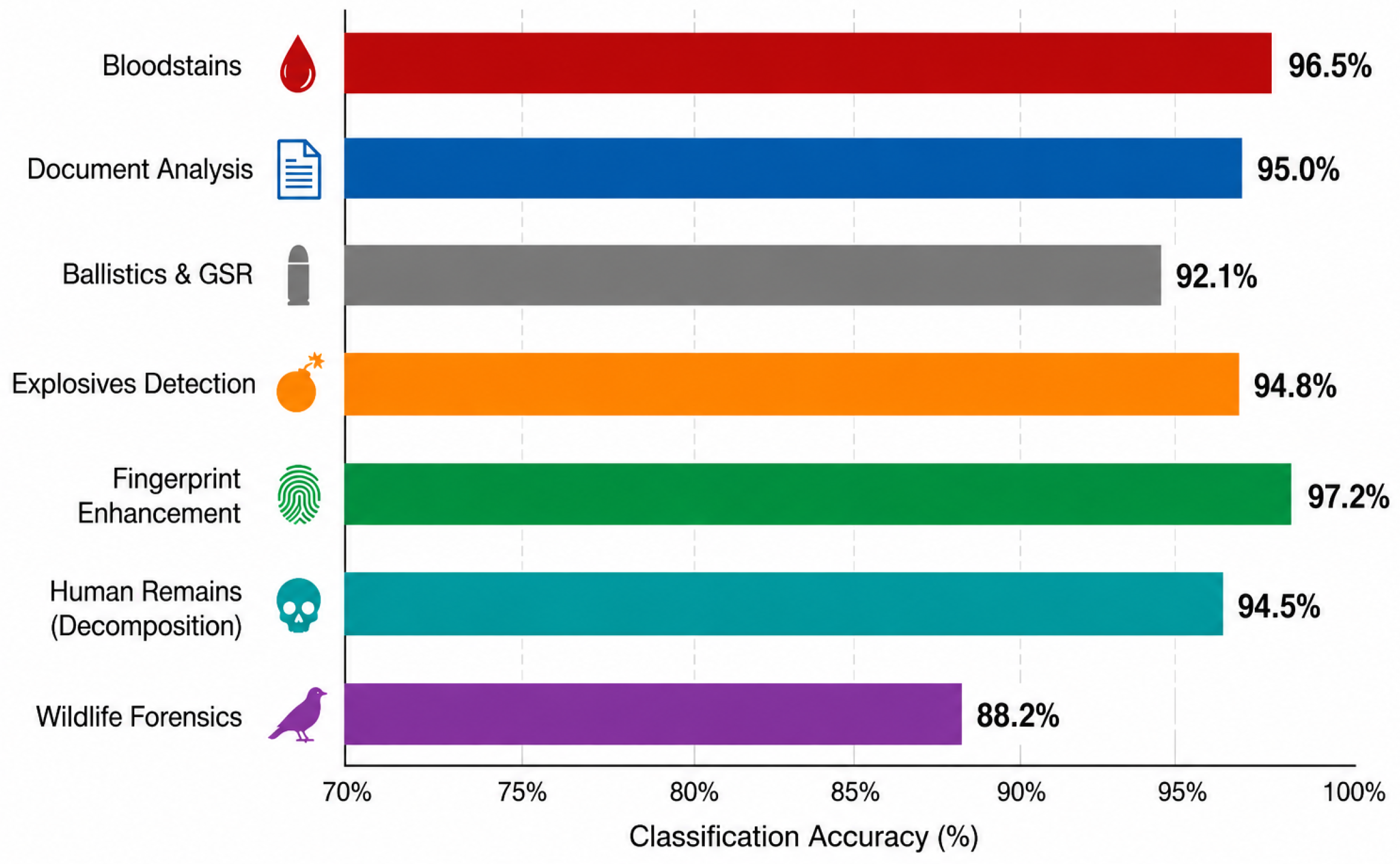
Figure 6 shows an overview of the reported accuracy of HSI in the different forensic application domains. The most developed areas in forensic HSI are blood stain examination, document examination, and biological fluid examination where several studies have been reported that achieve classification accuracies greater than 95%. These high accuracies are primarily due to the presence of distinguishable spectral signatures, controlled acquisition environment in laboratory settings, and well-defined preprocessing and chemometric methodologies.
Alternatively, newly proposed applications such as gunshot residue analysis, PMI estimation, bruise age estimation, and trace evidence examination are less mature forensic HSI areas. Their accuracies are somewhat lower because their spectral signatures are greatly dependent on the substrate composition, environmental effects, aging processes, and the lack of annotated forensic data sets. Most importantly, the prediction of PMI depends on many biological and environmental factors.
The comparative analysis of the machine-learning approaches also demonstrates that the traditional chemometric models like PCA, Partial Least Squares Discriminant Analysis (PLS-DA), Linear Discriminant Analysis (LDA), and SVMs are popular due to their strong performance in smaller forensic datasets. New deep-learning models like CNNs provide better results by using the spectral and spatial information together but need more annotated datasets and computational capabilities as well as standardization of the imaging protocol to work properly. The novel architecture of ViTs, GNNs, spectral–spatial attention networks, and self-supervised learning show great potential for model generalization but have not been widely used in forensic HSI yet.
There are some issues that affect all forensic areas equally and include unavailability of benchmark data, variability because of different imaging devices and lighting, lack of standardization of the acquisition protocol, lack of external validation, and low interpretability of the machine-learning models for forensic purposes. To address these issues and facilitate the use of HSI in forensics, standardized spectral libraries, multi-center forensic datasets, and explainable artificial intelligence should be implemented.

13. Challenges, Limitations, and Standardization

13.1. Technical Challenges

The processing of hyperspectral images leads to the generation of spectral data in high dimensions, which necessitates special methodologies to analyze them properly. One such method that has gained a lot of recognition in the analysis of hyperspectral images is anomaly detection as it helps in the detection of spectrally unique objects regardless of their known features. One of the most basic methods used for anomaly detection is Reed-Xiaoli (RX) detector proposed by Reed and Yu [102].
In most forensic HSI applications, substrate interference is considered a difficult problem. Substrate materials can have spectral signatures similar to those of the target evidence, leading to low accuracy rates and making automatic detection a tough job. In forensic applications of HSI, substrate interference is important due to the necessity of finding evidence in a variety of different substrates. The so-called “one-class classification” problem, in which there is sufficient characterization of only the target class but not the background material, has gained increased interest in the study of forensic HSI. Recent works using anomaly detection and one-class classifiers such as the one-class SVM (OC-SVM) have shown very promising results in blood stain detection in hyperspectral scenes [20,103]. Anomaly detection has become a potential approach for the one-class classification task in forensic HSI due to challenges associated with obtaining representative background samples. Anomaly detection algorithms aim to capture the properties of the target object while considering all other objects as anomalous. Recently, it was shown that Isolation Forest-based approaches can be successfully used in hyperspectral anomaly detection tasks. In particular, Li et al. [104] introduced a Kernel Isolation Forest (KIF) method that exploits the non-linear structure of hyperspectral data and, thus, enhances anomaly detection. Similarly, Song et al. [105] proposed an enhanced spectral–spatial Isolation Forest algorithm which takes into account both spectral and spatial information for anomaly detection. Along with other one-class learning techniques like One-Class Support Vector Machines (OC-SVMs), Autoencoders, and Variational Autoencoders (VAEs), they allow detecting anomalous spectral signatures without need in background samples. This makes them a promising choice for forensic applications, e.g., for blood stain detection, latent fingerprints enhancement, gunshot residue detection, and trace evidence analysis in heterogeneous environments. Thus, while anomaly detection approaches have shown great promise in analyzing hyperspectral data, their use within the context of forensic HSI is still limited and needs to be explored in more detail through the analysis of extensive forensic data sets collected from crime scenes. Environmental variables may impact the spectral features of materials and, thus, may influence the reliability of HSI-based material identification/classification model performance. Such variables as moisture content, changes in temperature, oxidation, and lighting may impact the spectral behavior of materials and negatively impact the generalization ability of the predictive models. In addition, differences between different HSI systems, as well as between different calibrations and acquisition parameters, may create variability in spectra and complicate the task of transferring the models between different systems and environments [106].

13.2. Validation and Standardization

Perhaps one of the key hurdles hindering the implementation of forensic HSI into everyday use is the lack of standardized and validated analytical protocols. Unlike DNA profiling methods, which are supported by international standards of validation procedures and proficiency testing, HSI-based forensic techniques have yet to undergo proper validation processes in terms of forensic quality assurance guidelines. These include developmental validation, internal validation, reproducibility testing, error rate estimation, and blind inter-laboratory proficiency testing [107].
The second important barrier is the lack of comprehensive and standardized spectral libraries. The training of machine-learning and classification algorithms relies on good quality spectral data. Variation in imaging devices, acquisition parameters, substrates, and conditions might affect spectral fingerprints to a considerable degree and make it impossible to construct transferable models. Thus, the creation of standardized spectral libraries with various types of evidences, substrates, and acquisition parameters is considered to be a key point towards the improvement of HSI-based analysis [44,108].

13.3. Legal and Courtroom Admissibility

For HSI-derived evidence to be admissible in legal proceedings, it must satisfy established scientific reliability standards. In the United States, the Daubert standard requires that scientific evidence be based on testable methodologies with known error rates, peer-reviewed validation, and general acceptance within the relevant scientific community. Similar admissibility standards apply in the United Kingdom and other Commonwealth jurisdictions. Even though there has been an increasing body of peer-reviewed literature available on forensic HSI in recent times, validation studies, inter-laboratory testing and statistical analysis of the analytical capabilities have been scarce. National Research Council has stressed that any forensic technique which is used for legal purposes should be validated scientifically and should adhere to certain standards of reliability and uncertainty [107]. Therefore, additional research would be needed to define the general standards of performance for HSI techniques in forensics. International validation standards and reporting guidelines should be developed for HSI techniques to make their use in forensics more common.

13.4. Evaluation Metrics for Forensic HSI

Even though classification accuracy is the most common performance metric mentioned in the literature, this metric may not sufficiently reflect the reliability of the forensic HSI systems. False positives could mean that some of the evidence will be falsely linked to a certain individual or object, and false negatives would mean that some critical pieces of evidence will be missed. Thus, using only accuracy as an indicator can give misleading results when dealing with imbalanced class distributions. Some other evaluation metrics such as precision, recall (sensitivity), specificity, F1 score, receiver operating characteristic (ROC) curve, area under the ROC curve (AUC), and false positive rate (FPR) help to evaluate the performance of classifiers. While precision helps to estimate what percentage of true positive instances was detected from all of the predicted positives, recall allows assessing how well the classifier detects the relevant forensic evidence. F1 score is helpful when dealing with imbalanced classes since this metric combines precision and recall. Also, both specificity and FPR are crucial for the forensic application of models because they estimate the ability of the classifier to correctly identify non-target pieces of evidence. From the review of the literature, it can be seen that many of the early HSI research studies in forensics evaluated their models using classification accuracy alone; however, it has become common practice in recent years for studies to evaluate models using other measures like precision, recall, F1-measure, confusion matrices, and ROC-AUC analysis as well.

14. Future Directions and Research Priorities

14.1. Portable and Field-Deployable Systems

Field-ready HSI systems that are strong and economical are among the most significant technological developments for the successful implementation of HSI technology in real-life situations. Classical HSI devices usually tend to be costly and bulky. Moreover, they require specific acquisition conditions that cannot always be provided in practice. The technological developments such as sensor miniaturization and compact camera design including Fabry–Pérot cameras and mosaic cameras have allowed designing portable HSI cameras. These technical developments might help to perform data acquisition quicker and make HSI implementation more effective [109].
In order to be useful in practice beyond the laboratory setting, HSI systems should be portable, power efficient, fast at data acquisition, and resilient to changing conditions. Miniaturization of sensors, speed of imaging, and integration of the components involved have enabled the creation of small-sized HSI systems that can be used in real-time and field conditions. The innovations can lead to broader application of HSI in scenarios where quick and non-invasive analysis is necessary and laboratory-based imaging systems are not feasible.

14.2. Integration with Artificial Intelligence

The integration of HSI with advanced artificial intelligence (AI) methodologies represents one of the most transformative future research directions in forensic science. Modern deep-learning architectures, including convolutional neural networks (CNNs) and transformer-based models, have demonstrated exceptional performance in hyperspectral image classification tasks within remote sensing and biomedical imaging domains [110]. However, their application to forensic scenarios remains comparatively underdeveloped.
Several issues are currently restraining the implementation of deep-learning approaches for the analysis of hyperspectral images. One such issue is the relatively low amount of labeled data compared to the amounts of data used in training deep-learning models. Another issue is the high dimensionality and spectra of the hyperspectral data that makes overfitting and non-generalization likely when the data used for training are limited. The third issue is variation in data acquisition conditions that have a negative impact on the classification performance and necessitate using advanced techniques like transfer learning and data augmentation [111]. XAI methods become more and more popular in increasing the transparency and explainability of ML algorithms. Attention visualization, saliency mapping, and feature attribution are some methods that might assist in detecting spectral and spatial features that play role in model predictions and hence provide an understanding of model behavior [112,113]. In the context of forensics, the capability of explaining and justifying conclusions is especially significant since scientific evidence employed during legal procedures needs to be transparent, reliable, and scientifically justified [107]. The implementation of XAI approaches in forensic HSI tools can be beneficial in terms of both methodological transparency and trust in automated forensic analysis results.

14.3. Multi-Modal Analytical Platforms

However, no one approach to data analysis will be able to fully describe a complex material or piece of evidence. Thus, it is likely that future analysis procedures will be increasingly combining the use of hyperspectral imagery with other sensing technologies to increase the reliability of the identification process. The ability to integrate spatial spectral data with other types of data will allow one to achieve better performance when classifying samples and conducting their comprehensive characterization. Integration of complementary sources of data for hyperspectral imagery and pattern recognition is an ongoing research direction [114].
Correlative multi-modal imaging has the potential to significantly improve the reliability and specificity of forensic evidence interpretation. For example, HSI may rapidly localize suspicious regions across a large evidence surface, while Raman spectroscopy or XRF may subsequently provide molecular or elemental confirmation. The development of automated integrated platforms capable of performing sequential multi-modal analysis without manual evidence transfer represents an important future research direction with significant implications for laboratory efficiency, evidence preservation, and analytical reproducibility.

14.4. Database Development and International Harmonization

Development of curated, high-quality, and publicly available spectral libraries for different types of forensic evidence could greatly enhance the practical application and standardization of forensic HSI technology. The development of spectral databases should include a broad range of forensic evidence such as blood, bodily fluids, inks, paints, fibers, explosives, drugs, and biological materials obtained under different environmental conditions on various instruments. Likewise, it is important to coordinate and standardize methods of forensic analysis on the international level, in order to ensure consistency, replicability, and compatibility of the methods used in different labs. As previous experience of ENFSI shows, cooperation is beneficial for setting the standards and validation procedure for forensic science [115]. Regarding the use of HSI for forensics, cooperation between forensic labs, universities, and instrument manufacturers would help develop unified spectral libraries, calibration, data processing, and reporting procedure.

15. Conclusions

In this review, a complete study of HSI for forensic purposes has been carried out via the critical evaluation of over 50 publications related to forensic science from nine major fields. Based on the results obtained, it can be stated that HSI has become an advanced non-destructive technique able to provide spectral and spatial data during a forensic investigation. For instance, mature forensic science applications such as document examination, blood stain analysis, and biological fluids identification achieved up to 99% of classification accuracy. Emerging techniques like fingerprint enhancement, GSR detection, trace evidence analysis, and PMI estimation continue to develop significantly. Moreover, the role of machine-learning and deep-learning methods in further increasing the precision of HSI analysis was emphasized. SVM, PLS-DA, and CNN algorithms became popular in this respect. Nevertheless, a number of important issues still need to be addressed. Namely, the need for standards and forensic spectral database development, high price of necessary hardware, and legal questions about the admission of HSI data in courts should be mentioned. Thus, future works should concentrate on the development of portable HSI, explainable AI, multi-modality analytical approaches, and spectral library standardization.


Author Contributions

Conceptualization, J.S. and V.M.M.; methodology, J.S. and V.M.M.; literature survey, J.S.; investigation, J.S.; formal analysis, J.S.; data curation, J.S.; visualization, J.S.; writing—original draft preparation, J.S.; writing—review and editing, V.M.M. and J.S.; supervision, V.M.M.; project administration, V.M.M. All authors have read and agreed to the published version of the manuscript.

Funding

This research was funded by SRM University-AP, Amaravati 522240, Andhra Pradesh, India.

Institutional Review Board Statement

Not applicable.

Informed Consent Statement

Not applicable.

Data Availability Statement

Data will be made available on request.

Conflicts of Interest

The authors declare no conflicts of interest. The funders had no role in the design of the study; in the collection, analyses, or interpretation of data; in the writing of the manuscript; or in the decision to publish the results.

References

  1. Pallocci, M.; Treglia, M.; Passalacqua, P.; De Luca, L.; Zanovello, C.; Mazzuca, D.; Guarna, F.; Gratteri, S.; Marsella, L.T. Forensic applications of hyperspectral imaging technique: A narrative review. Med. Sci. Law 2022, 62, 277–287. [Google Scholar] [CrossRef] [Scilit] [PubMed]
  2. Virkler, K.; Lednev, I.K. Analysis of Body Fluids for Forensic Purposes: From Laboratory Testing to Non-Destructive Rapid Confirmatory Identification at a Crime Scene. Forensic Sci. Int. 2009, 188, 1–17. [Google Scholar] [CrossRef] [Scilit] [PubMed]
  3. Goetz, A.F.H. Three decades of hyperspectral remote sensing of the Earth: A personal view. Remote Sens. Environ. 2009, 113, S5–S16. [Google Scholar] [CrossRef] [Scilit]
  4. Edelman, G.; Gaston, E.; van Leeuwen, T.; Cullen, P.; Aalders, M. Hyperspectral imaging for non-contact analysis of forensic traces. Forensic Sci. Int. 2012, 223, 28–39. [Google Scholar] [CrossRef] [Scilit] [PubMed]
  5. Lu, G.; Fei, B. Medical hyperspectral imaging: A review. J. Biomed. Opt. 2014, 19, 010901. [Google Scholar] [CrossRef] [Scilit] [PubMed]
  6. Pradeep, A.S.; Babu, J.; Sandana, J.S.; Deivalakshmi, S. Innovations in forensic science: Comprehensive review of hyperspectral imaging for bodily fluid analysis. Forensic Sci. Int. 2024, 364, 112227. [Google Scholar] [CrossRef] [Scilit] [PubMed]
  7. Hong, D.; Li, C.; Yokoya, N.; Zhang, B.; Jia, X.; Plaza, A.; Gamba, P.; Benediktsson, J.A.; Chanussot, J. Hyperspectral Imaging. Nat. Rev. Methods Prim. 2026, 6, 19. [Google Scholar] [CrossRef] [Scilit]
  8. Cheng, M.F.; Mukundan, A.; Karmakar, R.; Valappil, M.A.; Jouhar, J.; Wang, H.C. Modern Trends and Recent Applications of Hyperspectral Imaging: A Review. Technologies 2025, 13, 170. [Google Scholar] [CrossRef] [Scilit]
  9. Zhang, G.; Abdulla, W. Transformers Meet Hyperspectral Imaging: A Comprehensive Study of Models, Challenges and Open Problems. Eng. Appl. Artif. Intell. 2026, 169, 113947. [Google Scholar] [CrossRef] [Scilit]
  10. Zhao, X.; Ma, J.; Wang, L.; Zhang, Z.; Ding, Y.; Xiao, X. A Review of Hyperspectral Image Classification Based on Graph Neural Networks. Artif. Intell. Rev. 2025, 58, 172. [Google Scholar] [CrossRef] [Scilit]
  11. Edelman, G.; Manti, V.; van Ruth, S.M.; van Leeuwen, T.; Aalders, M. Identification and age estimation of blood stains on colored backgrounds by near infrared spectroscopy. Forensic Sci. Int. 2012, 220, 239–244. [Google Scholar] [CrossRef] [Scilit] [PubMed]
  12. Mariotti, K.d.C.; Ortiz, R.S.; Ferrão, M.F. Hyperspectral imaging in forensic science: An overview of major application areas. Sci. Justice 2023, 63, 387–395. [Google Scholar] [CrossRef] [Scilit] [PubMed]
  13. Yagi, Y.; Murase, T.; Kagawa, S.; Tsuruya, S.; Nakahara, A.; Yamamoto, T.; Umehara, T.; Ikematsu, K. Immunohistochemical detection of CD14 and combined assessment with CD32B and CD68 for wound age estimation. Forensic Sci. Int. 2016, 262, 113–120. [Google Scholar] [CrossRef] [Scilit] [PubMed]
  14. Álvarez, Á.; Yáñez, J. Screening of Gunshot Residue in Skin Using Attenuated Total Reflection Fourier Transform Infrared (ATR FT-IR) Hyperspectral Microscopy. Appl. Spectrosc. 2020, 74, 400–407. [Google Scholar] [CrossRef] [Scilit] [PubMed]
  15. Khan, Z.; Shafait, F.; Mian, A. Automatic Ink Mismatch Detection for Forensic Document Analysis. Pattern Recognit. 2015, 48, 3615–3626. [Google Scholar] [CrossRef] [Scilit]
  16. de Carvalho, M.A.; Talhavini, M.; Pimentel, M.F.; Amigo, J.M.; Pasquini, C.; Alves Junior, S.; Weber, I.T. NIR hyperspectral images for identification of gunshot residue from tagged ammunition. Anal. Methods 2018, 10, 4711–4717. [Google Scholar] [CrossRef] [Scilit]
  17. Konrad, H.; Jürgens, L.; Hartung, B.; Poetsch, M. More than just blood, saliva, or sperm—Setup of a workflow for body fluid identification by DNA methylation analysis. Int. J. Leg. Med. 2023, 137, 1683–1692. [Google Scholar] [CrossRef] [Scilit] [PubMed]
  18. Malegori, C.; Alladio, E.; Vincenti, M.; Garofano, P.; Berti, A.; Oliveri, P. Identification of Invisible Biological Traces in Forensic Evidences by Hyperspectral NIR Imaging Combined with Chemometrics. Talanta 2020, 215, 120911. [Google Scholar] [CrossRef] [Scilit] [PubMed]
  19. Schalike, R.; Illes, M. A review of spectroscopic methods applied to bloodstain pattern analysis. J. Multidiscip. Res. 2020, 2, 90–104. [Google Scholar]
  20. Acampora, G.; Santoro, M.; Vitiello, A. A Comparative Study of One-Class Classification Methods for Bloodstain Detection in Hyperspectral Forensic Imaging. Neural Comput. Appl. 2026, 38, 52. [Google Scholar] [CrossRef] [Scilit]
  21. Workman, J., Jr.; Weyer, L. Practical Guide and Spectral Atlas for Interpretive Near-Infrared Spectroscopy, 2nd ed.; CRC Press: Boca Raton, FL, USA, 2012. [Google Scholar] [CrossRef] [Scilit]
  22. Chang, C.I. Hyperspectral Data Processing: Algorithm Design and Analysis; Wiley: Hoboken, NJ, USA, 2013. [Google Scholar] [CrossRef] [Scilit]
  23. Hege, E.K.; O’Connell, D.; Johnson, W.; Basty, S.; Dereniak, E.L. Hyperspectral imaging for astronomy and space surveillance. In Proceedings of the SPIE; SPIE: Bellingham, WA, USA, 2004; Volume 5159, pp. 380–391. [Google Scholar] [CrossRef] [Scilit]
  24. Prasad, S.; Chanussot, J. (Eds.) Hyperspectral Image Analysis: Advances in Machine Learning and Signal Processing; Springer: Cham, Switzerland, 2020. [Google Scholar] [CrossRef] [Scilit]
  25. Brekke, C.; Solberg, A.H.S. Oil Spill Detection by Satellite Remote Sensing. Remote Sens. Environ. 2005, 95, 1–13. [Google Scholar] [CrossRef] [Scilit]
  26. Eismann, M.T. Hyperspectral Remote Sensing; SPIE Press: Bellingham, WA, USA, 2012. [Google Scholar] [CrossRef] [Scilit]
  27. Specim, Spectral Imaging Ltd. Specim FX Series Hyperspectral Cameras; Specim, Spectral Imaging Ltd.: Oulu, Finland, 2026. [Google Scholar]
  28. Nakamura, A.; Okuda, H.; Nagaoka, T.; Akiba, N.; Kurosawa, K.; Kuroki, K.; Ichikawa, F.; Torao, A.; Sota, T. Portable Hyperspectral Imager with Continuous Wave Green Laser for Identification and Detection of Untreated Latent Fingerprints on Walls. Forensic Sci. Int. 2015, 254, 100–105. [Google Scholar] [CrossRef] [Scilit] [PubMed]
  29. Rinnan, Å.; van den Berg, F.; Engelsen, S.B. Review of the most common pre-processing techniques for near-infrared spectra. TrAC Trends Anal. Chem. 2009, 28, 1201–1222. [Google Scholar] [CrossRef] [Scilit]
  30. Barnes, R.J.; Dhanoa, M.S.; Lister, S.J. Standard normal variate transformation and de-trending of near-infrared diffuse reflectance spectra. Appl. Spectrosc. 1989, 43, 772–777. [Google Scholar] [CrossRef] [Scilit]
  31. Green, A.A.; Berman, M.; Switzer, P.; Craig, M.D. A transformation for ordering multispectral data in terms of image quality with implications for noise removal. IEEE Trans. Geosci. Remote Sens. 1988, 26, 65–74. [Google Scholar] [CrossRef] [Scilit]
  32. Hyvärinen, A.; Oja, E. Independent component analysis: Algorithms and applications. Neural Netw. 2000, 13, 411–430. [Google Scholar] [CrossRef] [Scilit] [PubMed]
  33. Camps-Valls, G.; Bruzzone, L. Kernel-based methods for hyperspectral image classification. IEEE Trans. Geosci. Remote Sens. 2005, 43, 1351–1362. [Google Scholar] [CrossRef] [Scilit]
  34. Melgani, F.; Bruzzone, L. Classification of hyperspectral remote sensing images with support vector machines. IEEE Trans. Geosci. Remote Sens. 2004, 42, 1778–1790. [Google Scholar] [CrossRef] [Scilit]
  35. Książek, K.; Romaszewski, M.; Głomb, P.; Grabowski, B.; Cholewa, M. Blood stain classification with hyperspectral imaging and deep neural networks. Sensors 2020, 20, 6666. [Google Scholar] [CrossRef] [Scilit] [PubMed]
  36. Li, S.; Song, W.; Fang, L.; Chen, Y.; Ghamisi, P.; Benediktsson, J.A. Deep learning for hyperspectral image classification: An overview. IEEE Trans. Geosci. Remote Sens. 2019, 57, 6690–6709. [Google Scholar] [CrossRef] [Scilit]
  37. Audebert, N.; Le Saux, B.; Lefèvre, S. Deep learning for classification of hyperspectral data: A comparative review. IEEE Geosci. Remote Sens. Mag. 2019, 7, 159–173. [Google Scholar] [CrossRef] [Scilit]
  38. Chen, J.; Li, W.; El-Askary, H. Spectral–Spatial Transformer With Multiscale Convolutional Attention for Hyperspectral Image Classification. IEEE J. Sel. Top. Appl. Earth Obs. Remote Sens. 2025, 18, 24223–24241. [Google Scholar] [CrossRef] [Scilit]
  39. Zhang, W.T.; Bai, Y.; Zheng, S.D.; Cui, J.; zhen Huang, Z. Tensor Transformer for Hyperspectral Image Classification. Pattern Recognit. 2025, 163, 111470. [Google Scholar] [CrossRef] [Scilit]
  40. Shi, H.; Luo, Z.; Ma, Y.; Zhu, G.; Dai, X. SSGTN: Spectral–Spatial Graph Transformer Network for Hyperspectral Image Classification. Remote Sens. 2026, 18, 199. [Google Scholar] [CrossRef] [Scilit]
  41. Wen, T.; Wang, H.; Wang, L. Dual-Branch Spatial–Spectral Transformer with Similarity Propagation for Hyperspectral Image Classification. Remote Sens. 2025, 17, 2386. [Google Scholar] [CrossRef] [Scilit]
  42. Wang, X.; Huang, S.; Chen, G.; Wang, D.; Yan, J. CSFRT: Cross-Spectral Feature-Refined Transformer for Hyperspectral Image Classification. IET Image Process. 2025, 19, e70161. [Google Scholar] [CrossRef] [Scilit]
  43. Bremmer, R.H.; Edelman, G.; Vegter, T.D.; Bijvoets, T.; Aalders, M.C. Remote spectroscopic identification of bloodstains. J. Forensic Sci. 2011, 56, 1471–1475. [Google Scholar] [CrossRef] [Scilit] [PubMed]
  44. Zulfiqar, M.; Ahmad, M.; Sohaib, A.; Mazzara, M.; Distefano, S. Hyperspectral imaging for bloodstain identification. Sensors 2021, 21, 3045. [Google Scholar] [CrossRef] [Scilit] [PubMed]
  45. Zhang, J.; Zhang, X.; Lin, C.; Liang, Y.; Ren, H.; Zhu, H.; Peng, X.; Wang, J.; Shang, Y.; Peng, C.; et al. Identification of Bloodstains by Species Using Extreme Learning Machine and Hyperspectral Imaging Technology. Appl. Spectrosc. 2024, 78, 942–950. [Google Scholar] [CrossRef] [Scilit] [PubMed]
  46. Pereira, J.F.; Silva, C.S.; Vieira, M.J.L.; Pimentel, M.F.; Braz, A.; Honorato, R.S. Evaluation and Identification of Blood Stains with Handheld NIR Spectrometer. Microchem. J. 2017, 133, 561–566. [Google Scholar] [CrossRef] [Scilit]
  47. Dalal, A.A.; Al-Qaness, M.A. Enhancing Forensic Blood Detection Using Hyperspectral Imaging and Advanced Preprocessing Techniques. Talanta 2025, 283, 127097. [Google Scholar] [CrossRef] [Scilit] [PubMed]
  48. Edelman, G.; van Leeuwen, T.G.; Aalders, M.C.G. Hyperspectral imaging for the age estimation of blood stains at the crime scene. Forensic Sci. Int. 2012, 223, 72–77. [Google Scholar] [CrossRef] [Scilit] [PubMed]
  49. Bastide, B.; Porter, G.; Renshaw, A. The Effects of Heat on the Physical and Spectral Properties of Bloodstains at Arson Scenes. Forensic Sci. Int. 2021, 325, 110891. [Google Scholar] [CrossRef] [Scilit] [PubMed]
  50. Li, B.; Beveridge, P.; O’Hare, W.T.; Islam, M. The Age Estimation of Blood Stains up to 30 Days Old Using Visible Wavelength Hyperspectral Image Analysis and Linear Discriminant Analysis. Sci. Justice 2013, 53, 270–277. [Google Scholar] [CrossRef] [Scilit] [PubMed]
  51. Lin, H.; Zhang, Y.; Wang, Q.; Li, B.; Huang, P.; Wang, Z. Estimation of the Age of Human Bloodstains under the Simulated Indoor and Outdoor Crime Scene Conditions by ATR-FTIR Spectroscopy. Sci. Rep. 2017, 7, 13254. [Google Scholar] [CrossRef] [Scilit] [PubMed]
  52. Braz, A.; López-López, M.; García-Ruiz, C. Raman spectroscopy for forensic analysis of inks in questioned documents. Forensic Sci. Int. 2013, 232, 206–212. [Google Scholar] [CrossRef] [Scilit] [PubMed]
  53. Exline, D.L.; Wallace, C.; Roux, C.; Lennard, C.; Nelson, M.P.; Treado, P.J. Forensic Applications of Chemical Imaging: Latent Fingerprint Detection Using Visible Absorption and Luminescence. J. Forensic Sci. 2003, 48, 1047–1053. [Google Scholar] [CrossRef] [Scilit]
  54. Causin, V.; Casamassima, R.; Marruncheddu, G.; Lenzoni, G.; Peluso, G.; Ripani, L. The discrimination potential of diffuse-reflectance ultraviolet-visible-near infrared spectrophotometry for the forensic analysis of paper. Forensic Sci. Int. 2012, 216, 163–167. [Google Scholar] [CrossRef] [Scilit] [PubMed]
  55. Khan, Z.; Shafait, F.; Mian, A.S. Towards automated hyperspectral document image analysis. In Proceedings of the AFHA 2013, Washington, DC, USA, 22–23 August 2013; pp. 41–45. [Google Scholar]
  56. Khan, M.J.; Yousaf, A.; Khurshid, K.; Abbas, A. Deep Learning for Automated Forgery Detection in Hyperspectral Document Images. J. Electron. Imaging 2018, 27, 053001. [Google Scholar] [CrossRef] [Scilit]
  57. Abrar, A.; Iqbal, H. Quantitative ink analysis: Estimating the number of inks in documents through hyperspectral imaging. arXiv 2023, arXiv:2306.05784. [Google Scholar]
  58. Jaiswal, G.; Sharma, A.; Yadav, S. DFD-SS: Document Forgery Detection Using Spectral–Spatial Features for Hyperspectral Images. J. Vis. Commun. Image Represent. 2022, 89, 103690. [Google Scholar] [CrossRef] [Scilit]
  59. Braz, A.; Chaves, A.B.; Pimentel, M.F. Evaluating the Potential of Near Infrared Hyperspectral Imaging Associated with Multivariate Data Analysis for Examining Crossing Ink Lines. Forensic Sci. Int. 2019, 298, 169–176. [Google Scholar] [CrossRef] [Scilit] [PubMed]
  60. Brauns, E.B.; Dyer, R.B. Fourier Transform Hyperspectral Visible Imaging and the Nondestructive Analysis of Potentially Fraudulent Documents. Appl. Spectrosc. 2006, 60, 833–840. [Google Scholar] [CrossRef] [Scilit] [PubMed]
  61. Reed, G.; Savage, K.; Edwards, D.; Nic Daeid, N. Hyperspectral Imaging of Gel Pen Inks: An Emerging Tool in Document Analysis. Sci. Justice 2014, 54, 71–80. [Google Scholar] [CrossRef] [Scilit] [PubMed]
  62. Hedjam, R.; Cheriet, M. Historical Document Image Restoration Using Multispectral Imaging System. Pattern Recognit. 2013, 46, 2297–2312. [Google Scholar] [CrossRef] [Scilit]
  63. Hollaus, F.; Diem, M.; Sablatnig, R. Binarization of MultiSpectral Document Images. In Proceedings of the Computer Analysis of Images and Patterns (CAIP 2015); Springer: Berlin/Heidelberg, Germany, 2015; Volume 9257, pp. 109–120. [Google Scholar] [CrossRef] [Scilit]
  64. López-Baldomero, A.B.; Buzzelli, M.; Moronta-Montero, F.; Martínez-Domingo, M.Á.; Valero, E.M. Ink Classification in Historical Documents Using Hyperspectral Imaging and Machine Learning Methods. Spectrochim. Acta Part A Mol. Biomol. Spectrosc. 2025, 335, 125916. [Google Scholar] [CrossRef] [Scilit] [PubMed]
  65. Singh, P.S.; Karthikeyan, S.; Upadhayay, G.M.; Sadhukhan, S.; Soni, P.K.; Malche, T. Unsupervised Deep Autoencoder-Based Reconstruction for Ink Mismatch Detection in Hyperspectral Document Images. Discov. Comput. 2025, 28, 333. [Google Scholar] [CrossRef] [Scilit]
  66. Payne, G.; Reedy, B.J.; Lennard, C. A Further Study to Investigate the Detection and Enhancement of Latent Fingerprints Using Visible Absorption and Luminescence Chemical Imaging. Forensic Sci. Int. 2005, 150, 33–51. [Google Scholar] [CrossRef] [Scilit] [PubMed]
  67. Cucci, C.; Delaney, J.K.; Picollo, M. Reflectance Hyperspectral Imaging for Investigation of Works of Art: Old Master Paintings and Illuminated Manuscripts. Acc. Chem. Res. 2016, 49, 2070–2079. [Google Scholar] [CrossRef] [Scilit] [PubMed]
  68. Sodhi, G.; Kaur, J. Powder method for detecting latent fingerprints: A review. Forensic Sci. Int. 2001, 120, 172–176. [Google Scholar] [CrossRef] [Scilit] [PubMed]
  69. Tahtouh, M.; Despland, P.; Shimmon, R.; Kalman, J.R.; Reedy, B.J. The Application of Infrared Chemical Imaging to the Detection and Enhancement of Latent Fingerprints: Method Optimization and Further Findings. J. Forensic Sci. 2007, 52, 1089–1096. [Google Scholar] [CrossRef] [Scilit] [PubMed]
  70. Nakamura, A.; Akiba, N.; Hibino, K.; Kakuda, H.; Kawada, K.; Karasawa, M.; Sakai, M.; Sota, T. Independent Component Analysis of Hyperspectral Data Measured from Overlapping Latent Fingermarks: Forensic Potential of Independent Component Images. Forensic Sci. Int. 2023, 343, 111549. [Google Scholar] [CrossRef] [Scilit] [PubMed]
  71. Carneiro, C.R.; Silva, C.S.; Weber, I.T. A Preliminary Study of Fingerprint Aging Using Near Infrared Hyperspectral Imaging (NIR-HSI). Anal. Methods 2023, 15, 6451–6459. [Google Scholar] [CrossRef] [Scilit] [PubMed]
  72. Bradshaw, R.; Wilson, G.; Denison, N.; Francese, S. Application of MALDI MS Imaging after Sequential Processing of Latent Fingermarks. Forensic Sci. Int. 2021, 319, 110643. [Google Scholar] [CrossRef] [Scilit] [PubMed]
  73. Cadd, S.; Li, B.; Beveridge, P.; O’Hare, W.T.; Campbell, A.; Islam, M. A comparison of visible wavelength reflectance hyperspectral imaging and Acid Black 1 for the detection and identification of blood stained fingerprints. Sci. Justice 2016, 56, 247–255. [Google Scholar] [CrossRef] [Scilit] [PubMed]
  74. Romolo, F.S.; Margot, P. Identification of gunshot residue: A critical review. Forensic Sci. Int. 2001, 119, 195–211. [Google Scholar] [CrossRef] [Scilit] [PubMed]
  75. Dalzell, K.A.; Ledergerber, T.; Tymitz, M.; Trejos, T.; Arroyo, L.E. Assessment of risk factors and preventive measures for inorganic and organic GSR secondary transfer in arrest scenarios. J. Forensic Sci. 2026, 71, 721–744. [Google Scholar] [CrossRef] [Scilit] [PubMed]
  76. Głomb, P.; Romaszewski, M.; Cholewa, M.; Domino, K. Application of hyperspectral imaging and machine learning methods for the detection of gunshot residue patterns. Forensic Sci. Int. 2018, 290, 227–237. [Google Scholar] [CrossRef] [Scilit] [PubMed]
  77. Khandasammy, S.R.; Rzhevskii, A.; Lednev, I.K. A Novel Two-Step Method for the Detection of Organic Gunshot Residue for Forensic Purposes: Fast Fluorescence Imaging Followed by Raman Microspectroscopic Identification. Anal. Chem. 2019, 91, 11731–11737. [Google Scholar] [CrossRef] [Scilit] [PubMed]
  78. Fambro, L.A.; Vandenbos, D.D.; Rosenberg, M.B.; Dockery, C.R. Laser-induced breakdown spectroscopy for the rapid characterization of lead-free gunshot residues. Appl. Spectrosc. 2021, 75, 670–681. [Google Scholar] [CrossRef] [Scilit] [PubMed]
  79. Edelman, G.J.; van Leeuwen, T.G.; Aalders, M.C.G. Hyperspectral Imaging of the Crime Scene for Detection and Identification of Blood Stains. In Proceedings of the SPIE; SPIE: Bellingham, WA, USA, 2013; Volume 8743, p. 87430A. [Google Scholar] [CrossRef] [Scilit]
  80. Claybourn, M.; Ansell, M. Using Raman spectroscopy to solve crime: Inks, questioned documents and fraud. Sci. Justice 2000, 40, 261–271. [Google Scholar] [CrossRef] [Scilit] [PubMed]
  81. Ferreira, K.; Oliveira, A.; Gonçalves, A.; Gomes, J. Evaluation of hyperspectral imaging visible/near infrared spectroscopy as a forensic tool for automotive paint distinction. Forensic Chem. 2017, 5, 46–52. [Google Scholar] [CrossRef] [Scilit]
  82. Almirall, J.; Trejos, T.; Lambert, K. Interpol review of glass and paint evidence 2016–2019. Forensic Sci. Int. Synerg. 2020, 2, 404–415. [Google Scholar] [CrossRef] [Scilit] [PubMed]
  83. Liang, H. Advances in Multispectral and Hyperspectral Imaging for Archaeology and Art Conservation. Appl. Phys. A 2012, 106, 309–323. [Google Scholar] [CrossRef] [Scilit]
  84. Bala, R.; Sharma, A.; Sharma, V. Animal Family Discrimination from Hair Using ATR-FTIR and Machine Learning Methods for Applications in Illegal Wildlife Trafficking. Sci. Nat. 2024, 111, 74. [Google Scholar] [CrossRef] [Scilit] [PubMed]
  85. Kranenburg, R.F.; Ou, F.; Sevo, P.; Petruzzella, M.; de Ridder, R.; van Klinken, A.; Hakkel, K.D.; van Elst, D.M.; van Veldhoven, R.; Pagliano, F.; et al. On-site illicit-drug detection with an integrated near-infrared spectral sensor: A proof of concept. Talanta 2022, 245, 123441. [Google Scholar] [CrossRef] [PubMed]
  86. Penido, C.; Pacheco, M.; Lednev, I.; Silveira, L. Raman spectroscopy in forensic analysis: Identification of cocaine and other illegal drugs of abuse. J. Raman Spectrosc. 2016, 47, 28–38. [Google Scholar] [CrossRef] [Scilit]
  87. Almeida, M.R.; Correa, D.N.; Zacca, J.J.; Logrado, L.P.L.; Poppi, R.J. Detection of explosives on the surface of banknotes by Raman hyperspectral imaging and independent component analysis. Anal. Chim. Acta 2015, 860, 15–22. [Google Scholar] [CrossRef] [Scilit] [PubMed]
  88. Demirel, G.; Öztürk, Y.E.; Yeter, O.; Aslıyüksek, H. Detection and quantification of drugs on banknotes by LC–MS/MS with a fast and non-destructive sample preparation: A comparison of three cities. Forensic Toxicol. 2025, 43, 217–225. [Google Scholar] [CrossRef] [Scilit] [PubMed]
  89. Wilczyński, S.; Koprowski, R.; Marmion, M.; Duda, P.; Błońska-Fajfrowska, B. The use of hyperspectral imaging in the VNIR (400–1000 nm) and SWIR range (1000–2500 nm) for detecting counterfeit drugs with identical API composition. Talanta 2016, 160, 1–8. [Google Scholar] [CrossRef] [Scilit] [PubMed]
  90. Haglund, W.D.; Sorg, M.H. Advances in Forensic Taphonomy; CRC Press: Boca Raton, FL, USA, 2002. [Google Scholar] [CrossRef] [Scilit]
  91. Shipman, P.; Foster, G.; Schoeninger, M. Burnt Bones and Teeth: An Experimental Study of Color, Morphology, Crystal Structure and Shrinkage. J. Archaeol. Sci. 1984, 11, 307–325. [Google Scholar] [CrossRef] [Scilit]
  92. Schmidt, V.M.; Zelger, P.; Wöss, C.; Fodor, M.; Hautz, T.; Schneeberger, S.; Huck, C.W.; Arora, R.; Brunner, A.; Zelger, B.; et al. Handheld hyperspectral imaging as a tool for the post-mortem interval estimation of human skeletal remains. Heliyon 2024, 10, e25844. [Google Scholar] [CrossRef] [Scilit] [PubMed]
  93. Ellingham, S.; Thompson, T.; Islam, M.; Taylor, G. Estimating temperature exposure of burnt bone—A methodological review. Sci. Justice 2015, 55, 181–188. [Google Scholar] [CrossRef] [Scilit] [PubMed]
  94. Pilling, M.; Vanezis, P.; Perrett, D.; Johnston, A. Visual assessment of the timing of bruising by forensic experts. J. Forensic Leg. Med. 2010, 17, 143–149. [Google Scholar] [CrossRef] [Scilit] [PubMed]
  95. Hughes, V.K.; Ellis, P.S.; Burt, T.; Langlois, N.E.I. The practical application of reflectance spectrophotometry for the demonstration of haemoglobin and its degradation in bruises. J. Clin. Pathol. 2004, 57, 355–359. [Google Scholar] [CrossRef] [Scilit] [PubMed]
  96. Grossman, S.E.; Johnston, A.; Vanezis, P.; Perrett, D.I. Can We Assess the Age of Bruises? An Attempt to Develop an Objective Technique. Med. Sci. Law 2011, 51, 170–176. [Google Scholar] [CrossRef] [Scilit] [PubMed]
  97. Cooney, G.S.; Köhler, H.; Chalopin, C.; Babian, C. Discrimination of Human and Animal Bloodstains Using Hyperspectral Imaging. Forensic Sci. Med. Pathol. 2024, 20, 490–499. [Google Scholar] [CrossRef] [Scilit] [PubMed]
  98. Ostrum, R.B. Application of Hyperspectral Imaging to Forensic Document Examination Problems. J. Am. Soc. Quest. Doc. Exam. 2006, 9, 85–93. [Google Scholar] [CrossRef] [Scilit]
  99. Bamburde, H.; Goutam, M.; Paikrao, H. Decipherment of Text on Foxed Documents by Hyperspectral Imaging on VSC 6000. J. Forensic Doc. Exam. 2023, 31, 5–12. [Google Scholar] [CrossRef] [Scilit]
  100. Huang, S.Y.; Mukundan, A.; Tsao, Y.M.; Kim, Y.; Lin, F.C.; Wang, H.C. Recent Advances in Counterfeit Art, Document, Photo, Hologram, and Currency Detection Using Hyperspectral Imaging. Sensors 2022, 22, 7308. [Google Scholar] [CrossRef] [Scilit] [PubMed]
  101. Kalacska, M.; Bell, L.S.; Sanchez-Azofeifa, G.A.; Caelli, T. Detection of Single Graves by Airborne Hyperspectral Imaging. Forensic Sci. Int. 2014, 245, 17–23. [Google Scholar] [CrossRef] [Scilit] [PubMed]
  102. Reed, I.S.; Yu, X. Adaptive Multiple-Band CFAR Detection of an Optical Pattern with Unknown Spectral Distribution. IEEE Trans. Acoust. Speech Signal Process. 1990, 38, 1760–1770. [Google Scholar] [CrossRef] [Scilit]
  103. Nascimento, J.M.P.; Bioucas-Dias, J.M. Does independent component analysis play a role in unmixing hyperspectral data? IEEE Trans. Geosci. Remote Sens. 2005, 43, 175–187. [Google Scholar] [CrossRef] [Scilit]
  104. Li, S.; Zhang, K.; Duan, P.; Kang, X. Hyperspectral Anomaly Detection With Kernel Isolation Forest. IEEE Trans. Geosci. Remote Sens. 2020, 58, 319–329. [Google Scholar] [CrossRef] [Scilit]
  105. Song, X.; Aryal, S.; Ting, K.M.; Liu, Z.; He, B. Spectral–Spatial Anomaly Detection of Hyperspectral Data Based on Improved Isolation Forest. IEEE Trans. Geosci. Remote Sens. 2022, 60, 1–16. [Google Scholar] [CrossRef] [Scilit]
  106. Gowen, A.A.; O’Donnell, C.P.; Cullen, P.J.; Downey, G.; Frias, J.M. Hyperspectral Imaging—An Emerging Process Analytical Tool for Food Quality and Safety Control. Trends Food Sci. Technol. 2007, 18, 590–598. [Google Scholar] [CrossRef] [Scilit]
  107. National Research Council. Strengthening Forensic Science in the United States: A Path Forward; National Academies Press: Washington, DC, USA, 2009. [Google Scholar] [CrossRef] [Scilit]
  108. Amigo, J.M. Practical Issues of Hyperspectral Imaging Analysis of Solid Dosage Forms. Anal. Bioanal. Chem. 2020, 412, 893–915. [Google Scholar]
  109. Stuart, M.B.; McGonigle, A.J.S.; Willmott, J.R. Hyperspectral Imaging in Environmental Monitoring: A Review of Recent Developments and Technological Advances in Compact Snapshot Sensors. Sensors 2019, 19, 3071. [Google Scholar] [CrossRef] [Scilit] [PubMed]
  110. Zhong, Z.; Li, J.; Luo, Z.; Chapman, M. Spectral-spatial residual network for hyperspectral image classification: A 3-D deep learning framework. IEEE Trans. Geosci. Remote Sens. 2018, 56, 847–858. [Google Scholar] [CrossRef] [Scilit]
  111. Wambugu, N.; Chen, Y.; Xiao, Z.; Tan, K.; Wei, M.; Liu, X.; Li, J. Hyperspectral Image Classification on Insufficient-Sample and Feature Learning Using Deep Neural Networks: A Review. Int. J. Appl. Earth Obs. Geoinf. 2021, 105, 102603. [Google Scholar] [CrossRef] [Scilit]
  112. Guidotti, R.; Monreale, A.; Ruggieri, S.; Turini, F.; Giannotti, F.; Pedreschi, D. A Survey of Methods for Explaining Black Box Models. ACM Comput. Surv. 2018, 51, 93. [Google Scholar] [CrossRef] [Scilit]
  113. Adadi, A.; Berrada, M. Peeking Inside the Black-Box: A Survey on Explainable Artificial Intelligence (XAI). IEEE Access 2018, 6, 52138–52160. [Google Scholar] [CrossRef] [Scilit]
  114. Ghamisi, P.; Plaza, J.; Chen, Y.; Li, J.; Plaza, A. Advanced Spectral Classifiers for Hyperspectral Images: A Review. IEEE Geosci. Remote Sens. Mag. 2017, 5, 8–32. [Google Scholar] [CrossRef] [Scilit]
  115. Welch, L.; Gill, P.; Phillips, C.; Ansell, R.; Morling, N.; Parson, W.; Palo, J.; Bastisch, I. European Network of Forensic Science Institutes (ENFSI): Evaluation of new commercial STR multiplexes that include the European Standard Set (ESS) of markers. Forensic Sci. Int. Genet. 2012, 6, 819–826. [Google Scholar] [CrossRef] [Scilit] [PubMed]
Figure 1. Evolution of HSI in forensic science from 2012 to 2026, illustrating key developments in forensic applications and AI-assisted analysis [4,12,15,16,17,18].
Algorithms 19 00629 g001
Figure 2. Major forensic applications of HSI.
Algorithms 19 00629 g002
Figure 3. PRISMA 2026 flow diagram illustrating the literature search and study selection process.
Algorithms 19 00629 g003
Figure 4. Hyperspectral data cube representation (x, y, λ) and corresponding reflectance spectrum.
Algorithms 19 00629 g004
Figure 5. General workflow of HSI in forensic analysis. Different colors indicate the major workflow stages.
Algorithms 19 00629 g005
Figure 6. Comparison of reported performance across major forensic HSI application domains.
Algorithms 19 00629 g006
Table 1. Comparative summary of selected HSI studies on blood stain analysis.
AuthorsRangeMethodApplicationKey FindingPerformance
Edelman et al. [11] (2012)NIRPLSBlood ID and agingAge estimation on coloured surfaces13.4% error
Książek et al. [35] (2020)VNIRCNN/SVMBlood classification3D-CNN performed best74–100% Acc.
Bremmer et al. [43] (2011)VISReflectanceBlood detectionRemote detection (2 m)>90% Acc.
Zulfiqar et al. [44] (Year)VNIRSVM/ANN/RFBlood identificationMulti-substrate detectionHigh accuracy
Zhang et al. [45] (2024)VIS–NIRELMSpecies identificationHuman vs. animal blood>96% Acc.
Pereira et al. [46] (2017)NIRKNN/LDAPortable blood IDField deployment feasible81–94% Acc.
Al-Alimi et al. [47] (2025)VIS–NIRFE + ETRBlood detectionReduced spectral redundancy and substrate effectsHigher accuracy & faster computation
Li et al. [50] (2013)VISLDAAge estimationReliable up to 30 days>90% Acc.
Table 2. Comparative summary of selected HSI studies on document examination.
AuthorsRangeMethodApplicationKey FindingPerformance
Causin et al. [54] (2012)UV–VIS–NIRDiffuse reflectancePaper discriminationPaper type differentiationHigh accuracy
Khan et al. [15] (2015)VNIRSpectral matchingInk mismatch detectionAutomatic ink mismatch detectionHigh accuracy
Abrar et al. [57] (2023)HSIClusteringInk quantificationEstimated number of inksImproved estimation
Jaiswal et al. [58] (2022)HSIDFD-SSForgery detectionSpectral–spatial featuresHigh accuracy
Braz et al. [59] (2019)NIRPLS-DACrossing ink linesWriting sequence determinationExcellent classification
Brauns et al. [60] (2006)VISFT-HSIFraudulent documentsNon-destructive analysisHigh discrimination
Reed et al. [61] (2014)VNIRHSIGel pen inksInk discriminationImproved contrast
López-Baldomero et al. [64] (2025)VIS–NIRDL + SVMHistorical ink classificationAccurate spectral ink mapping98% F1-score
Singh et al. [65] (2025)HSIAutoencoderInk mismatch detectionUnsupervised forgery detectionImproved robustness
Table 3. Comparative summary of HSI-based fingerprint detection and enhancement studies.
Authors (Year)Spectral RangeMethodApplicationKey FindingOutcome
Payne et al. [66] (2005)Visible imagingAbsorption and luminescence imagingFingerprint enhancementImproved latent ridge visibilityBetter detection after optimization
Cucci et al. [67] (2016)VIS–NIRReflectance HSIArtwork and document analysisNon-destructive material identificationDemonstrated HSI for material characterization
Sodhi and Kaur [68] (2001)VisiblePowder method reviewLatent fingerprint detectionReviewed powder development methodsSummarized conventional fingerprint techniques
Tahtouh et al. [69] (2007)Infrared imagingIR chemical imagingLatent fingerprint enhancementOptimized IR imaging protocolImproved fingerprint recovery
Nakamura et al. [70] (2023)VNIR HSIIndependent Component Analysis (ICA)Overlapping latent fingerprintsSeparated mixed fingerprints using ICAEnhanced donor discrimination
Carneiro et al. [71] (2023)NIR HSISpectral aging analysisFingerprint age estimationAge-related spectral changes identifiedPotential for age estimation
Cadd et al. [73] (2016)Visible HSIHSI vs. Acid Black 1Blood-stained fingerprintsComparable performance without chemicalsNon-destructive alternative
Table 4. Comparative summary of HSI-based GSR analysis studies.
AuthorsRangeMethodApplicationKey FindingOutcome
Álvarez et al. [14] (2020)ATR–FTIRHS microscopyGSR on skinChemical mappingNon-destructive
Dalzell et al. [75] (2026)Transfer analysisSecondary GSRTransfer assessmentForensic guidance
Głomb et al. [76] (2018)VNIRHSI + MLIGSR patternsBetter than RGBHigh detection
Khandasammy et al. [77] (2019)FluorescenceImaging + RamanOrganic GSRFast screeningHigh specificity
de Carvalho et al. [16] (2018)NIR HSIChemometricsTagged GSRReliable identificationField potential
Table 5. Comparative summary of HSI-based biological fluid identification studies.
Authors (Year)RangeMethodApplicationKey FindingOutcome
Edelman et al. [4] (2012)VIS–NIRReflectance HSIBlood/body fluid tracesRapid contact-free detectionCrime-scene screening
Pradeep et al. [6] (2024)VIS–NIRReviewBody fluid analysisHSI + AI integrationFuture forensic potential
Mariotti et al. [12] (2023)VIS–NIRReviewForensic applicationsSummarized HSI advancesEmerging forensic tool
Konrad et al. [17] (2023)VIS–NIRHSI workflowMultiple body fluidsRapid pre-screeningReduced destructive tests
Malegori et al. [18] (2020)NIRHSI + ChemometricsBlood, semen and urineDistinct spectral signaturesNon-destructive discrimination
Table 6. Comparative summary of HSI-based trace evidence analysis studies.
Authors (Year)RangeMethodApplicationKey FindingOutcome
Ferreira et al. [81] (2017)VIS/NIRPCA + HSIAutomotive paintsMost paint classes separatedHigh classification accuracy
Almeida et al. [87] (2015)Raman HSIICA analysisExplosive residuesTNT/RDX residues mappedSpatial localization
Demirel et al. [88] (2025)LC–MS/MSChemical analysisDrug-contaminated currencyDrug traces quantifiedForensic applicability
Wilczyński et al. [89] (2016)VNIR/SWIRHSI classificationCounterfeit drugsGenuine and counterfeit drugs differentiatedAuthentication achieved
Table 7. Cross-domain overview of HSI applications in forensic science.
Forensic DomainSpectral RangeML/Analysis MethodPerformanceStudiesMaturityMain Limitation
Document examinationVNIR; UV–SWIRSVM, CNN, Clustering92–98%∼12MatureSimilar inks; aged documents
Blood stain analysisVIS–NIRPLS, SVM, CNN, FE+ETR80–100%∼12AdvancedSubstrate variability aging effects
Biological fluidsVIS–NIRPLS-DA, SVM, PCA90–97%∼8AdvancedMixed fluids; textile effects
Fingerprint detectionUV–VIS–NIRSpectral unmixingHigh contrast∼8DevelopingDonor variability; aging
Trace evidenceVIS/NIR; SWIRPCA, LDA, SVMHigh∼8DevelopingDark samples; limited datasets
Drug and explosive detectionNIR; RamanICA, MCR, ML>98%∼8DevelopingTrace-level spectral overlap
Gunshot residueVNIR; MIRSVM, MCR-ALSHigh∼6EarlyLead-free GSR; limited datasets
PMI estimationVIS–NIRCNN, ML, Spectral analysisPromising∼5EarlyLimited clinical validation
Bruise age estimationVISReflectance spectroscopyObjective assessment∼5EarlySkin tone; healing variability
Disclaimer/Publisher’s Note: The statements, opinions and data contained in all publications are solely those of the individual author(s) and contributor(s) and not of MDPI and/or the editor(s). MDPI and/or the editor(s) disclaim responsibility for any injury to people or property resulting from any ideas, methods, instructions or products referred to in the content.