


Abstract
The incidence of fifth metatarsal fracture is somewhat common in sports and can be complicated in nature. Fractures of the fifth metatarsal can occur at a number of locations. Although some of these fractures respond well to conservative treatment, others have been notoriously hard to heal, with high rates of nonunions and other complications. Foot orthotic devices are commonly used as aids in the treatment of foot problems. In our case, we considered the combined effect of the surgical treatment and application of the custom-made foot orthoses. Special attention was taken with adjustments to the orthotic devices along and beneath the affected regions of the foot for adequate pain management and quick recovery to return to normal sports activities. Requirements for computer aided design/computer aided manufacturing orthotic design and manufacturing in this case were specific and considerably different from the usual procedure. (J Am Podiatr Med Assoc 99(6): 529-535, 2009)
Fractures of the fifth metatarsal are often problematic because they are prone to nonunion and refracture. One of the main reasons for these problems is its anatomy. The fifth metatarsal consists of a base, the tuberosity (styloid process), the diaphysis (the shaft), the neck, and the head. The plantar fascia and the peroneus brevis tendon insert on the tuberosity, and the peroneus tertius tendon inserts on the shaft. These areas of tendon insertions are the most potential points of fifth metatarsal fracture as a cause of overload trauma. The second reason is probably weak blood supply to the metaphyseal bone region.[1]
Stress fractures of the fifth metatarsal are a common injury in athletics that involve vigorous repetitive motion.[2,3] Stress fractures account for up to 10% of all injuries in athletes. Most patients are 18 to 25 years of age.[4] Approximately 95% of these fractures occur in the lower extremities, most commonly of the metatarsals (and tibia).[5] Stress fractures are a consequence of an increased amount of exercise over a prolonged period of time, which may result in microfractures. If the stress continues, bone reconstruction mechanisms are unable to repair the damage, so as the new microfractures occur, the macrofracture is inevitable.[6]
Many classification systems have attempted to simplify fractures of the fifth metatarsal, especially its proximal part. Fractures of the proximal fifth metatarsal can be generally classified into two groups. The two basic types are tuberosity avulsion fractures and fractures of the metatarsal diaphysis within 1.5 cm of the tuberosity. The latter type when acute is commonly referred to as the Jones fracture.[5]
One of the commonly used classifications is Stewart’s classification. Fractures are classified based on their anatomical site and articular involvement. Type I fractures correspond to the Jones fracture. Type II is an intra-articular fracture of the base of the metatarsal. Type III is an extra-articular fracture of the tuberosity. Type IV is an intra-articular comminuted fracture of the metatarsal. Type V is an injury to the apophysis in children.[7]
Another classification is Torg’s classification, which is based on radiographic findings. It has three stages. Stage I is an acute fracture with periosteal reaction, a planar-based fracture line, and no medullary sclerosis. Stage II is somewhat similar to stage I, but with medullary sclerosis and widening of the fracture line, which can be seen in delayed unions. Stage III shows complete obliteration of the medullary canal consistent with nonunions.[8] Healing times for acute and stress metatarsal fractures are highly variable.[8]
Treatment options are either 3 to 6 months of casting in a short nonweightbearing cast or intramedullary screw fixation with or without bone grafting. Fernandez Fairen et al,[9] in their study of 17 basketball players with fractures of the fifth metatarsal, concluded that even if the nonoperative treatment is able to provide a good result, it does not guarantee healing. Furthermore, players cannot train for 3 to 6 months. Therefore, percutaneous screw fixation presents itself as a good solution for basketball players.
The refracture after intramedullary screw fixation is also possible. Wright et al[10] studied six professional athletes with refractures and made four recommendations: 1) screw fixation with a large-diameter screw should be given careful consideration for patients with large body mass and for whom early return to activity is important; 2) functional bracing, shoe modification, or an orthosis should be considered for return to play; 3) if refracture occurs, exchange with a larger screw may allow return to play in the same season; and 4) alternative imaging should be considered to help document complete healing.
Foot orthotic devices are commonly used as aids in the treatment of foot problems. There are generally two broad rationales regarding the design of foot orthotic devices. The first theory has been called the total contact approach and assumes that uniform distribution of the weight on all structures of the foot will limit stresses and therefore improve and protect the foot. The second rationale is known as the biomechanical approach, and it postulates that most maladies of the foot and ankle are related to an imbalance in the supporting structures of the foot.[11,12]
In our case, we considered the combined effect of surgical treatment and application of custom-made foot orthotic devices. Special attention was taken with adjustments to the orthoses along and beneath the affected regions of the foot for adequate pain management and quick return to normal sports activities. Requirements for computer aided design/computer aided manufacturing (CAD/CAM) orthotic design and manufacturing in this case were specific and considerably different from the usual procedure.
Case Report
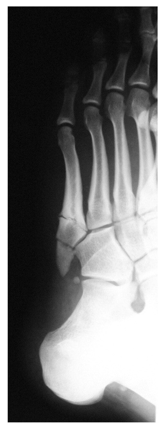
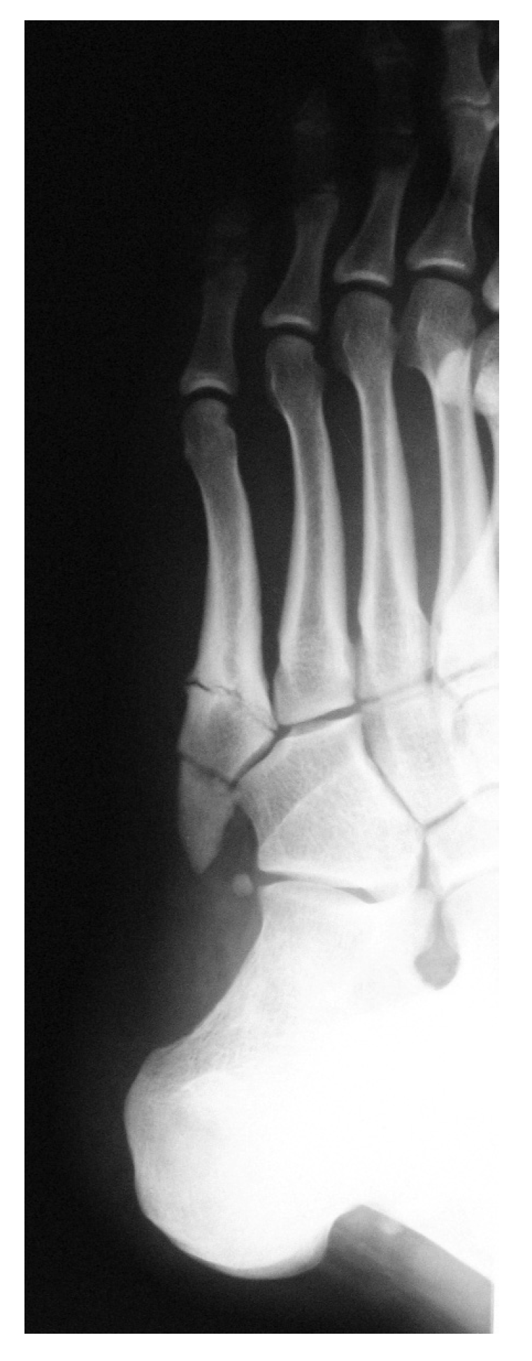
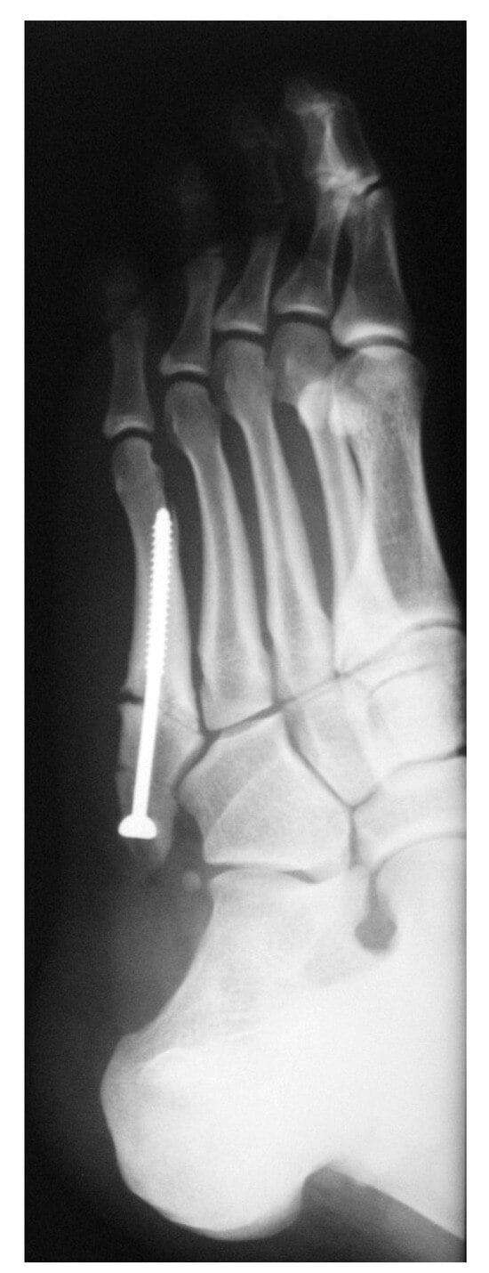
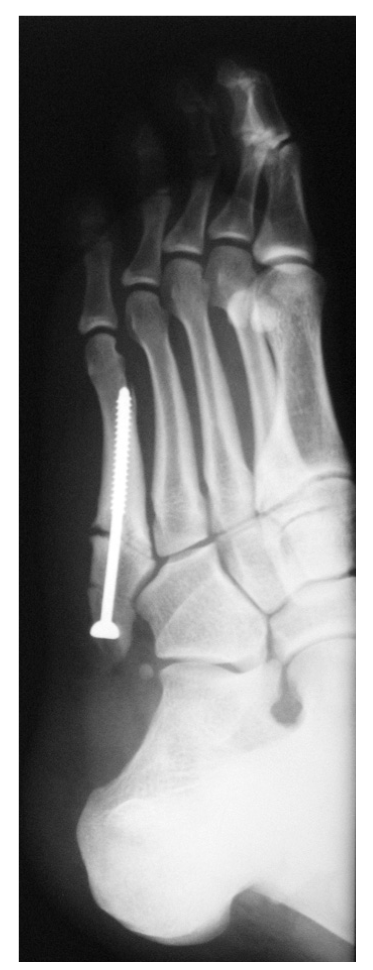
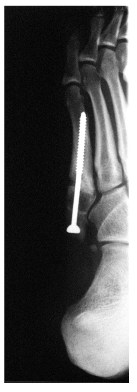
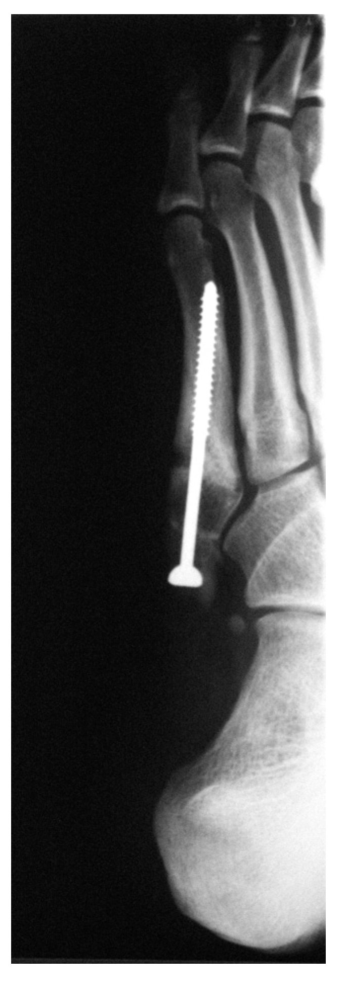
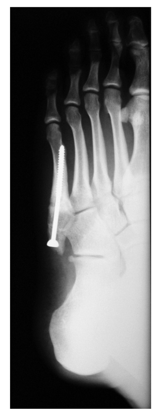
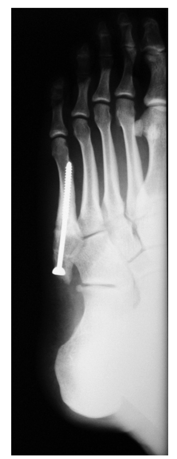
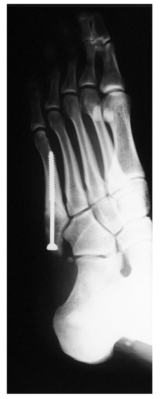
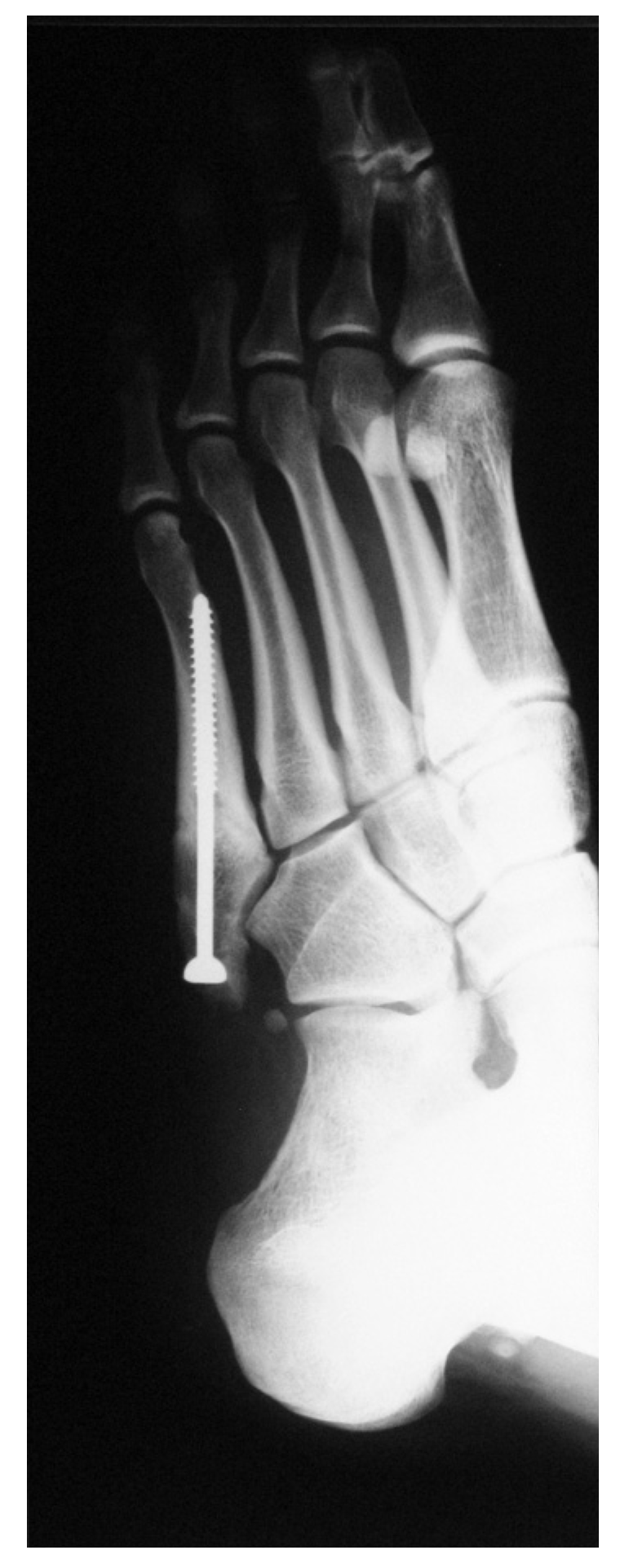
An 18-year-old basketball player (weight, 75 kg; height, 187 cm) presented to the Department of Traumatology, General Hospital Varazdin, Croatia, with persistent, sharp pain in his left foot. In the previous 6 months during his training and matches, he repeatedly had pain in his left foot localized around the base of the fifth metatarsal. After a jump, he felt sudden, sharp pain and was taken to our hospital. He was clinically and radiologically examined. Clinically, he was found to have a bow-leg deformity with oversupination of both feet. A series of standard radiographs of the left foot showed a stress fracture of the proximal third of the fifth metatarsal and an acute tuberosity avulsion fracture (Figure 1). After meeting the criteria for surgical intervention, the operative treatment of percutaneous intramedullary screw fixation with a 4.5-mm malleolar screw was undertaken (Figure 2). The physical therapy started at the first postoperative day in the ward and continued in the infirmary for the next 10 weeks. During the first 2 weeks following the operation, the patient was advised to avoid weightbearing on the injured leg and to use crutches. The patient followed-up 10 weeks after the surgery. Radiography showed good healing (Figure 3), and the patient was allowed to start with low-intensity training. Training intensity was gradually increased during the following 3 weeks, until full sports activity was approved. After 13 weeks, radiographs showed that both fractures healed completely (Figure 4). The final evaluation took place 1 year postoperatively, and there was no clinical or radiologic sign of nonunion or refracture (Figure 5).
Figure 1.
Preoperative radiograph of the injured foot.
Figure 2.
Postoperative radiograph on day 1.
Figure 3.
Radiograph 8 weeks postoperatively.
Figure 4.
Radiograph 12 weeks postoperatively.
Figure 5.
Radiograph 1 year postoperatively.
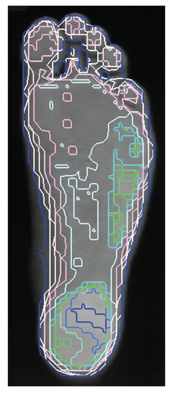
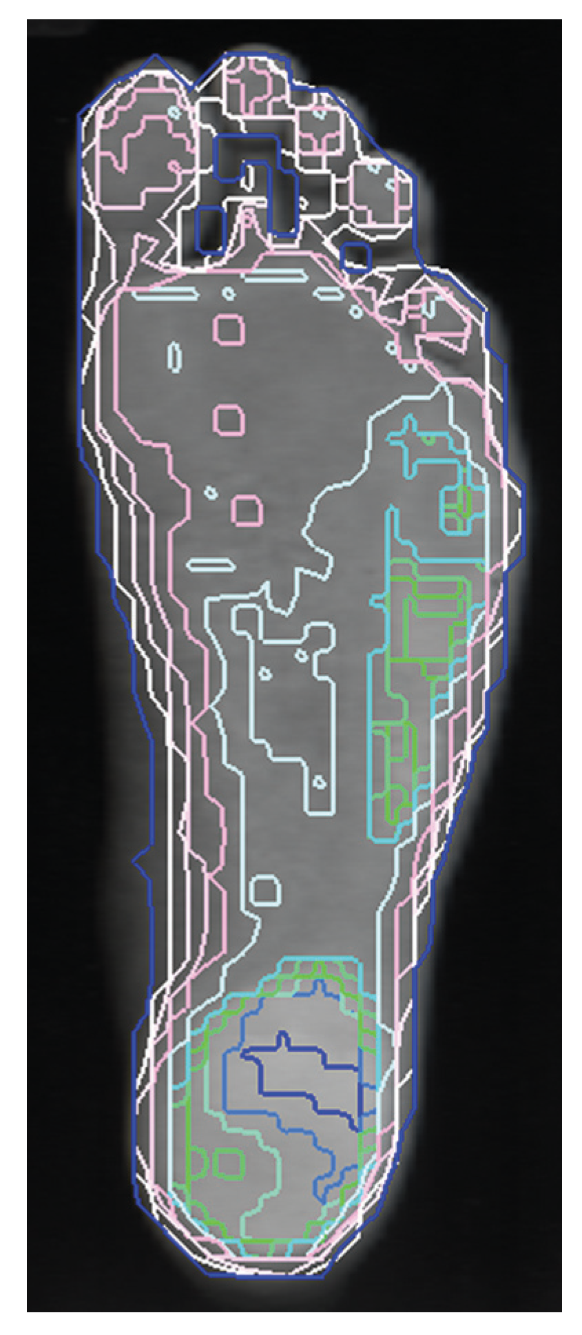
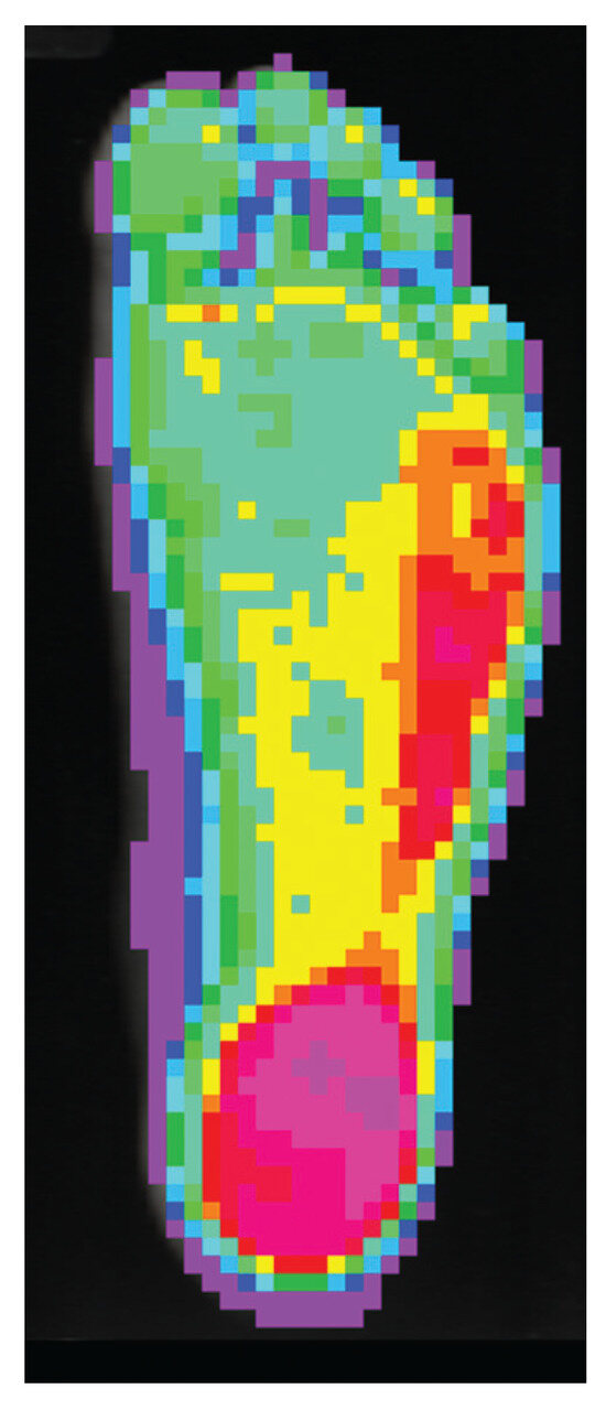
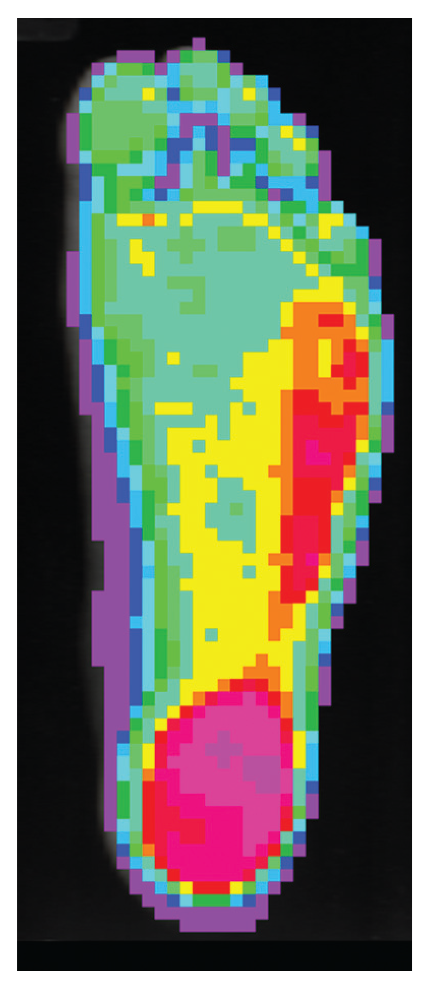
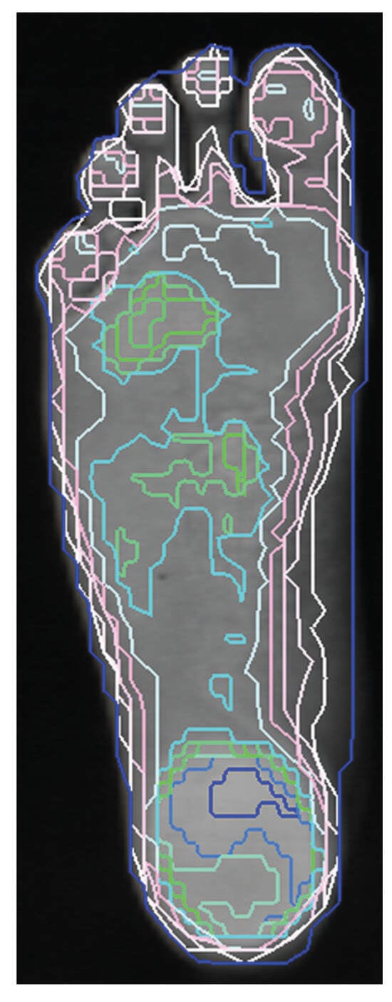
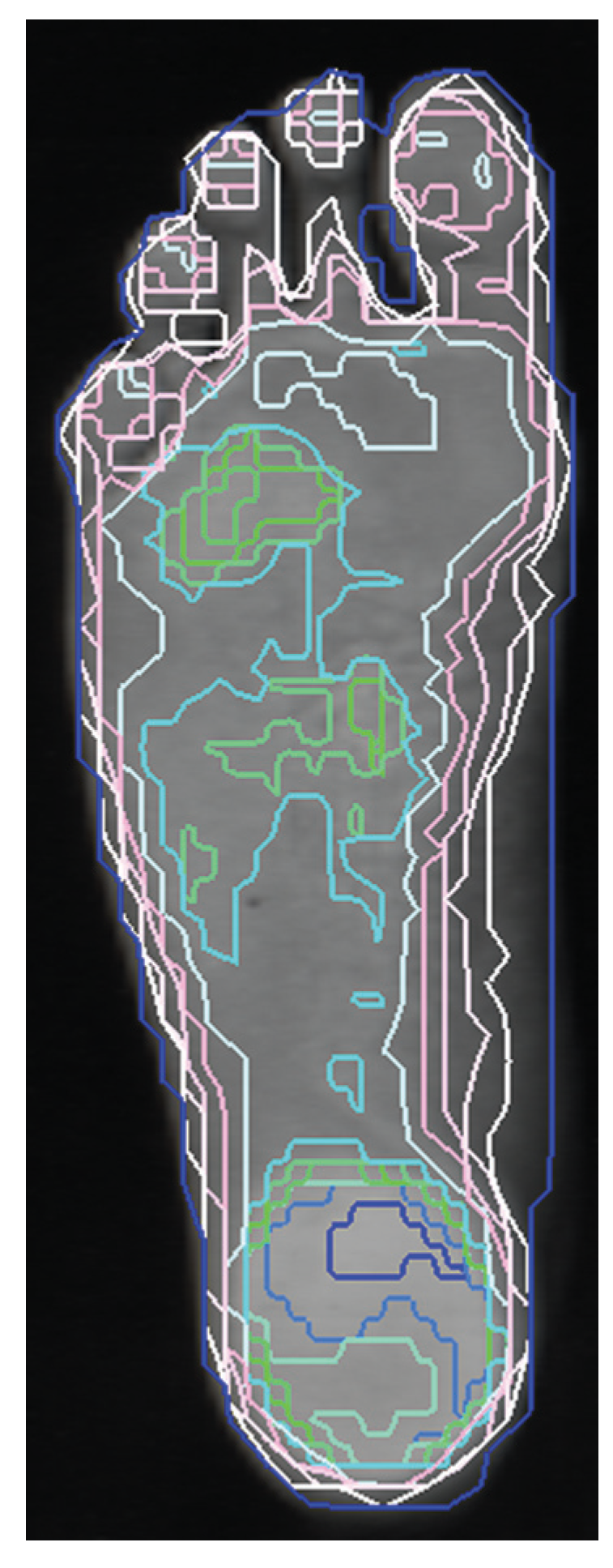
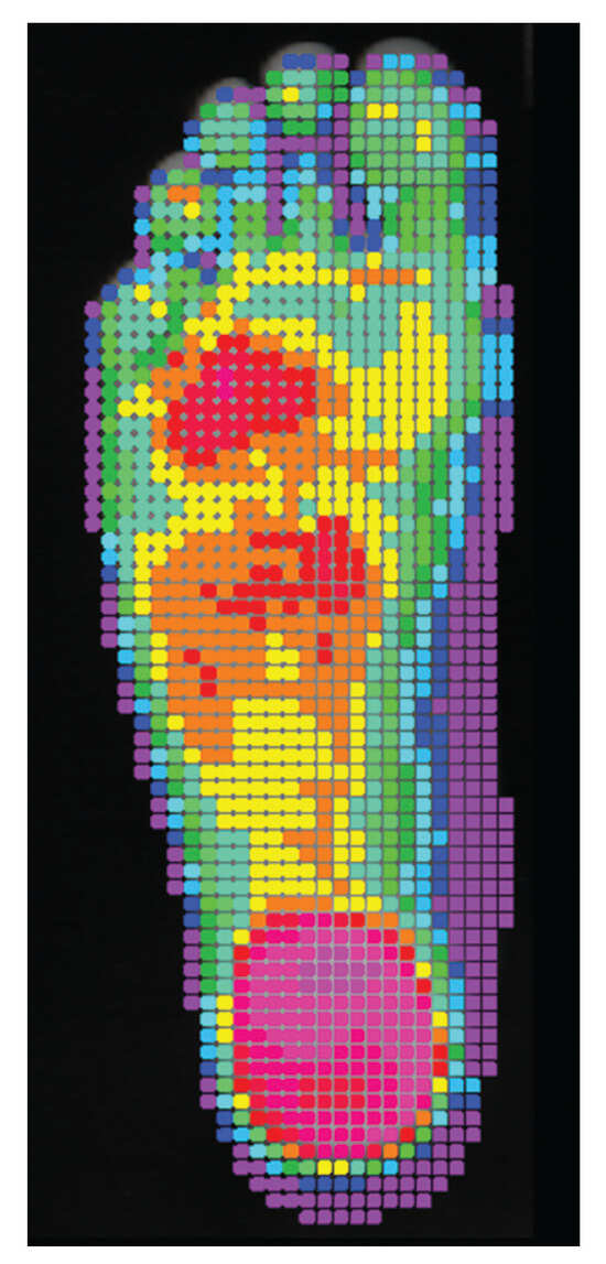
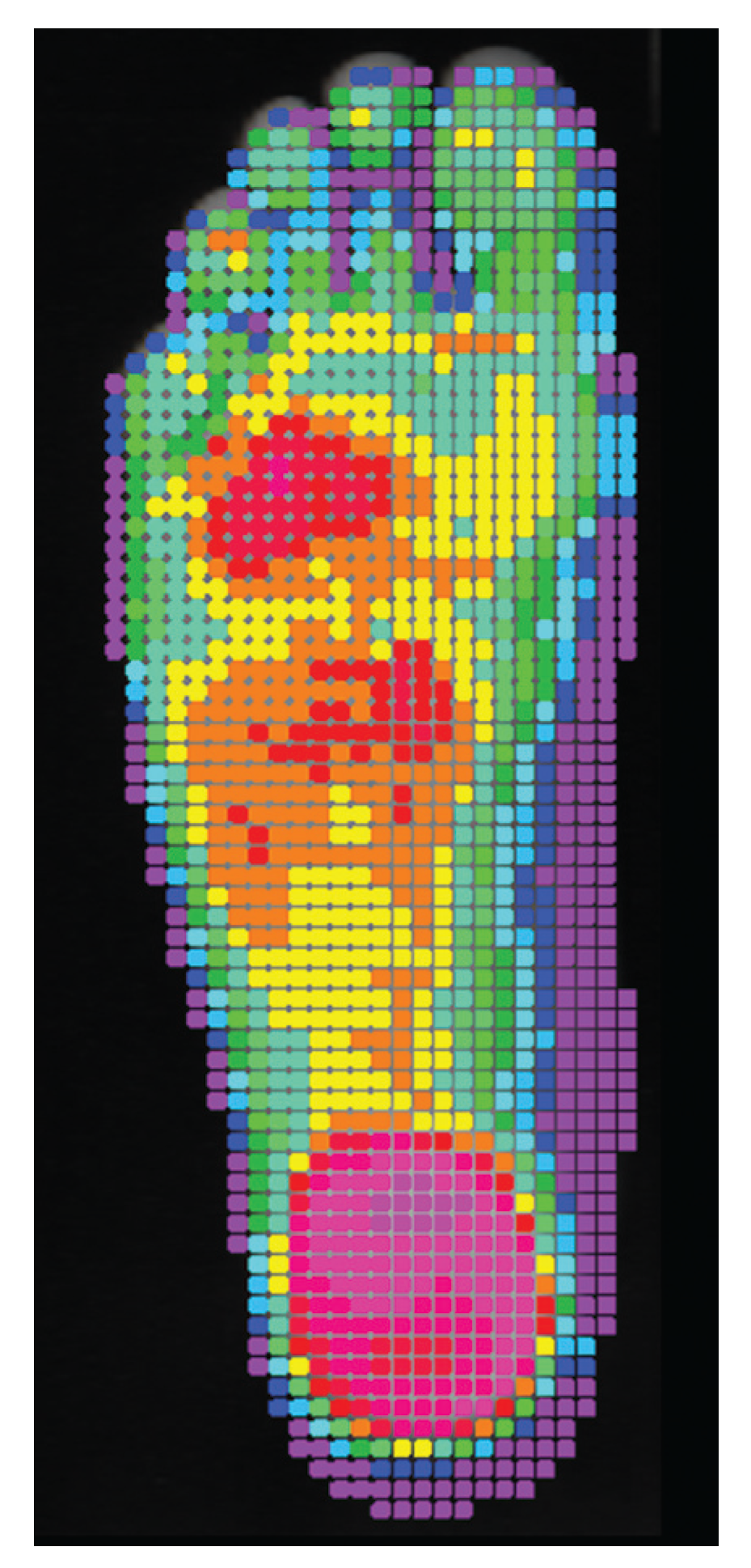
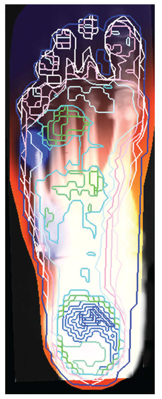
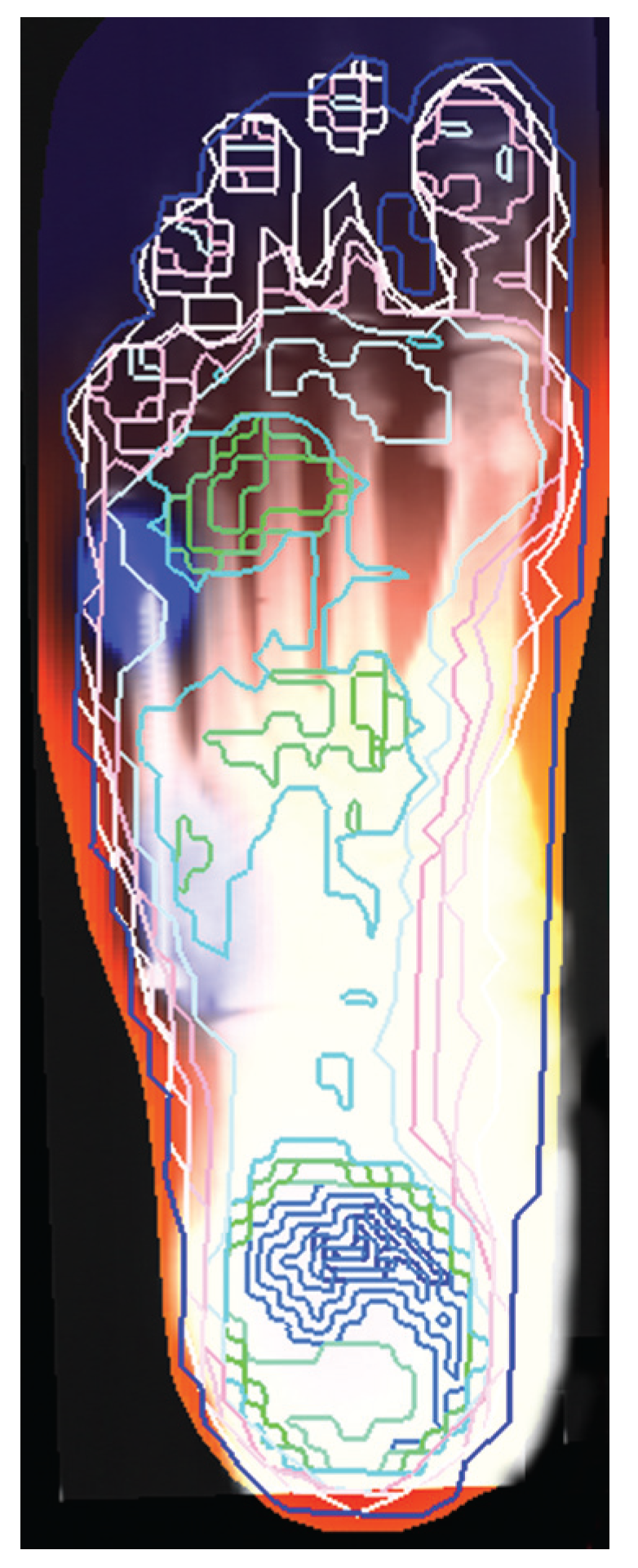
At the completion of his rehabilitation, the patient proceeded to the orthotics and prosthetics laboratory (Corkitdoo, Zagreb, Croatia) where additional tests were conducted. Custom-made functional foot orthotic devices were recommended. The static foot pressure measurement was made with the optical pedobarograph and Insole Modeller CAD/CAM system (Next Step Footcare, Houston, Texas). This system generates a relative semi-quantitative pressure map from the feet images and scan. The pressure pattern on the right foot clearly shows an increased load beneath the head of the fifth metatarsal bone, and overall increased pressures along the plantar aspect of the fifth ray. There is also a prominent pressure spot under the base of the fifth metatarsal bone (Figure 6 and Figure 7). Since the patient has bow-leg deformity, the oversupinated foot condition was actually expected to be an adjacent condition.[13] On the contrary, the pressure pattern of the surgically treated left foot showed slightly increased pressures beneath the heads of the third and the fourth metatarsal bone. Although the patient still favors the lateral side of the foot from being overloaded (from surgery), there is significant pressure relief under the fifth ray plantar projection, when compared to the right side. High pressure spots under the base and the head of the fifth metatarsal bone were absent on the left but visible on the right side (Figure 8 and Figure 9). Natural range of movements in the subtalar joints are preserved bilaterally. The contours of the feet were symmetric, with no other major aberrations. Mobility of the small joints are maintained to the full extent. The given region shows no pain, swelling, skin changes, or sounds during active or passive movements. For the orthotic support, we chose functional orthotic devices with a neutral heel post, heel cushion, and deep heel cup as a design objective.
Figure 6.
Static foot pressure measurement showing connected isobars view of the right foot.
Figure 7.
Static foot pressure measurement showing map of pressure values of the right foot.
Figure 8.
Static foot pressure measurement showing connected isobars view of the surgically treated left foot.
Figure 9.
Static foot pressure measurement showing map of pressure values of the surgically treated left foot.
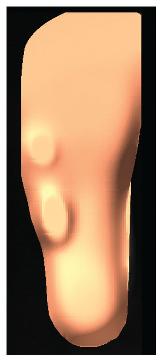
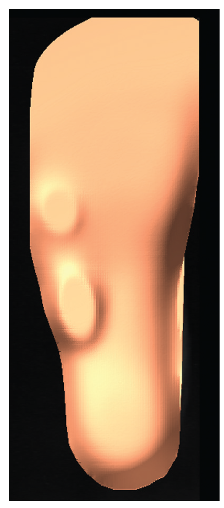
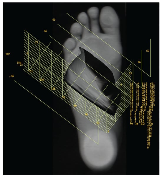
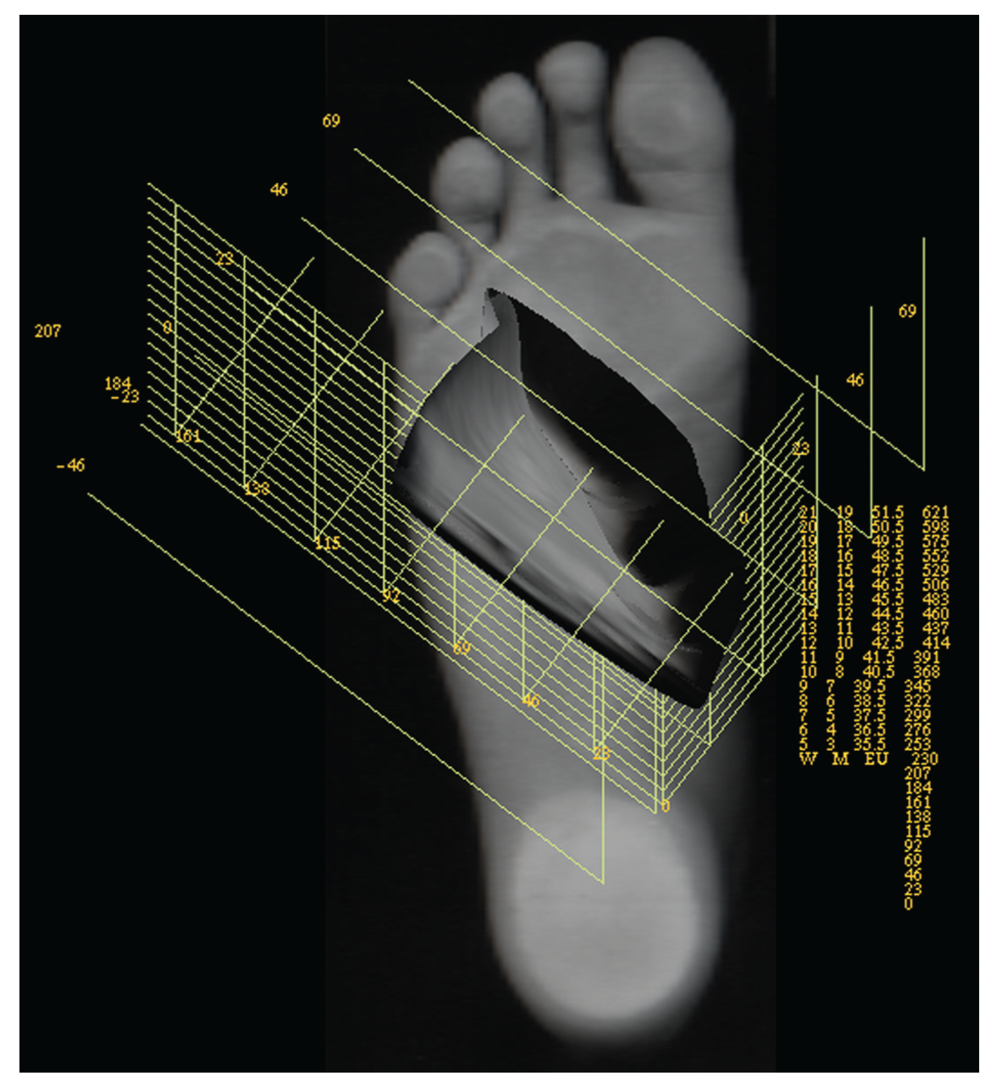
Special attention was given to the fracture location. The proprietary software allows the designer to import the digitized radiographic actual-size foot image (dorsoplantar and laterolateral aspect) into the design module. The image was superimposed over the surface acquired by the 3-D digitizer with transparency set to 50%. In this way, the alignment of the image over the surface was simplified, while all of the features on the surface and radiographic image were maintained and visible. This procedure dramatically increased accuracy of the pressure relief placement (Figure 10). This method also allowed an overview for positioning the intramedullary screw in regard to foot orthosis, so design could be adjusted in order to reduce its stress (Figure 11).
Figure 10.
Computer aided design showing reliefs on insole surface, navigated by radiograph and pressure image in background, with surface transparency turned on.
Figure 11.
Computer aided design showing adjusted insole surface with accomodations.
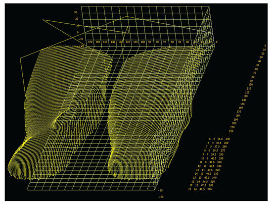
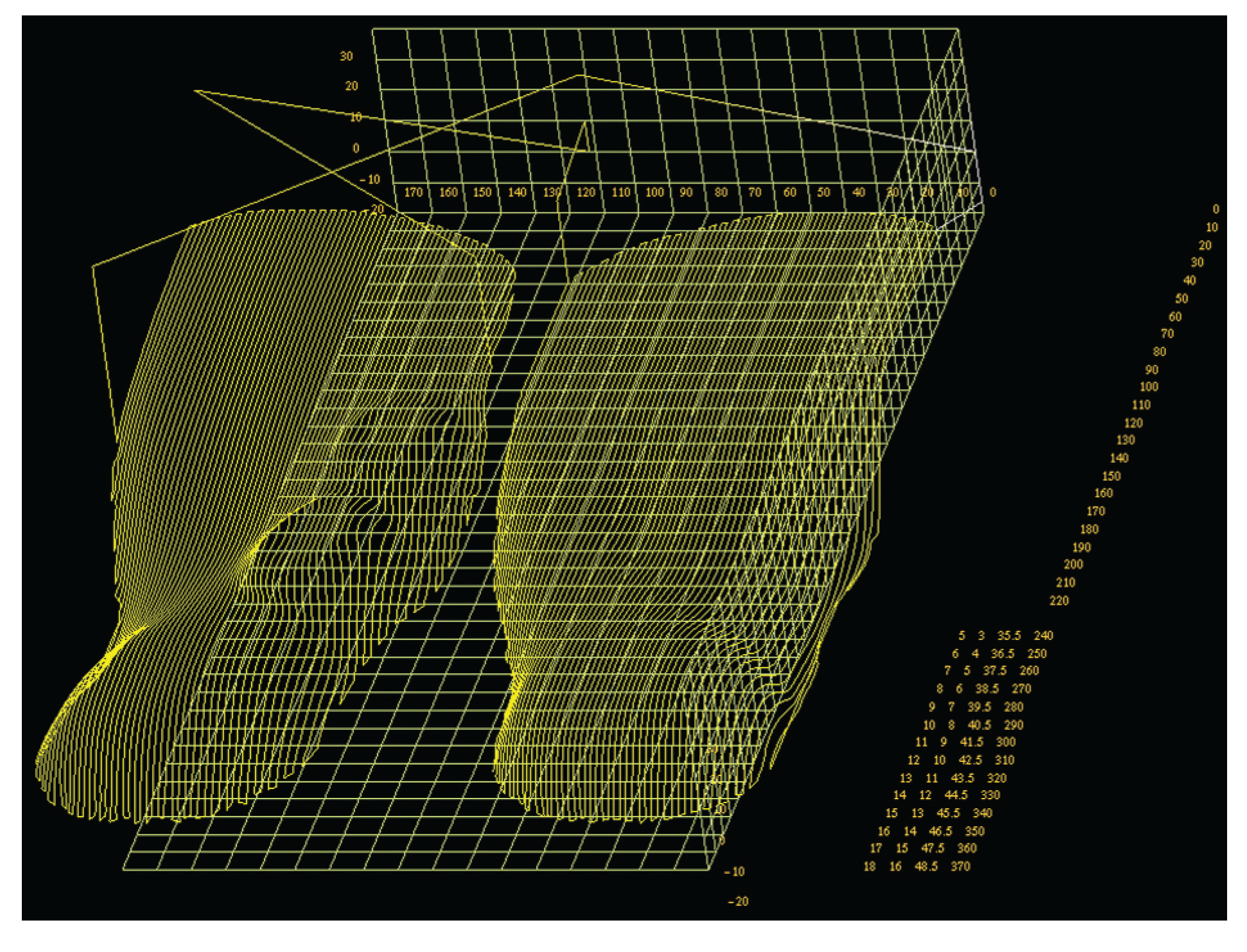
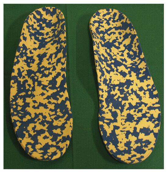
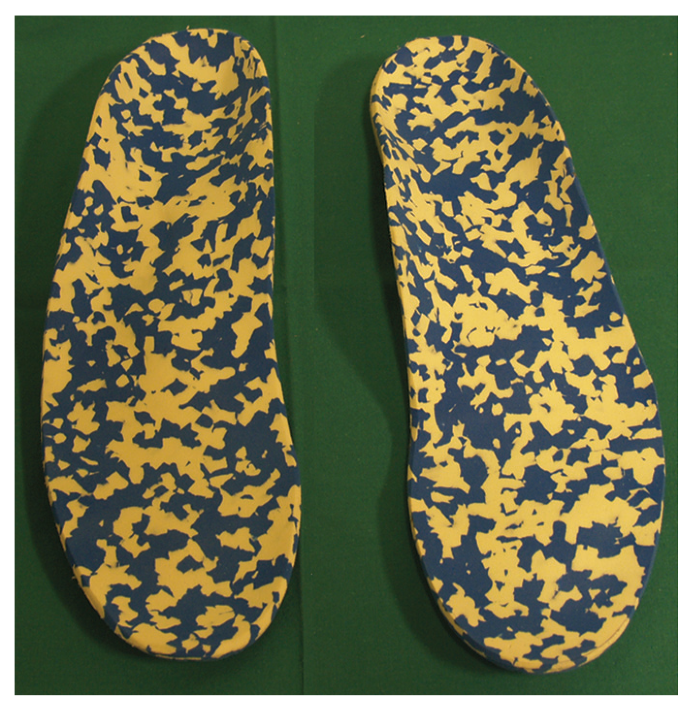
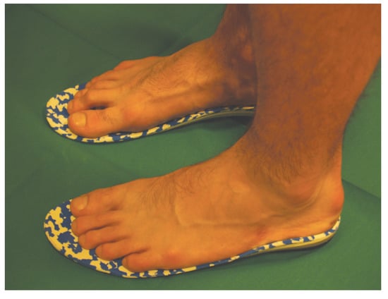
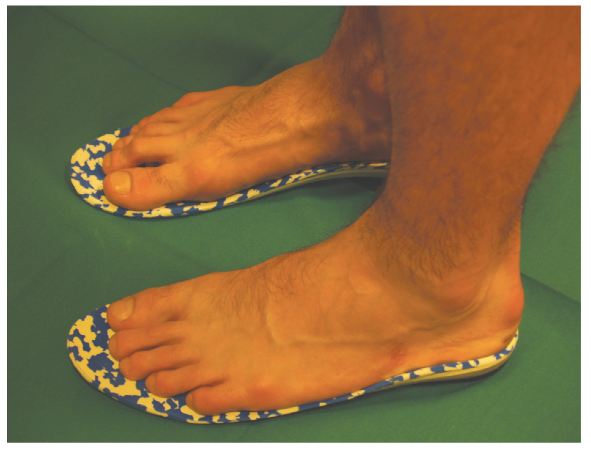
The orthotic base material was homopolymerpolypropylene (Otto-Bock Healthcare GmbH, Duderstadt, Germany) with trilaminate top cover. With the design complete, the tool path for the numerical controlled milling machine was generated (Figure 12). We used a boiled beech wood as a base mold material. The molding process was interrupted when the plastic was cooled down to room temperature. The plastic shell was posted with 65° ShA neoprene rubber neutral post (Figure 13). The patient acclimated very quickly to the orthotic device and noticed a decrease in pain and an increase in activity compared to a noorthotic condition (Figure 14). Two years postoperatively, he is an active basketball player and has no symptoms.
Figure 12.
Computer aided design showing generated computer numerical controlled milling machine tool path for a pair of the foot orthotic devices.
Figure 13.
Photograph showing the manufactured orthoses.
Figure 14.
Photograph showing the patient standing on the orthoses.
Discussion
According to general classifications, our patient had both of the two basic types of fractures: acute tuberosity avulsion fracture (Stewart’s Type III, Torg’s Stage I) and stress fracture of the metatarsal diaphorthotic devices. ysis within 1.5 cm of the tuberosity (Jones fracture, Stewart’s Type I, Torg’s Stage III).[5,7,8]
Between several conservative and operative treatment options, we chose a surgical treatment with intramedullary screw fixation. Using a percutaneous approach, we performed two of the most important procedures for two different types of fractures of the same bone. The first was drilling of the medullary cavity with removal of sclerotic bone. The second was making fracture fragments compression an imperative procedure for healing of both fractures. Next, we provided the possibility of shortening the nonweightbearing period and rapidly starting the training activities. At the end, as a prevention to avoid a possible refracture[14] or unexpected new fracture, we performed the orthotics and prosthetic laboratory additional tests that showed a better result of the operated foot than the nonoperated one. The patient was supplied with a custom-made functional foot orthotic device, and 1 year postoperatively, he is playing basketball without any sign of treatment complication.
The use of orthoses is an important part in prevention of injuries or possible refracture,[14] just as it is in treatment programs for foot and ankle injuries in sports.[11,15] In our experience with basketball players and the application of foot orthotic devices, both the total contact and biomechanical approaches have advantages and disadvantages. We combine both approaches in our praxes to reduce stress and maintain proper alignment to the foot structures. Our digital process to design and manufacture foot orthotic devices allows for accurate and adequate adjustments, a good digital database, and continuous follow-up.[12]
In the production of foot orthoses, we used a combination of homopolymer-polypropylene (Otto-Bock Healthcare GmbH) with trilaminate top cover as the orthotic base material because it is firm and is characterized as being significantly resistant to form loss and retains its functional properties even after prolonged exposure to stress and strain. It is also lightweight and available in thin sheets. We wanted to maintain accuracy during the production process to minimize outcome errors. Since homopolymer-polypropylene has a high thermal capacity, we needed to choose appropriate mold material that would not lose its shape under high temperature and pressure in the vacuum press. We decided to use boiled beech wood as a base mold material because of its excellent compressive strength and heat resistance. The main disadvantage of using this material is that on regular computer numerical controlled mill, more than one pass is required to create a smooth, moldable surface. A polyurethanebased material (like BioSculptor foam) requires only one pass and is approximately six times faster for mass production. After the orthotic device was formed in the vacuum press, the orthosis was not cooled in order to prevent plastic hardening, which makes orthoses more prone to breaking. When compared with initial measurements the mold height after almost an hour of exposure to high pressure and excessive heat from the melted plastic was negligible.
When compared with similar cases, we find the use of surgery combined with the application of custommade foot orthotic devices and precise protection of the affected foot region may improve the overall patient condition and reduce time needed for athletes to return to full-scale training. Additionally, we believe that the simultaneous use of the foot pressure map, foot picture (acquired from flat-bed scanner), and radiographic image to identify necessary adjustments in the orthotic design (Figure 15) increases accuracy and has a cumulative effect on the improvement of the orthotic functionality through computer aided design.
Figure 15.
Computer aided design showing textured view of the insole surface. Note an adjustment on the base of the fifth metatarsal bone.
This case report describes a stress fracture with a mechanical cause for propagation and development of a tuberosity fracture. On the basis of our experience it appears that, in the treatment of combined stress and tuberosity avulsion fractures of the same fifth metatarsal, percutaneous screw fixation with postoperative controlled rehabilitation, described pedobarographic testing, and application of the custommade foot orthoses before full intensity of training presents itself as a good solution for young, active athletes.
Financial Disclosure
None reported.
Acknowledgments
The Croatian Basketball Junior Men’s team player, for his cooperation and participation.
Conflict of Interest
None reported.
References
- HOCKENBURY, R.T. Forefoot problems in athletes. Med Sci Sports Exerc 1991, 31, 448. [Google Scholar] [CrossRef] [Scilit] [PubMed]
- PECINA, M.; BOJANIC, I.; DUBRAVCIC, S. Stress fractures in figure skaters. Am J Sports Med 1990, 18, 277. [Google Scholar] [CrossRef] [Scilit] [PubMed]
- MATHESON, G.O.; CLEMENT, D.B.; MCKENZIE, D.C. ET AL: Stress fractures in athletes: a study of 320 cases. Am J Sports Med 1987, 15, 46. [Google Scholar] [CrossRef] [Scilit] [PubMed]
- SALLIS, R.E.; JONES, K. Stress fractures in athletes: how to spot this underdiagnosed injury. Postgrad Med 1991, 89, 185. [Google Scholar] [CrossRef] [Scilit] [PubMed]
- MCBRYDE, A.M., JR. “Stress Fractures in Athletes,” in Prevention and Treatment of Running Injuries, ed by RD D’Ambrosia, D Drez Jr, p 21; Slack: Thorofare, NJ, 1982. [Google Scholar]
- KNAPP, T.P.; GARRETT WE, J.R. Stress fractures: general concepts. Clin Sports Med 1997, 16, 339. [Google Scholar] [CrossRef] [Scilit] [PubMed]
- STEWART, I.M. Jones’ fractures: fractures of the base of the fifth metatarsal. Clin Orthopedics 1960, 16, 190. [Google Scholar]
- TORG JS: Fractures of the base of the fifth metatarsal distal to the tuberosity. Orthopedics 1990, 13, 731. [CrossRef] [Scilit] [PubMed]
- FERNANDEZ FAIREN, M.; GUILLEN, J.; BUSTO, J.M. ET AL: Fractures of the fifth metatarsal in basketball players. Knee Surg Sports Traumatol Arthrosc 1999, 7, 373. [Google Scholar] [CrossRef] [Scilit]
- WRIGHT, R.W.; FISCHER, A.D.; SHIVELY, A.R.; et al. Refracture of proximal fifth metatarsal (Jones) fracture after intramedullary screw fixation in athletes. Am J Sports Med 2000, 28, 732. [Google Scholar] [CrossRef] [Scilit] [PubMed]
- JANISSE DJ: Indications and prescriptions for orthoses in sports. Orthop Clin North Am 1994, 25, 95. [CrossRef] [Scilit]
- UDILJAK, T.; GRANBERRY, W.M. OBROVAC K: A New Approach to Foot Orthosis Design and Manufacture. World Manufacturing Congress, Rochester, NY, April 2-5, 2002; Abstracts Book, 2001; p. 51. [Google Scholar]
- THE AMERICAN COLLEGE OF FOOT AND ANKLE ORTHOPEDICS AND MEDICINE. Prescription Foot Orthoses Practice Guidelines. Available online: http://www.acfaom.org/pg1103 (accessed on 11 September 2009).
- ROMANSKY, N.; BECKER, T. Essential insights on treating fifth metatarsal fractures. Podiatry Today 2006, 19, 76. [Google Scholar]
- GOODMAN A: Foot orthoses in sports medicine. South Med J 2004, 97, 267.
© 2009 American Podiatric Medical Association