


Abstract
Traumatic second metatarsophalangeal dislocation is extremely rare. This article reports a case of traumatic second metatarsophalangeal dislocation associated with fracture of the second metatarsal shaft. Anatomical reduction is indispensable to preserve the function of the metatarsophalangeal joint and to avoid joint ankylosis and chronic foot pain. The patient was treated with open reduction of the fracture without violating the joint to decrease postoperative stiffness. He was followed-up for 6 months. The patient resumed full activity 3 months after surgery, with his joint attaining full range of movement. He reported no pain or joint stiffness during his last visit. To our knowledge, no similar injury has been reported previously. (J Am Podiatr Med Assoc 99(6): 525-528, 2009)
Most traumatic metatarsophalangeal dislocations involve the first metatarsophalangeal joint.[1] Metatarsophalangeal dislocation of the lesser metatarsals usually involves more than one metatarsal.[1,2,3,4] Isolated traumatic second metatarsophalangeal dislocation is extremely rare.[5] It has been reported only once, by Murphy.[5] Dislocation of the metatarsophalangeal joint frequently occurs as a result of high-energy trauma.[2] The usual mechanism of injury is hyperextension of the proximal phalanx in a plantarflexed foot.[1–3,[5,6,7] The metatarsophalangeal joints play an important role in formation of the transverse arch of the foot and in plantarflexion in the immediate toeoff phase of walking.[2] Consequently, anatomical reduction is indispensable to preserve those functions, decrease joint ankylosis, and avoid chronic foot pain.[6] Closed reduction of dislocated first and second metatarsophalangeal joints was successful in a case reported by Gentless et al.[1] However, open reduction is commonly resorted to for treating this injury.[2,3,4,5] It can be performed through a dorsal[3,5] or plantar[4] approach. Ito et al[2] used both approaches in a case of dislocation involving all of the metatarsophalangeal joints of the right foot. A dorsal incision avoids injury to the neurovascular structures and a painful plantar scar.[6] However, the disrupted plantar plate is technically more difficult to repair through a dorsal incision compared with a plantar approach.
This article reports a case of traumatic second metatarsophalangeal dislocation associated with fracture of the shaft of the second metatarsal. A review of the literature did not reveal a similar injury previously reported.
Case Report
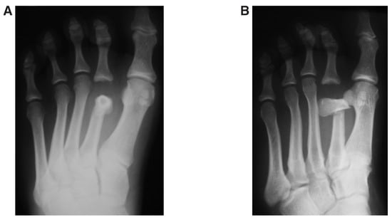
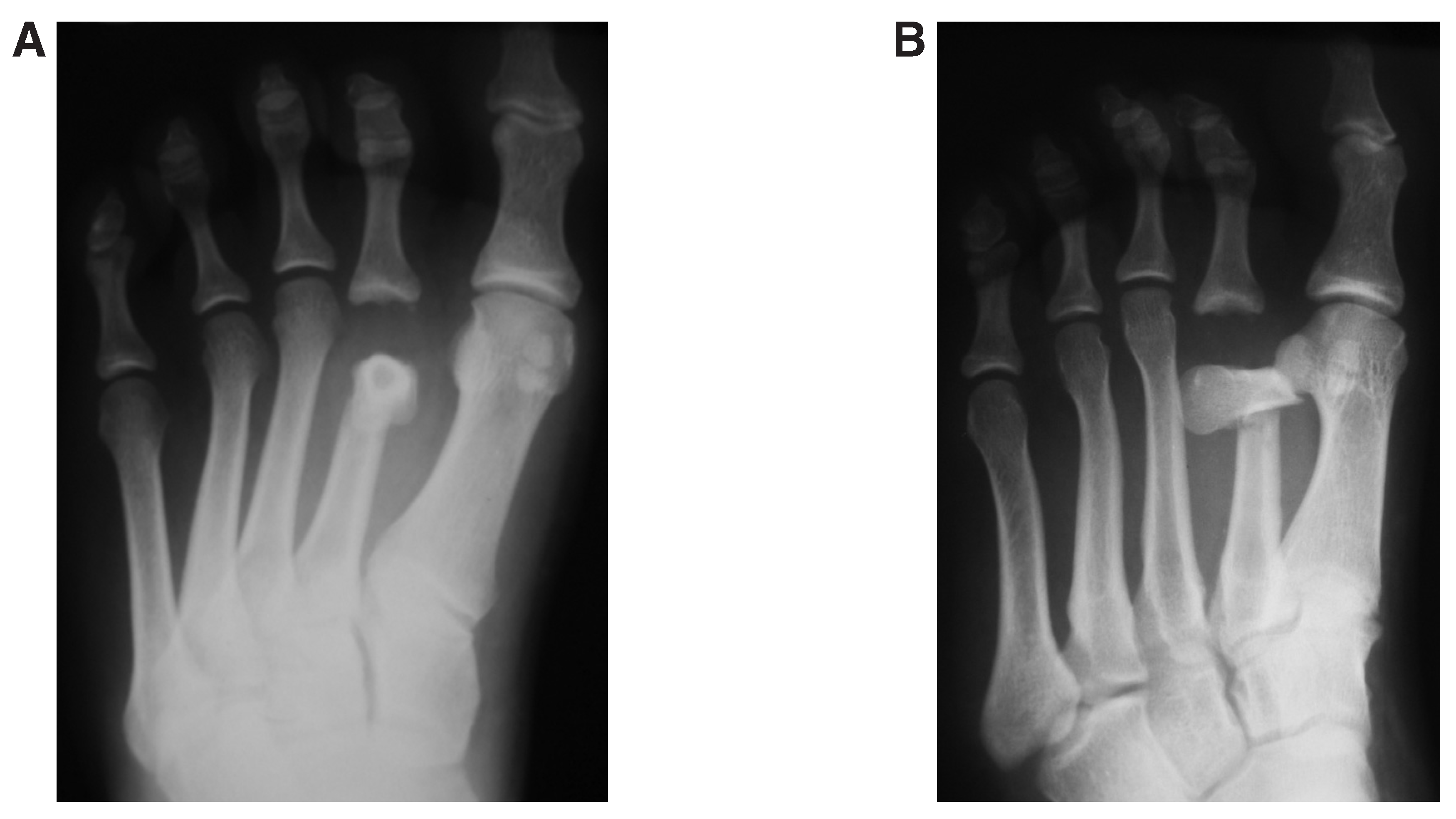
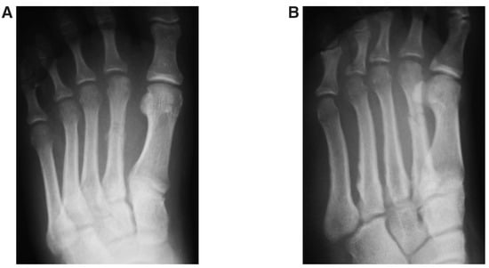
A 16-year-old boy presented to the emergency department 1 hour after kicking the ground while playing soccer barefoot. He had pain, swelling, and an inability to bear weight on his left forefoot. Findings from his medical history, system review, and family history were insignificant. He had minimal shortening of the second toe. A bony prominence was easily felt through the intact skin of the dorsal and plantar aspects of his left foot. No signs of ecchymosis or blistering were noticed. Nonweightbearing standard radiographs showed dorsal dislocation of the second metatarsophalangeal joint and fracture of the distal third of the second metatarsal shaft with 90° of plantar angulation (Figure 1).
Figure 1.
Preoperative nonweightbearing standard radiographs of the left foot. The anteroposterior (A) and oblique (B) views show dorsal dislocation of the second metatarsophalangeal joint associated with a fracture of the distal third of the second metatarsal shaft. The distal metatarsal fragment has 90° of plantar angulation.
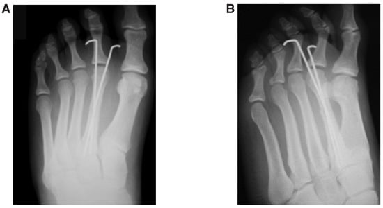
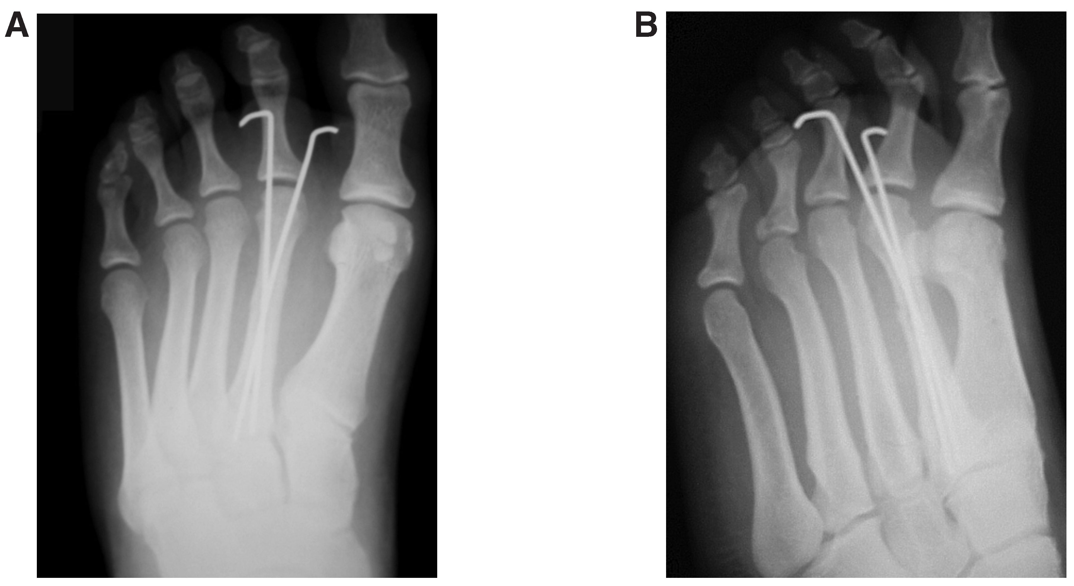
Three hours after the emergency presentation, he was taken to the operating room. With the patient under general anesthesia, closed reduction was attempted but was not successful. We, hence, performed an open reduction. Through a 4-cm dorsal skin incision, which was centered at the fracture site, the distal metatarsal fragment was exposed and was held with a pair of sharp bone-holding forceps. Traction was applied to the second toe by the assistant pulling on it, with counter traction applied through the bone-holding forceps. The metatarsophalangeal joint was reduced by manipulation of the attached bone-holding forceps, without any dissection through the periarticular region. Two crossed Kirschner wires were introduced percutaneously from the plantar aspect of the foot and were used for extra-articular fracture fixation (Figure 2). The joint was stable after reduction and fixation of the metatarsal fracture. A posterior slab was applied with the ankle in the neutral position.
Figure 2.
Immediate postoperative standard radiographs of the left foot. The anteroposterior (A) and oblique (B) views show anatomical reduction of the second metatarsophalangeal joint and the metatarsal fracture.
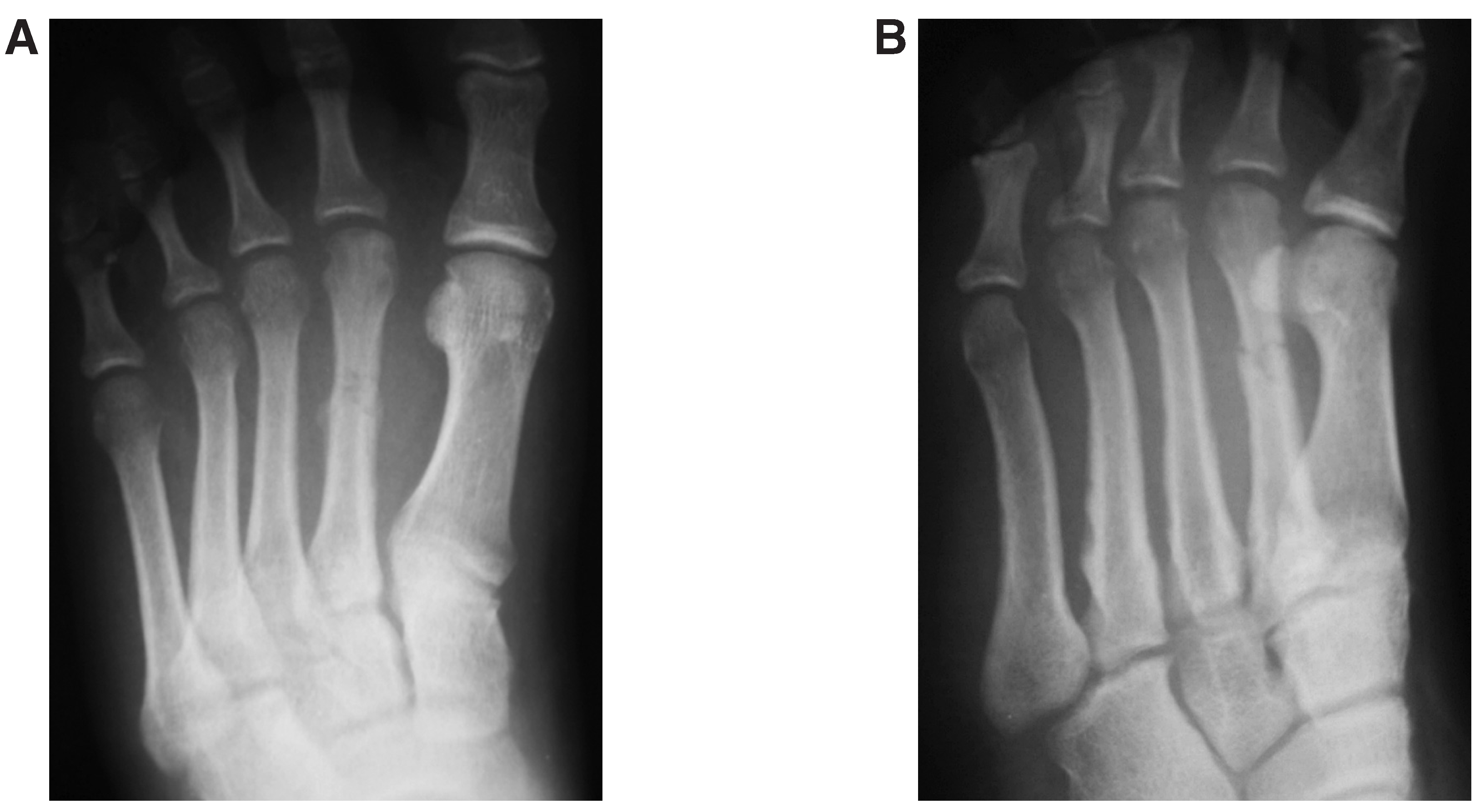
After foot elevation for 48 hours, a below-the-knee cast was applied, and the patient was discharged. The Kirschner wires were removed 5 weeks postoperatively, and partial weightbearing was started. Radiographs taken 7 weeks after surgery showed healing of the fracture with callus, with maintained reduction after weightbearing (Figure 3). No further radiographic assessment was performed after fracture healing, and further follow-up was based only on clinical assessment, owing to institutional guidelines that limit radiographic studies of pediatric patients. Full weightbearing was permitted 8 weeks after surgery. The patient was able to practice his sporting activities 3 months postoperatively. He was followed-up for 6 months after surgery, and he did not report any foot pain at his last visit. Digital strength and position on the ground and joint active and passive range of motion were similar to those of the normal contralateral side, with no crepitus or instability. The latter was assessed with the drawer test (the amount of dorsal translation of the proximal phalanx base on the metatarsal head when a dorsally directed force is applied to the proximal phalanx while concurrently stabilizing the metatarsal head) and valgus/varus stress tests.
Figure 3.
Standard radiographs of the left foot 7 weeks after surgery. The anteroposterior (A) and oblique (B) views show healing of the metatarsal fracture with callus formation. The anatomical reduction was maintained after Kirschner wire remova.
Discussion
Traumatic dislocation of the metatarsophalangeal joint frequently occurs as a result of high-energy trauma.[2] In this case, the dislocation was caused by a lowenergy sports injury. Similar to previously reported cases, the mechanism of injury was hyperextension of the proximal phalanx in a plantarflexed foot. However, the back pressure applied from the ground to the metatarsal head led to metatarsal fracture after metatarsophalangeal dislocation. The amount of plantar angulation of the distal metatarsal fragment is unusual; however, it indicates the possible spectrum of angular displacement. Failure of closed reduction in this case could be explained by the presence of the associated metatarsal fracture, so inline traction was ineffective without counter traction. When the latter was provided by sharp bone-holding forceps applied at the distal metatarsal fragment, reduction was successful. A dorsal incision was used in the management of this injury; however, reduction was obtained without dissection through the periarticular region. Achieving the reduction without violating the joint may limit intra-articular scarring and joint stiffness. With the dorsal approach, the disrupted plantar plate is unsutured. Nevertheless, the joint was stable after reduction in the patient reported by Murphy,[5] in which he used a dorsal incision. In the present case, the joint was stable after reduction and fully functional after fracture healing. Post-reduction joint instability seems to be related to multiple metatarsophalangeal dislocations and warrants transarticular pinning.[2,3] The outcome of the management of this injury has been similar to that of previously reported metatarsophalangeal dislocations involving the lesser metatarsals.[2,3,5,7,8] Irrespective of the direction of dislocation and the type of management, all of the reported metatarsophalangeal dislocations were anatomically reduced. In this patient, the rate of recovery and regain of full range of joint motion was faster than in the previous ones, probably because of his younger age. This might explain the shorter period needed for follow-up.
A literature review did not reveal any case of a single metatarsophalangeal dislocation associated with fracture of the involved metatarsal shaft. The present case is unique in being the first reported traumatic second metatarsophalangeal dislocation associated with fracture of the shaft of the second metatarsal. In addition, the degree of plantar angulation of the distal metatarsal fragment is unusual.
Financial Disclosure
None reported.
Conflicts of Interest
None reported.
References
- GENTLESS, J.; JOSHI, P.; CASELLI, M. ET AL: First and second metatarsophalangeal joint dislocation: a case report. JAPMA 1992, 82, 630. [Google Scholar] [CrossRef] [Scilit]
- ITO, M.M.; MURASE, K.; TANAKA, S. ET AL: Dislocation of all metatarsophalangeal joints caused by horse injury. J Trauma 2007, 63, 938. [Google Scholar] [PubMed]
- STEPHENSON, K.A.; BECK, T.L.; RICHARDSON, E.G. Plantar dislocation of the metatarsophalangeal joint: case report. Foot Ankle Int 1994, 15, 446. [Google Scholar] [CrossRef] [Scilit] [PubMed]
- NAKANO, Y.; MOGAMI, A.; KANEKO, K. ET AL: Irreducible dorsal MTP joint dislocation in the second and third toes. Injury 2003, 34, 870. [Google Scholar] [CrossRef] [Scilit] [PubMed]
- MURPHY JL: Isolated dorsal dislocation of the second metatarsophalangeal joint. Foot Ankle 1980, 1, 30. [CrossRef] [Scilit] [PubMed]
- BRUNET JA: Pathomechanics of complex dislocations of the first metatarsophalangeal joint. Clin Orthop Relat Res 1996, 332, 126. [CrossRef] [Scilit] [PubMed]
- RAO, J.P.; BANZON, M.T. Irreducible dislocation of the metatarsophalangeal joints of the foot. Clin Orthop Relat Res 1979, 145, 224. [Google Scholar] [CrossRef] [Scilit]
- KAMATH, S.U.; SAVUR, A.D.; PRAMOD, S. Irreducible traumatic dislocation of the fourth metatarsophalangeal joint: a case report. Injury Extra 2005, 36, 212. [Google Scholar] [CrossRef] [Scilit]
© 2009 American Podiatric Medical Association