


Abstract
Flexible pes planovalgus is a common condition with flattening of the medial longitudinal arch accompanied by hindfoot valgus. Severe cases of pes planovalgus may need surgery, and a technique that has gained popularity over the past decades is subtalar arthroereisis. An endoorthotic implant of various shapes is inserted in the sinus tarsus, which limits the excessive eversion of the subtalar joint present in flexible pes planovalgus. None of these implants, however, allow for easy control of the extent of talocalcaneal and talonavicular correction. The primary aim of this study was to describe our technique with the custom-built cone-shaped implant. Our secondary aim was to evaluate patient satisfaction, clinical and radiologic results, and complications with a minimal follow-up of 5 years. Between January 1992 and June 2002, 40 patients (80 feet) underwent subtalar arthroereisis for flexible pes planovalgus. After temporary sinus tarsi tenderness (12 feet), implant dislocation (two feet) was the most common complication. Questionnaires from 27 patients (54 feet) were analyzed and 44 feet were also clinically and radiographically evaluated. Thirteen patients were lost to follow-up. Mean (± SD) follow-up was 12.6 years (range, 5.9–16.1). Eighty-one percent of the patients were satisfied with the result. Clinically, normal alignment was present in 14 feet, and mild deformities remained in 26 feet. Radiographically, the average foot angle measurements were normal. We conclude that subtalar arthroereisis is a simple, minimally invasive operative option with satisfactory subjective and clinical results after mid- to long-term follow-up.
Flexible pes planovalgus (PPV) is a common condition with flattening of the medial longitudinal arch accompanied by hindfoot valgus. In children, flexible PPV is usually asymptomatic, and adult alignment may still develop up to the age of 10 years.[1–3] However, some patients with flexible PPV complain of pain, fatigue, or a cramping sensation in the foot. Parents may complain about the child’s clumsy gait or loss of activity.[4] In most cases, supportive footwear and insoles are successful.[5] Although controversial, operative treatment may be indicated for severe PPV or painful feet.[5–7] One technique that has gained popularity over the past decades is subtalar arthroereisis. An implant of various shapes—an endoorthosis—is inserted in the sinus tarsus, which limits excessive eversion of the subtalar joint present in flexible PPV.[8] This directly restores the talocalcaneal relations and the talonavicular subluxation and, by principles of the windlass mechanism, subsequently the medial arch.[9] In the growing skeleton, this method, in theory, facilitates remodeling with permanent realignment in adulthood.
Many publications have reported on subtalar arthroereisis in adults with either congenital or acquired flatfoot deformities.[10–14] Others addressed the treatment of flexible PPV in children.[4,6,15–18] For this, a wide variety of implants with different geometry and materials have been used. None of these implants, however, allow for easy control of the extent of talocalcaneal correction. Therefore, this study was intended to describe our technique of subtalar arthroereisis with the custom-built cone-shaped implant for flexible PPV in the pediatric population that we have used for more than 15 years. We performed a retrospective study to evaluate patient satisfaction and clinical and radiologic results with a minimal follow-up of 5 years. To the best of our knowledge, mid-to long-term results have not been reported.
Patients and Methods
Operative Technique
Surgery was performed under local anesthesia. Patients were placed supine with a tourniquet applied to the lower leg. After internally rotating the foot and localizing the sinus tarsus, an incision exposing the sinus was made (Fig. 1). After clearing excess fat (Fig. 2), the custom-built polyethylene implant (30 × 5 × 15 mm; Kuijpers Instruments, Groesbeek, the Netherlands), which is cone-shaped and partially flattened on the broad side (Fig. 3), was adjusted to the size of the foot by cutting off excess length on the narrow end (Fig. 4). When the appropriate length was realized, the implant was placed into the sinus tarsus in a lateral-to-medial direction with the flattened side flush with the sinus tarsi roof (Fig. 5). Fixation was achieved by a screw through the implant in a perpendicular direction to the implant advancing into the floor of the sinus tarsus (Fig. 6). Dorsal flexion was tested with the hindfoot in neutral position. If flexion was less than 10°, we performed additional percutaneous lengthening of the Achilles tendon. After closure of the skin in layers, a pressure bandage was applied. Correct positioning of the screw was checked radiographically (Fig. 7). Patients received a walking cast for 4 weeks and were allowed to mobilize weightbearing 1 week postoperatively. After removal of the walking cast, patients received insoles for 1 year. The surgeon who introduced the procedure chose to remove the implants routinely 1 year after surgery.
Patients
Data were collected from all patients who underwent subtalar arthroereisis for pediatric flexible PPV between January 1992 and June 2002 in our clinic, St. Maartenskliniek, Nijmegen, the Netherlands. Operative treatment was indicated for patients with severe PPV in which conservative therapy (insoles, stretching exercises of the calf muscles) had proven to be insufficient. The diagnosis of flexible PPV was confirmed by passive manual correction of the deformity and correction of both the medial arch and hindfoot valgus on toe stance. Radiologic evaluation was used to exclude causes of rigid flatfoot deformities. Exclusion criteria were PPV deformities of spastic or neurologic origin and PPV attributable to clubfoot deformities. Patients with previous hindfoot surgery were also excluded.
Evaluation
Questionnaire.
A patient-based questionnaire that we developed for the purpose of this study (Table 1) was sent to all patients. The questionnaire asked about pain, functionality, and satisfaction. Pain was classified as no, mild, moderate, or severe pain. Patient satisfaction was rated “satisfied,” “reasonably satisfied,” “moderately satisfied,” or “dissatisfied.”
Clinical evaluation.
We also invited these patients for a clinical and radiographic examination. On follow-up, alignment of the foot was scored as normal or as having a planus, valgus, or planovalgus deformity. Mild deformities were defined as a lowered medial arch, but still visible on weightbearing, a hindfoot valgus deformity of less than 10°, or both. A complete loss of the medial arch on weightbearing or a hindfoot valgus deformity of more than 10° were considered severe deformities.
Radiographic evaluation.
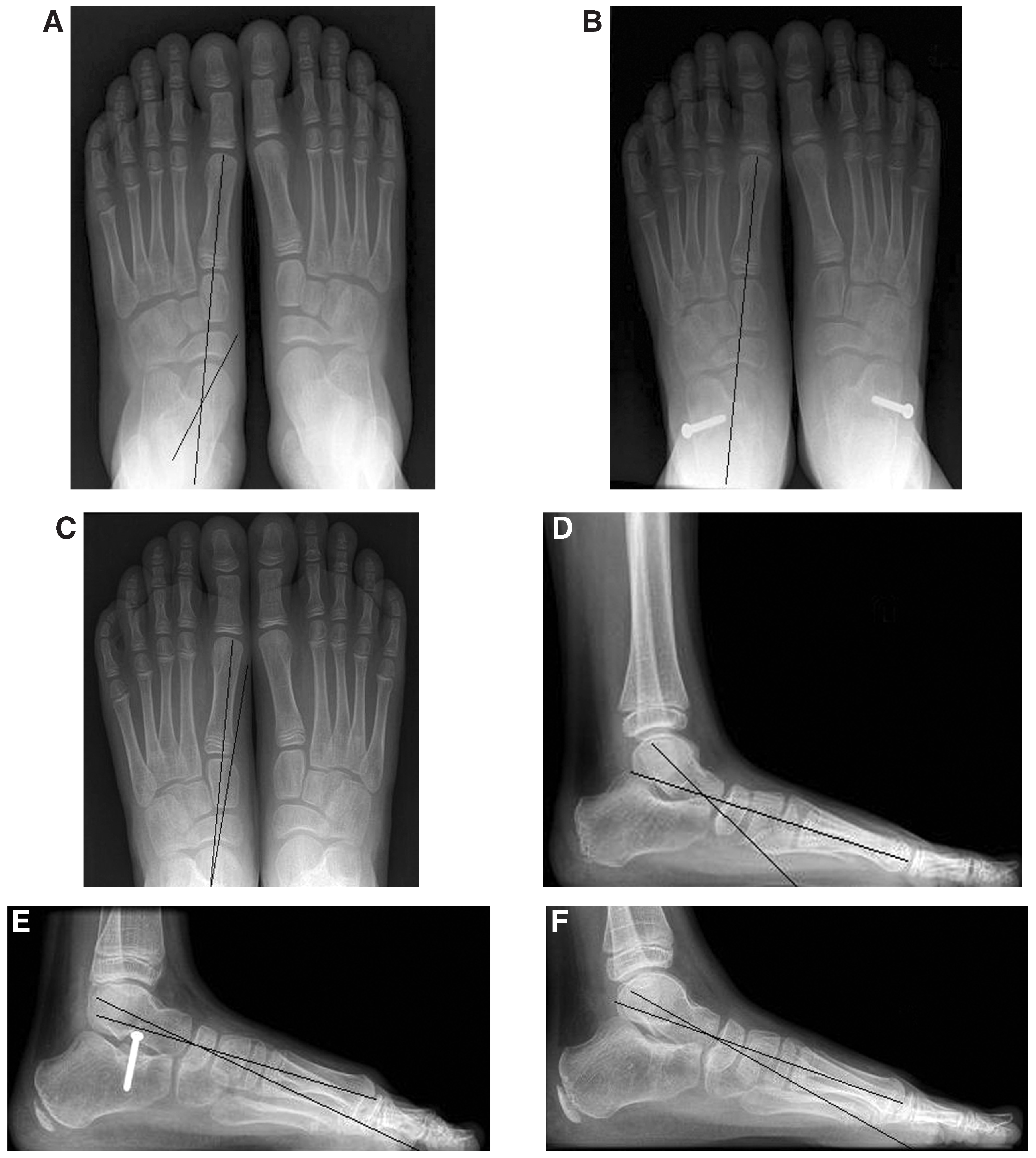
Weightbearing lateral radiographs were obtained. We evaluated the hindfoot planus by means of the calcaneal pitch; midfoot planus by the first talometatarsal angle; and hindfoot valgus was evaluated by measuring the talocalcaneal angle. Comparison with pre- and postoperative radiographs was not possible because standardized radiographs were not available for all feet. Both clinical and radiologic measurements were taken by the same author (P.M.K.). Finally, medical records were reviewed for postoperative results and complications.
Analysis.
Data were analyzed with SPSS version 12.0 (SPSS Corp, Chicago, Illinois). The data in Table 2 are presented as mean (SD; range).
Results
Complications
Dislocation of one of the implants occurred in two patients. This necessitated revision in one patient and early removal of the implant after 9 months in the other. Pain developed in one foot, which required revision of the implant and neurolysis of the nervus peroneus superficialis. Sinus tarsi tenderness was reported in 12 other feet, but it was not considered severe enough for early removal of the implant. The tenderness resolved in all cases after removal of the implants. Superficial wound infection led to impaired wound healing in one patient after implantation but resolved spontaneously, and the implant could be left in situ. No deep wound infections were observed.
Evaluation
A total of 80 feet of 40 patients were operated on for flexible PPV between 1992 and 2002. Thirteen patients with similar age and sex distribution to the study population were lost to follow-up, leaving questionnaires of 27 patients available for analysis. Four of these patients, however, did not visit the clinic because they felt it was unnecessary, and in one patient health problems hindered the visit. The 22 males and five females had surgery at a mean age of 8 years (SD ± 1.8; range, 4–11). The implant was in situ for a mean period of 1 year (SD ± 0.2; range, 8–16 months), and mean duration of follow-up was 12.6 years (SD ± 3.1; range 5.9–16.1).
Questionnaire.
Data are presented in Table 1. The five patients who did not attend our clinic all reported to be satisfied with the procedure, and none of the patients experienced any discomfort from their feet. In total, 22 patients (44 feet) were satisfied with the result, and 18 patients (36 feet) did not experience any discomfort.
Clinical examination.
Clinical evaluation of the 22 patients (44 feet) showed normal alignment in 14 feet. A remaining mild planus was present in 11 feet and mild valgus in four other feet. Both midfoot planus and hindfoot valgus, (PPV), was mild in 11 feet and severe in four feet. Medical records showed recurrence of the PPV after removal of the implant in ten feet (five patients) out of the total of 54 feet. On recent follow-up, one of these patients (two feet) had normal feet. In this particular case, the implants had been removed when the patient was 7 years and 11 months old, and the implant had been in situ for 13 months.
Radiographic evaluation.
Discussion
In general, surgery for the pediatric flexible PPV is seldom indicated. In the normal child, flat feet are a physiologic condition, and the adult foot model usually starts to develop at approximately 6 years of age.[19,20] There are no prospective studies with long-term follow-up on the natural course supporting prophylactic surgery for flexible pediatric flat feet. Also, the majority of symptomatic cases respond very well to conservative measures such as over-the-counter or custom-molded insoles,[21] although they do not influence the development of the feet.[21–23] If flatfoot is accompanied by contraction of the Achilles tendon, a rigorous stretching program is often beneficial.[1]
However, despite optimal conservative measures, severe PPV without any tendency toward normal alignment or that is accompanied by pain may justify operative procedures in an effort to correct the misalignment and consequently relieve pain. In this study, the majority of the patients (81%) were satisfied and the clinical results were considered satisfactory, with severe persistent PPV in only four feet. Generally, the hindfoot valgus and abduction was corrected better than the hindfoot and midfoot planus deformities. This is in concordance with other studies.[5,24] Radiologically, the mean values of all three measured foot angles fell within the normal range described by others.[19,25–27]
Technically, subtalar arthroereisis is relatively simple and leaves osseous and ligamentous tissue intact. Therefore, it does not compromise future operative procedures, like the Evans lateral column lengthening, which can be considered if the subtalar implant is not tolerated or if deformities persist after its removal. Some authors, however, consider the Evans osteotomy to be the preferred treatment for symptomatic PPV in children.[28–29] A second advantage of subtalar arthroereisis is that it limits excessive eversion of the subtalar joint, but subtalar joint motion is preserved and the hindfoot maintains its ability to adapt to uneven surfaces. Finally, the amount of correction can be easily controlled because of the implant’s cone-shaped configuration. The deeper the implant is inserted, the more distension created, thereby achieving correction. After achieving the right amount of correction with the foot in neutral position, the implant is simply fixated with a screw that advances through the sinus tarsi floor.
Few major complications were reported, with implant dislocation (two feet) being the most common. However, sinus tarsi tenderness was reported in 12 patients. We think that this is partially associated with the position of the implant. When properly inserted, the flattened side of the implant lies flush with the sinus tarsi roof. Otherwise, the edges on each side of the flattened surface may impinge on the talus. Overall, the major complication rate compares well to other studies, except for one study by Verheyden et al,[18] who reported an unacceptable high rate (22%) of implant dislocation; they no longer use this technique.
As arches may still develop up to the age of 10, it is conceivable that an unknown number of patients in our study might have developed normal feet, irrespective of subtalar arthroereisis. The same, of course, applies to most other studies on pediatric subtalar arthroereisis. This possibility was clearly illustrated in one of our patients in which the PPV had returned after removal of the implant at almost 8 years of age. On follow-up, however, the feet had a completely normal alignment. Despite this result, we and other authors believe that when conservative treatment has failed, severe PPV with talonavicular subluxation deserves operative intervention,[6,10,13,30] especially if these patients complain about pain located over the medially protruding talus that worsens with the use of insoles. Subtalar arthroereisis effectively restores the talonavicular subluxation, thereby relieving the discomfort. Furthermore, although supported by limited scientific evidence, uncorrected severe PPV may become progressively symptomatic in adolescence or adulthood, requiring more invasive measures.[10,31]
Another topic associated with the variable age of spontaneous correction of the foot is the timing of subtalar arthroereisis. On one hand, spontaneous development of a normal alignment is to be expected from around the age of 6 and up to the age of 10. On the other hand, if PPV persists after the ages of 10 to 12, little spontaneous correction is to be expected.[1–3] Therefore, we choose to perform subtalar arthroereisis for severe PPV around the age of 8. It is our experience that temporary subtalar arthroereisis has little lasting effect after the age of 10. If a younger child, for example 6 or 7 years of age, presents with hindering pain medially over the talus, we consider performing the procedure at an earlier age in an effort to relieve the symptoms. Therefore, we now consider leaving the implant in situ for more than 1 year but only if the implant is well tolerated. In general, the reason for removing the implant after 1 year was because of the relatively high rate of sinus tarsi tenderness. The rationale for temporary arthroereisis was also confirmed by the use of bioabsorbable implants used by Giannini et al.[15] These implants showed loss of integrity after 1 year, but reported results were good after a follow-up of 4 years. In children, the idea behind subtalar arthroereisis is secondary remodeling during further growth. To what extent this concerns osseous or soft-tissue remodeling is not known, but the phenomenon seems to be supported by other studies.[4,15] Therefore, randomized controlled trials are needed to examine the extent to which secondary remodeling (eg, subtalar arthroereisis) takes place.
Apart from the retrospective nature of the present study, we acknowledge several limitations. First, besides the clinical outcome, we were unable to compare the postoperative results to the preoperative situation as subjective (validated questionnaires) and radiologic data were unavailable. Therefore, to evaluate patients’ subjective benefit, we developed a questionnaire that would answer the most relevant items in our opinion: pain, functionality, and satisfaction. Secondly, mild deformities remained in 26 feet. However, arbitrary cut-off points were used, which meant that feet with only a minor degree of planus, valgus, or both were also classified as having mild deformities. We agree with Viladot et al[14] who already pointed out based upon their results, that it is not necessary to normalize radiographic angles to ensure the best outcome for the patient.
Conclusions
In conclusion, subtalar arthroereisis for pediatric flexible PPV is a simple and minimally invasive operative option with satisfactory subjective and clinical results after mid- to long-term follow-up. The advantage of the cone-shaped implant above other types of implants is that it allows a controllable correction of the foot, and when performed in childhood, some degree of permanent remodeling may take place.

Table 1.
Questionnaire Results on Follow-upa
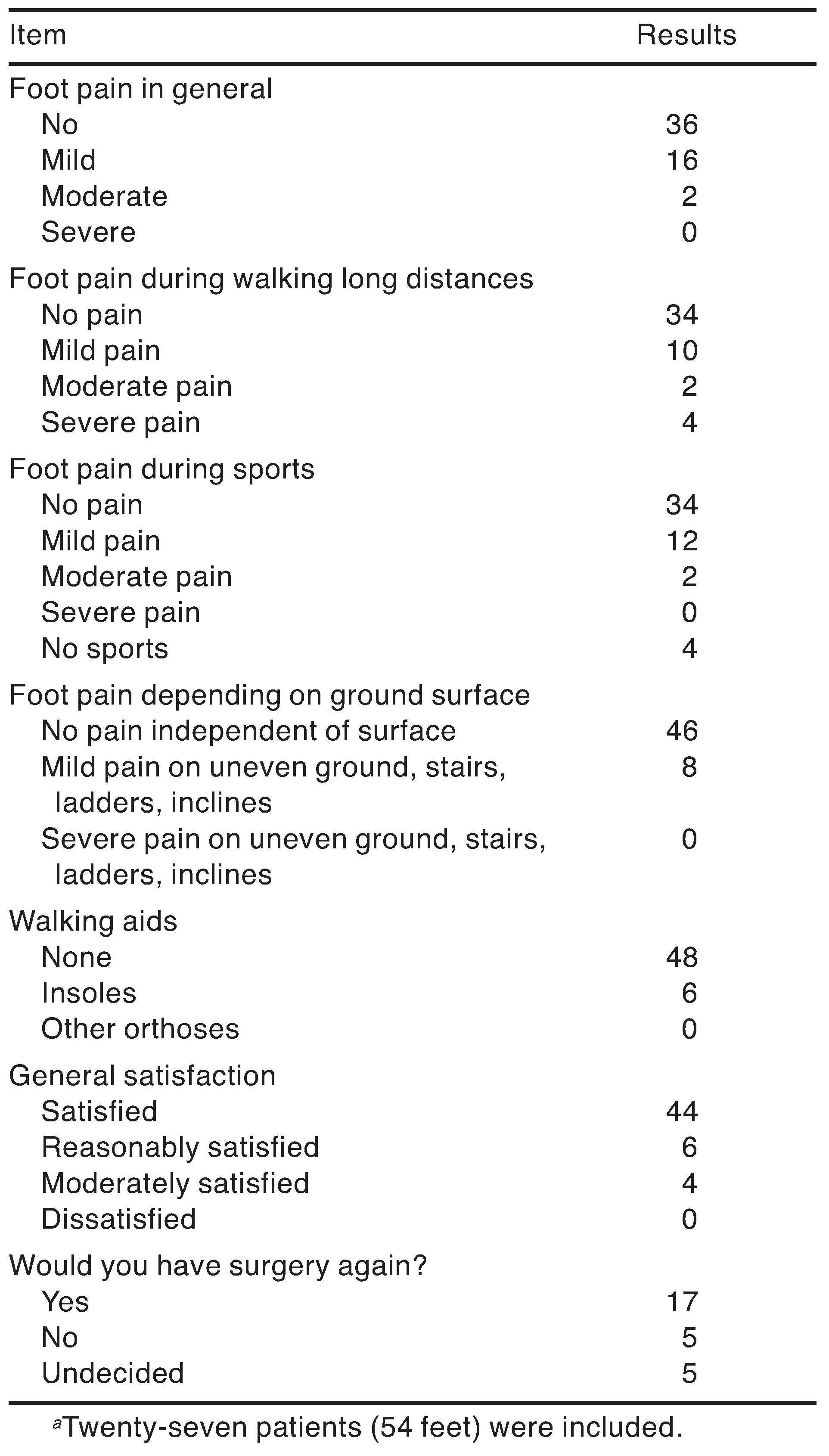
Table 2.
Radiographic Measurements on Follow-upa

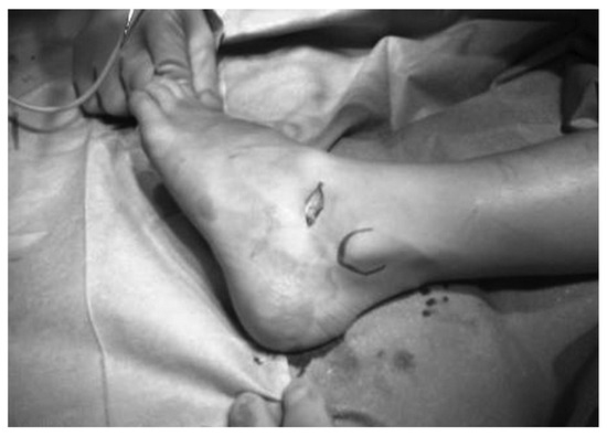
Figure 1.
Incision over the sinus tarsus.
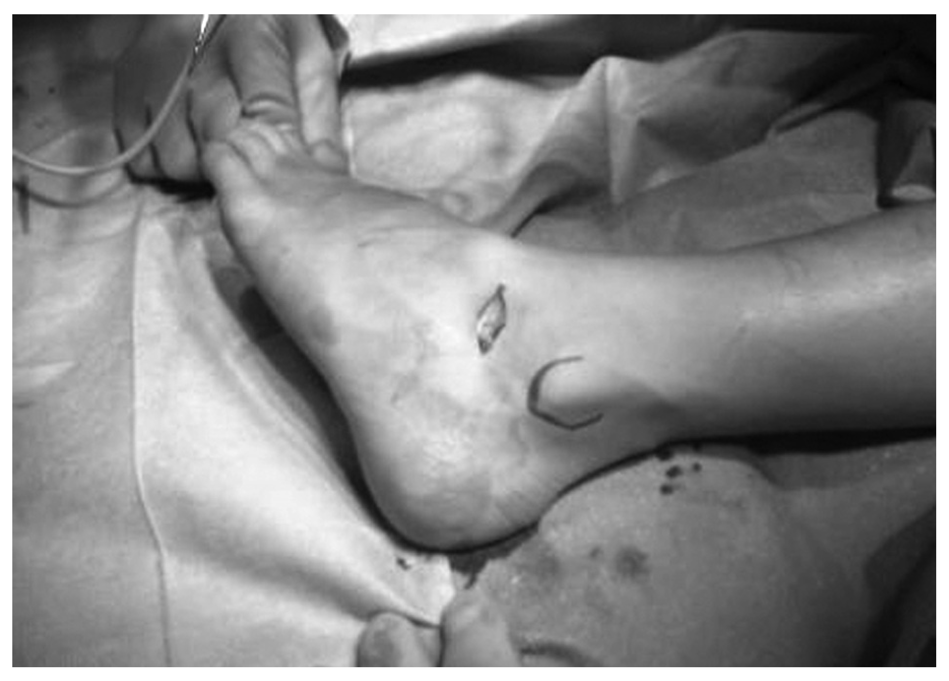
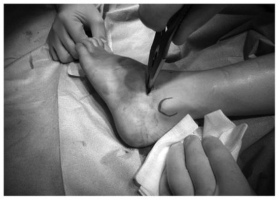
Figure 2.
Removal of sinus tarsus fat tissue.
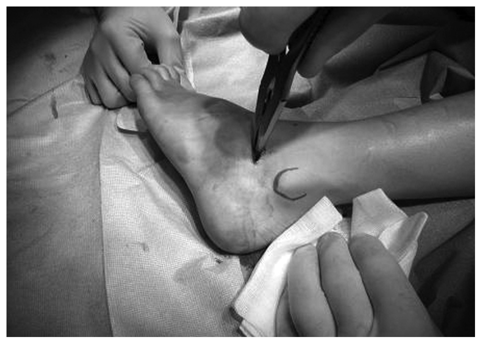
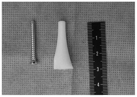
Figure 3.
The cone-shaped implant and fixating screw.
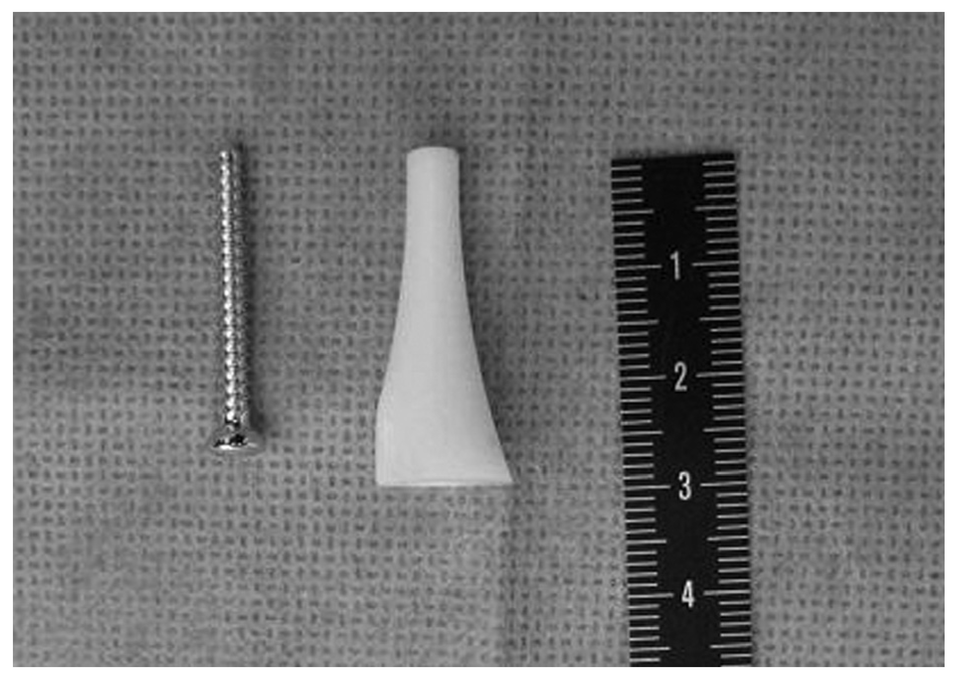
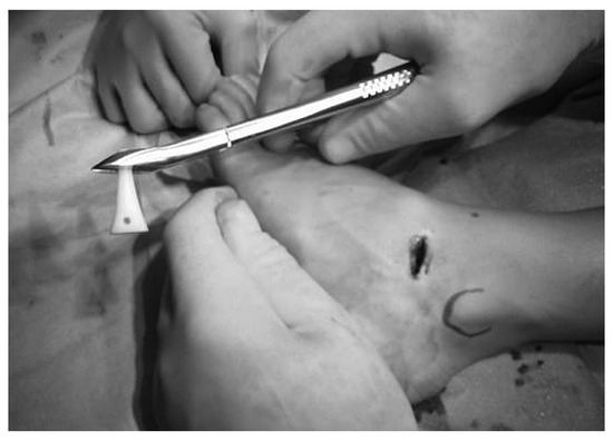
Figure 4.
The implant is customized to the size of the foot.
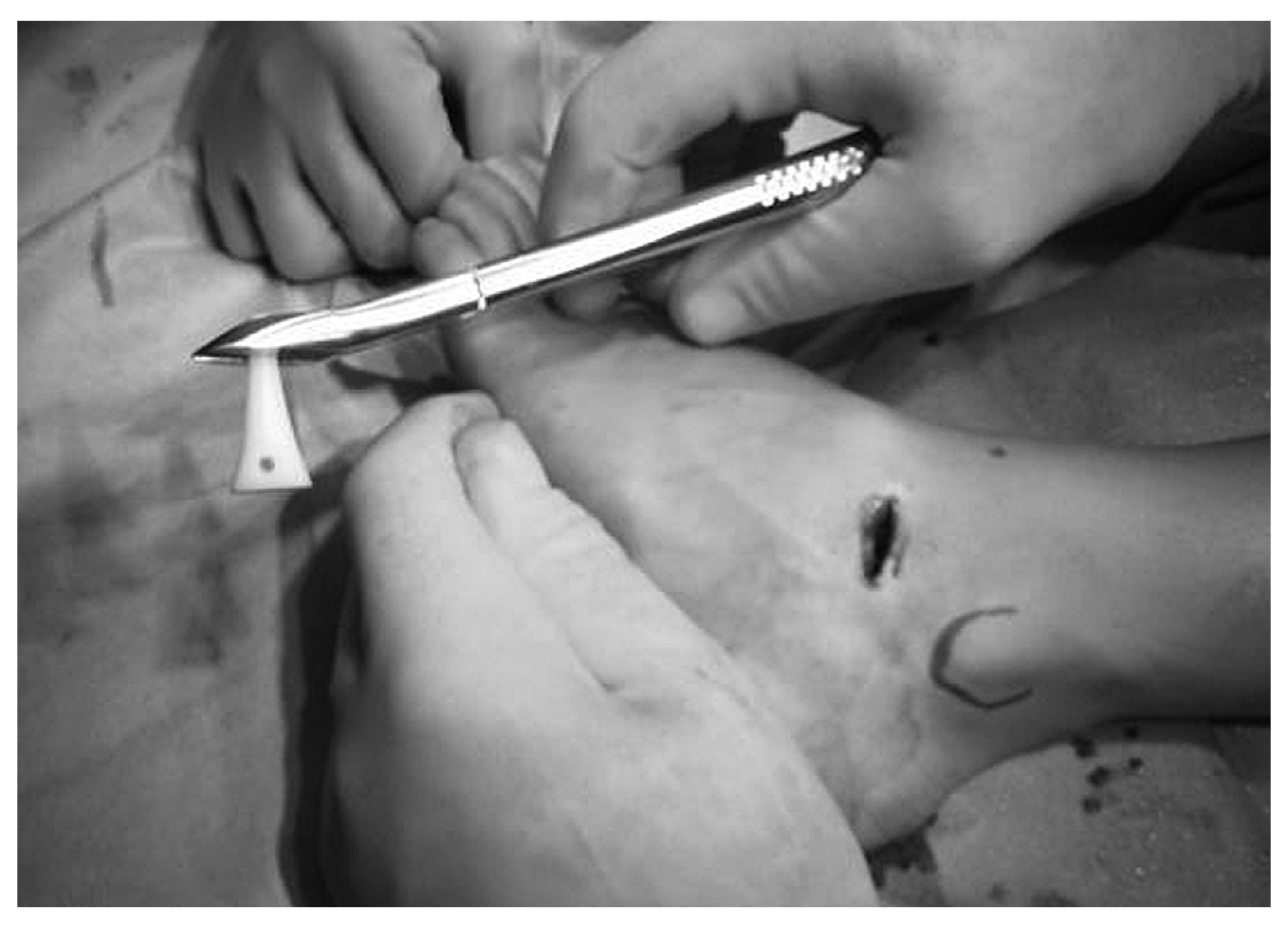
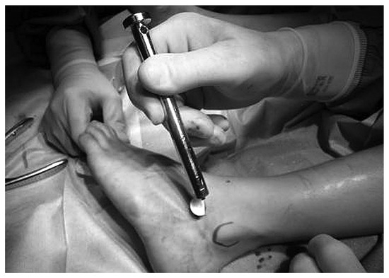
Figure 5.
The implant is drilled into the sinus tarsus.
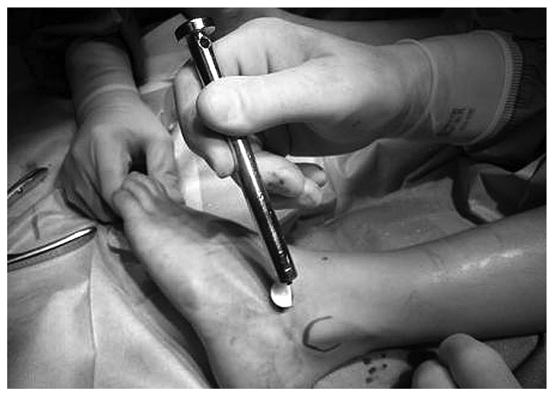
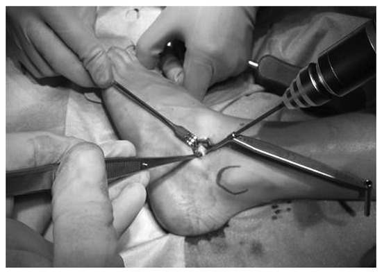
Figure 6.
The implant is fixated by a screw through the implant, advancing into the floor of the sinus.
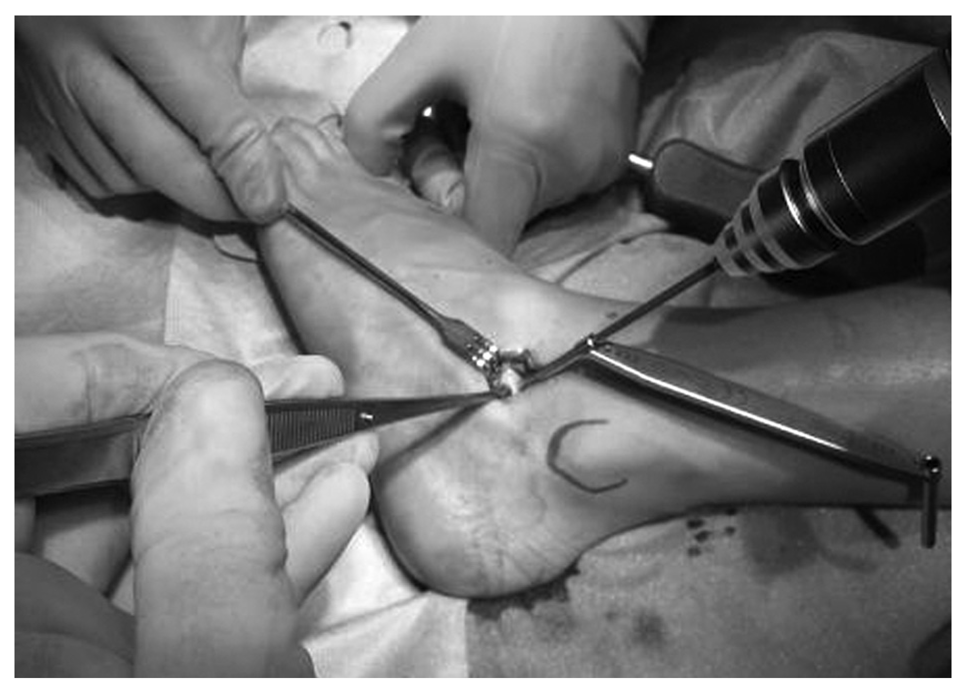
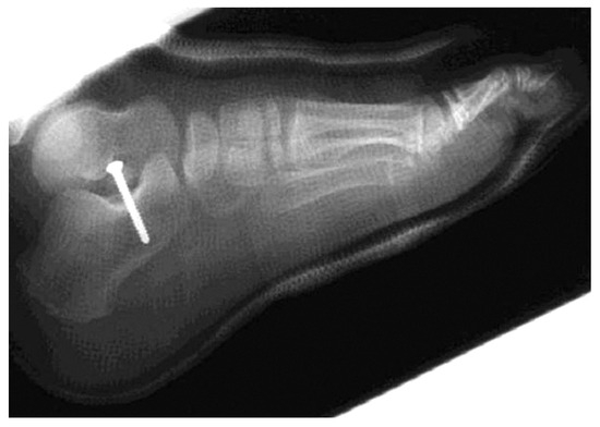
Figure 7.
Postoperative lateral radiograph showing correct position of the screw.
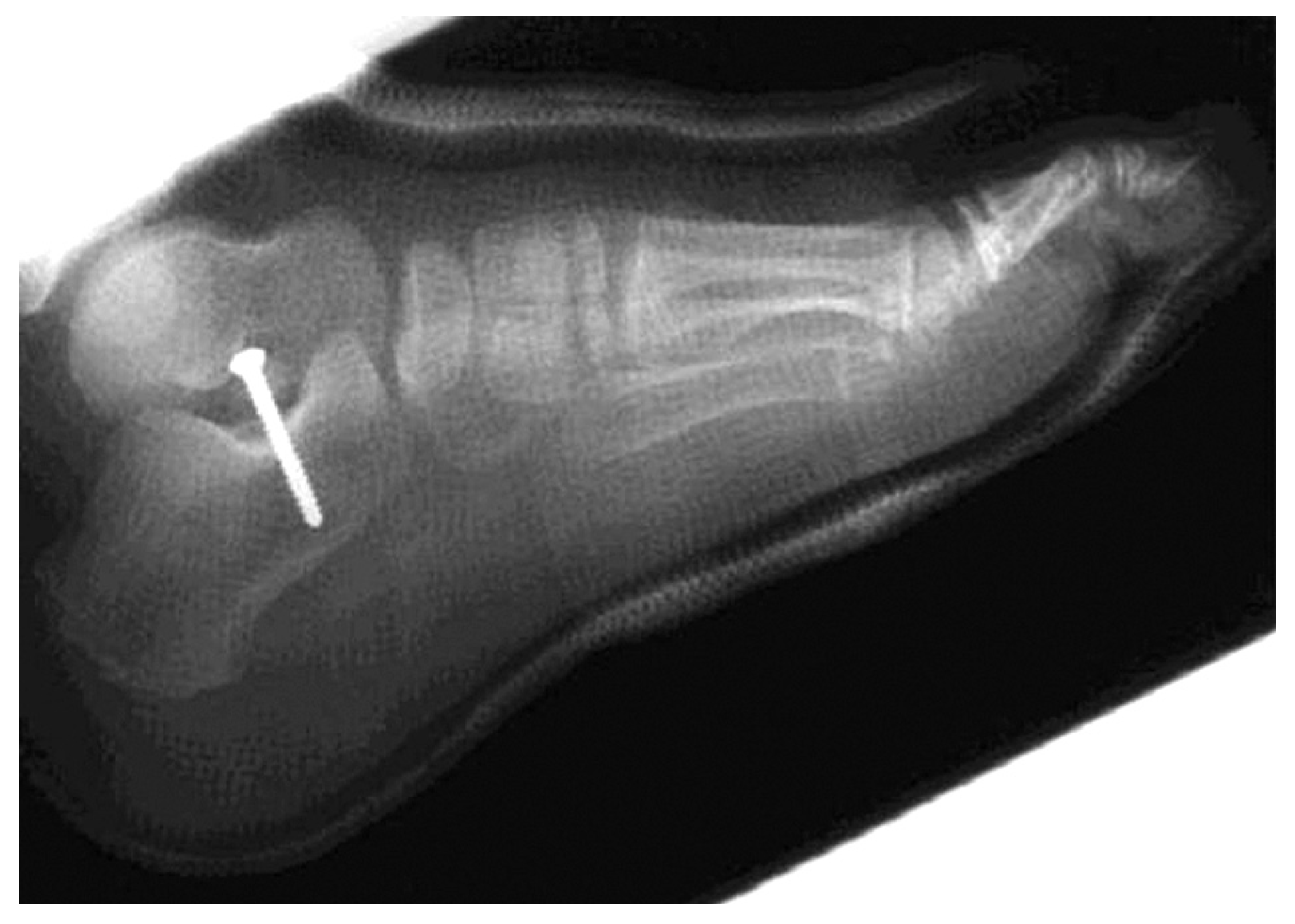
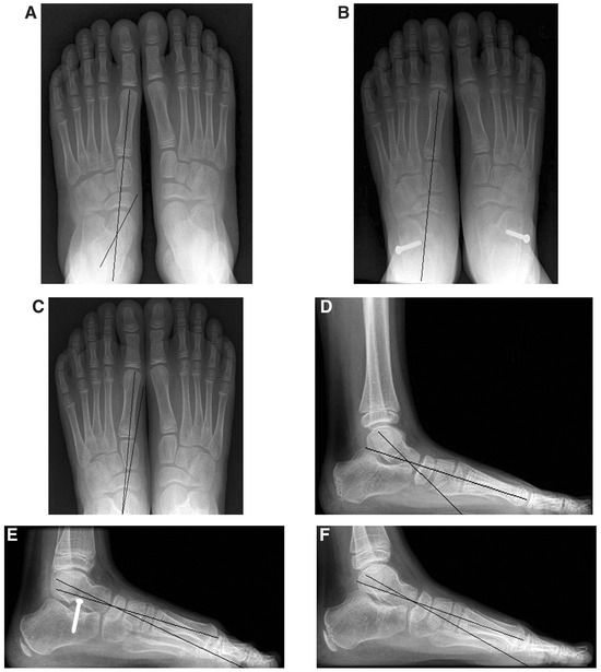
Figure 8.
Anteroposterior radiograph showing the feet before surgery (A), 5 months postoperatively (B), and 4 months after implant removal (C). D–F, Lateral radiographs of the feet seen in parts A, B, and C.
Financial Disclosure: None reported.
Conflict of Interest: None reported.
References
- Morrissy RT, Weinstein SL: Lovell and Winter’s Pediatric Orthopaedics, Lippincott Williams & Wilkins, London. ,2000. .
- Staheli LT, Chew DE, Corbett M: The longitudinal arch: a survey of eight hundred and eighty two feet in normal children and adults. .J Bone Joint Surg Am69::426. ,1987. .
- Welton EA: The Harris and Beath footprint: interpretation and clinical value. .Foot Ankle13::462. ,1992. .
- Smith SD, Millar EA: Arthrorisis by means of a subtalar polyethylene peg implant for correction of hindfoot pronation in children. .Clin Orthop Rel Res181::15. ,1983. .
- Soomekh DJ, Baravarian B: Pediatric and adult flatfoot reconstruction: subtalar arthroereisis versus realignment osteotomy surgical options. .Clin Podiatr Med Surg23::695. ,2006. .
- De Pellegrin M: Die subtalare schrauben-arthrorise beim kindlichen plattfuβ. .Orthopäde34::941. ,2005. .
- Needleman RL: Current topic review: subtalar arthroereisis for the correction of flexible flatfoot. .Foot Ankle Int26::336. ,2005. .
- Mann RA: “Biomechanics of the Foot and Ankle,” in Surgery of the Foot and Ankle, 6th ed, Vol 1, ed by RA Mann, MJ Coughlin, p 3, Mosby-Year Book, St. Louis. ,1993. .
- Mosca VS: “Flexible Flatfoot and Skewfoot,” in The Child’s Foot and Ankle, ed by JC Drennan, p 355, Raven, New York. ,1992. .
- Giannini, S: Operative treatment of the flexible flatfoot: why and how. .Foot Ankle Int19::52. ,1998. .
- Maxwell JR, Carro A, Sun C: Use of the Maxwell-Brancheau arthroereisis implant for the correction of posterior tendon tibial dysfunction. .Clin Podiatr Med Surg16::479. ,1999. .
- Maxwell JR, Knudson W, Cerniglia MW: “The MBA Arthroereisis Implant: Early Prospective Results,” in Reconstructive Surgery of Foot and Leg: Update ’97, p 13, ed by NS Vickers, SJ Miller, KT Mahan, The Podiatry Institute, Tucker, GA. ,1998. .
- Needleman RL: A surgical approach for flexible flatfeet in adults including a subtalar arthroereisis with the MBS sinus tarsi implant. .Foot Ankle Int27::9. ,2006. .
- Viladot R, Pons M, Alvarez F, et al: Subtalar arthroereisis for posterior tendon tibial dysfunction: a preliminary report. .Foot Ankle Int24::600. ,2003. .
- Giannini S, Ceccarelli F, Benedetti MG, et al: Surgical treatment of flexible flatfoot in children: a four year follow-up study. .J Bone Joint Surg83A: (supp 2, part 2):73. ,2001. .
- Giannini S, Girolami M, Ceccarelli F: The surgical treatment of the infantile flat foot: a new expanding endoorthotic implant. .Ital J Orthop Traumatol11::315. ,1985. .
- Gutierrez PR, Lara MH: Giannini prosthesis for flatfoot. .Foot Ankle Int26::918. ,2005. .
- Verheyden F, Vanlommel E, Van Der Bauwhede J, et al: The sinus tarsi spacer in the operative treatment of flexible flat feet. .Acta Orthop Belg63::305. ,1997. .
- Hefti, F: Kinderorthopädie in der Praxis, Springer, Heidelburg, Germany. ,1997. .
- Jani L: Der kindliche Knick-Senkfuβ. .Orthopäde15::199. ,1986. .
- Theologis T, Gordon C, Benson M: Heel seats and shoe wear. .J Pediatr Orthop18::760. ,1998. .
- Gould N, Moreland M, Alvarez R, et al: Development of the child’s arch. .Foot Ankle9::241. ,1989. .
- Wenger D, Mauldin D, Speck G, et al: Corrective shoes and inserts as treatment for flexible flatfoot in infants and children. .J Bone Joint Surg Am71::800. ,1989. .
- Nelson SC, Haycock DM, Little ER: Flexible flatfoot treatment with arthroereisis: radiographic improvement and child survey analysis. .J Foot Ankle Surg43::144. ,2004. .
- Davids JR, Gibson TW, Pugh LI: Quantitative segmental analysis of weight-bearing radiographs of the foot and ankle for children: normal alignment. .J Pediatr Orthop25::769. ,2005. .
- Ono K, Hayashi H: Residual deformity of treated congenital club foot: a clinical study employing frontal tomography of the hind part of the foot. .J Bone Joint Surg Am56::1577. ,1974. .
- Vanderwilde R, Staheli LT, Chew DE, et al: Measurements on radiographs of the foot in normal infants and children. .J Bone Joint Surg Am70::407. ,1988. .
- Mosca VS: “Flexible Flatfoot and Tarsal Coalition,” in Orthopaedic Knowledge Update: Pediatrics 2, ed by BS Richards, p 215, American Academy of Orthopaedic Surgeons, Rosemont, IL. ,2002. .
- Roye DP, Raimundo RA: Surgical treatment of child’s and adolescent’s flexible flatfoot. .Foot Ankle Clin3::593. ,1998. .
- Root ML, Orlen WP, Weed JH: Normal and Abnormal Function of the Foot, Vol 2, p 295, Clinical Biomechanics Corporation, Los Angeles. ,1977. .
- Dockery GL: Pediatric Foot & Ankle Surgery, WB Saunders, Philadelphia. ,1999. .
© 2009 American Podiatric Medical Association