


Abstract
The two-portal hindfoot endoscopy is a relatively new technique that is becoming increasingly popular. It gives excellent access to the posterior ankle compartment, the subtalar joint, and extra-articular structures. We report a 24-year-old man with a complex talus fracture involving the posterior part of the talar body and posterolateral process. He was treated endoscopically, with a two-portal posterior approach to the hindfoot. This approach allowed a better visualization and treatment of accompanying pathologies. Combined excision of the posterolateral process and fixation of the fracture was performed with the two-portal hindfoot endoscopy, which has not been previously described to our knowledge. The two-portal posterior endoscopic approach can be an attractive treatment alternative for the posterior part fractures of the talus, which can in turn, be a new indication for this technique.
Talar fractures have a high risk of complications. Classical treatment for these fractures is open reduction and internal fixation. Arthroscopic treatment may be used in the management of transchondral dome fractures.[1–4] A few cases have also been reported about the arthroscopically assisted internal fixation of talar body fractures using anterior portals.[5–7]
We present a complex talus fracture involving the posterior part of the talar body and posterolateral process, treated endoscopically, with a two-portal posterior approach to the hindfoot. This approach allowed a better visualization and treatment of accompanying pathologies. The two-portal posterior endoscopic approach can be an attractive treatment alternative for the posterior part fractures of the talus, which can in turn, be a new indication for this technique.
Case Report
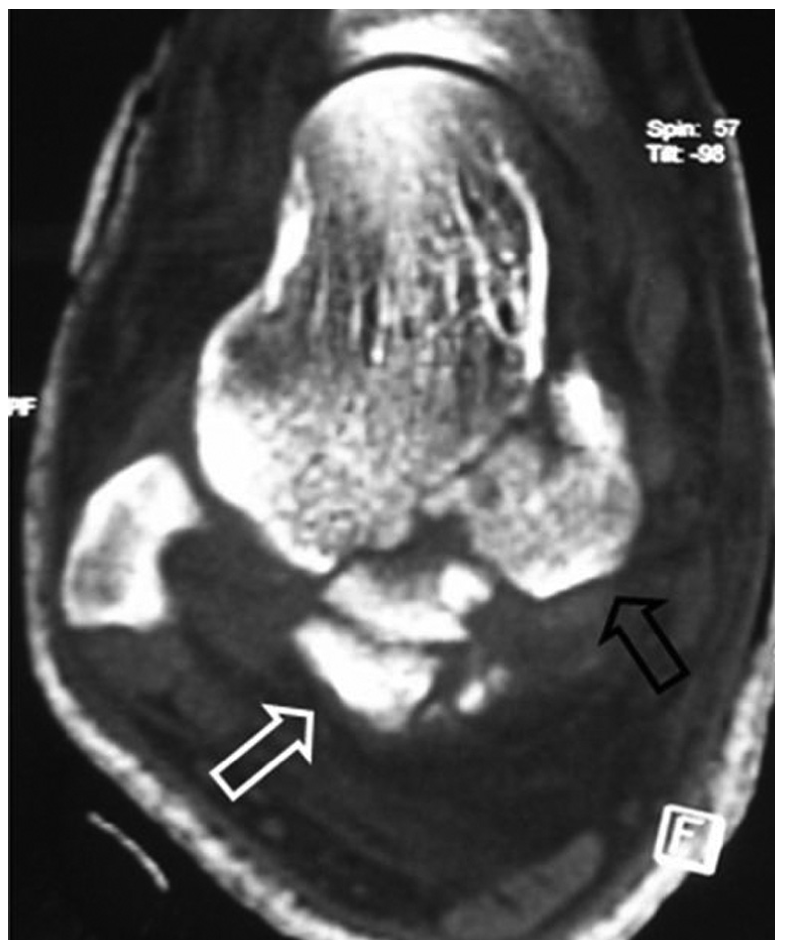
A 24-year-old man was admitted with right ankle and back pain after a traffic accident. The physical and radiologic examinations revealed L1 compression fracture and comminuted coronal plane fracture of the posterior part of the talar body also involving the posterolateral process (Fig. 1). The patient underwent surgery on the day of his admission. He was positioned prone on the operating table. Initially, the vertebra fracture was addressed and posterior instrumentation and fusion were performed. Following the closure of the lumbar incision, the left leg was draped separately with the patient still in the prone position. A folded surgical drape was placed beneath the ankle to facilitate the manipulation of the foot. A 4.0-mm arthroscope was introduced through the posterolateral portal, and a 4.0-mm full-radius shaver was introduced through the posteromedial portal, which were made adjacent to the Achilles tendon just above a line drawn from the tip of the lateral malleolus, parallel to the foot sole. Gentle gravity flow was used for irrigation to avoid extravasation.
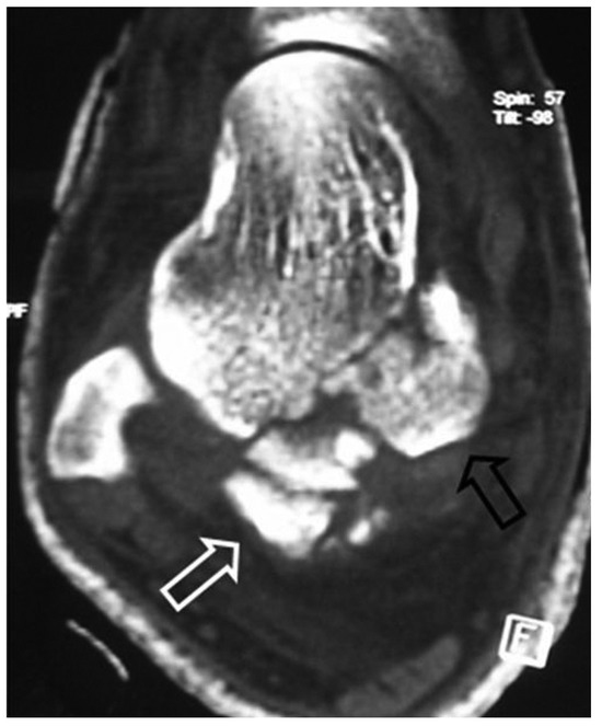
Figure 1.
Axial computed tomography scan. Note the posterolateral process fracture (white arrow) and the large fracture fragment at the posteromedial part of the talar body (black arrow).
The torn flexor retinaculum fibers were debrided. The joint capsule and fatty tissues were removed. The muscular part of the flexor hallucis longus (FHL) was identified and the shaver was kept lateral to it. Care was taken to protect the neurovascular structures prior to the exposure of the FHL tendon. The posterolateral process was broken and floating, attached to the posterior talofibular ligament and adjacent soft tissues. As the posterior talofibular ligament was loose, its release and debridement were not as easy as in the classical os trigonum excision, where the detachment of a tight ligament is technically not so demanding. After the release of the posterior talofibular ligament, the posterolateral process was removed. To gain better access to the tibiotalar joint space and to facilitate manipulation, the tibial slip was also released. The FHL tendon was identified. Its sheath was entered with the scope and released further with a punch to expose its fibro-osseous tunnel. The small osteochondral fragments were detected adjacent to the FHL and were removed. During the entire procedure, care was taken to remain lateral to the FHL tendon. Small osteochondral debris were also detected in the subtalar joint space, and they were washed out.
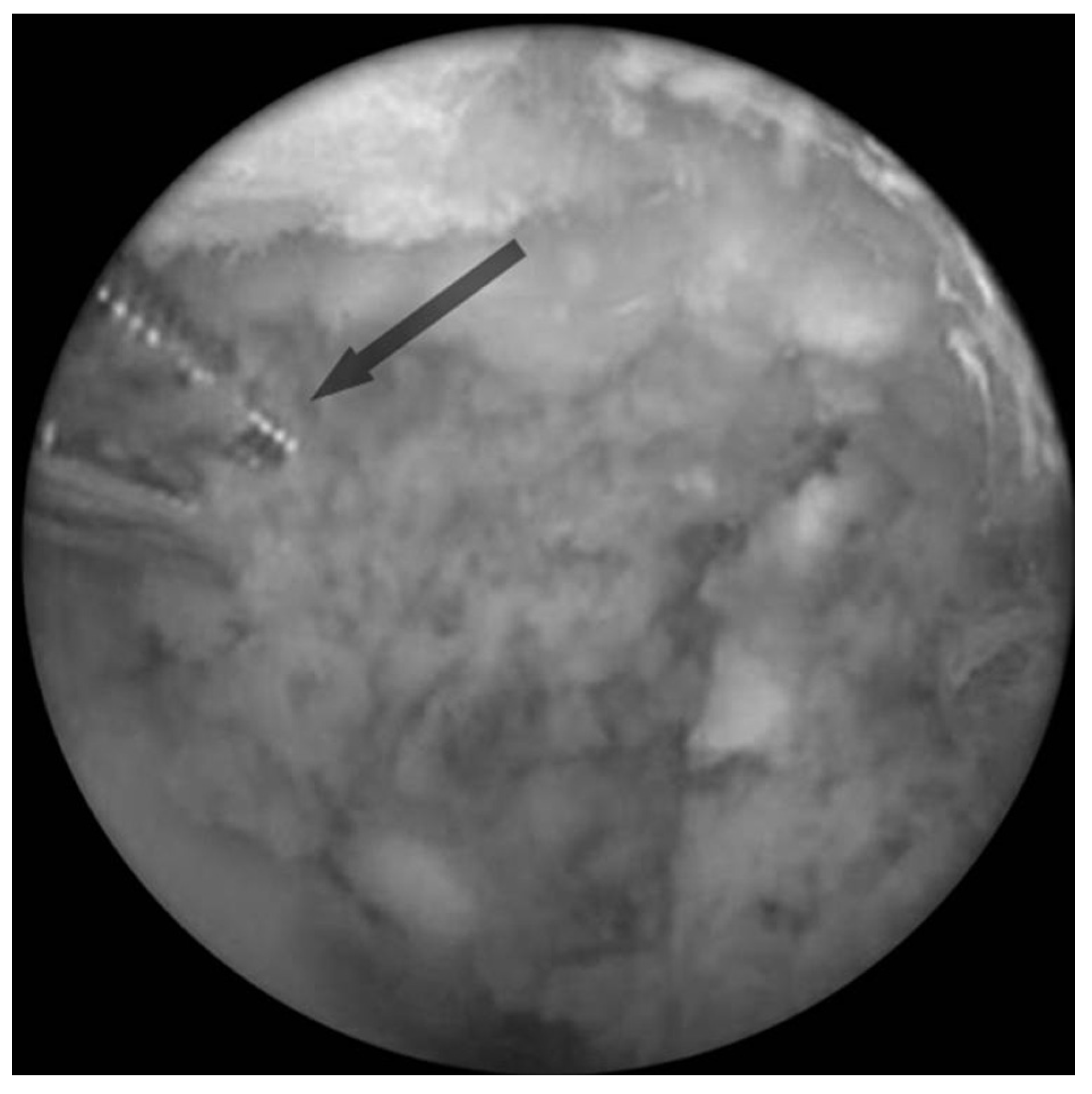
Under arthroscopic and fluoroscopic control, a partially threaded Kirschner wire was passed percutaneously, perpendicular to the large fragment (Fig. 2) from the posteromedial aspect of the ankle. The reduction was achieved by pivoting with the Kirschner wire and manipulation with an arthroscopic probe, and then the wire was advanced to fix the fragment. A second percutaneous Kirschner wire was passed to complete the fixation (Fig. 3).
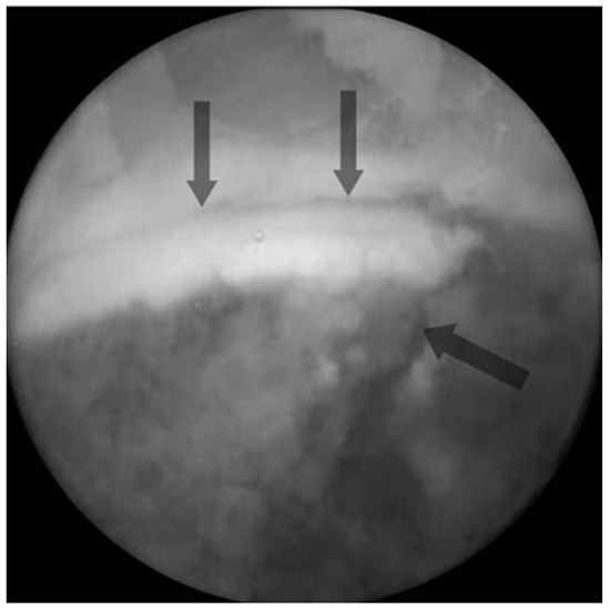
Figure 2.
Arthroscopic view of the large fracture fragment at the posterolateral part of the talar body. The fracture line is delineated by arrows.
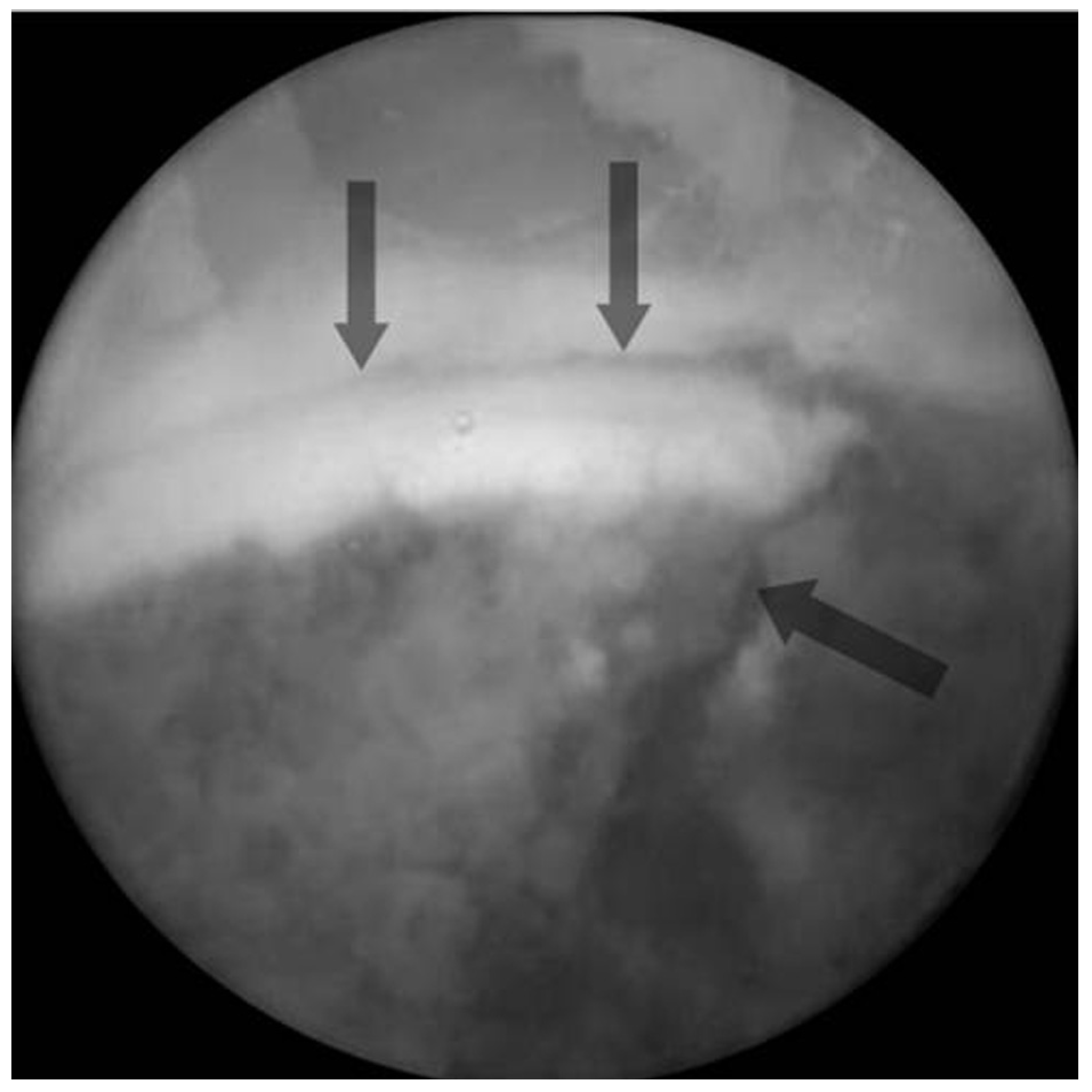
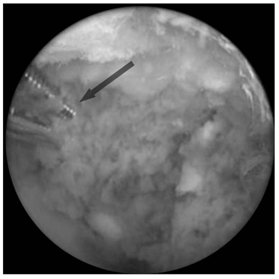
Figure 3.
The fixation of the fracture was achieved with two percutaneous Kirschner wires (arrow).
Postoperatively, a below-the-knee plaster cast splint was applied with a compressive bandage (Fig. 4). The foot was elevated until the soft-tissue swelling settled. Ten days after the injury, operative portals healed without complication. By 7 weeks, the splint and Kirschner wires were removed, and range-of-motion exercises began. Progressive weightbearing was allowed after 10 weeks. In 12 months, the patient had resumed normal activities and returned to his daily style of living with no gait abnormality or ankle complaint. The control computed tomography scan at 12 months revealed that the posteromedial fracture fragment was healed with a solid union (Fig. 5). At his final follow-up, the patient had 15° of active dorsiflexion and 55° of active plantarflexion of the ankle; there was no paresthesia on the foot and ankle.
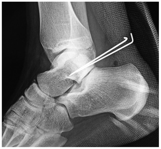
Figure 4.
Postoperative lateral radiograph.
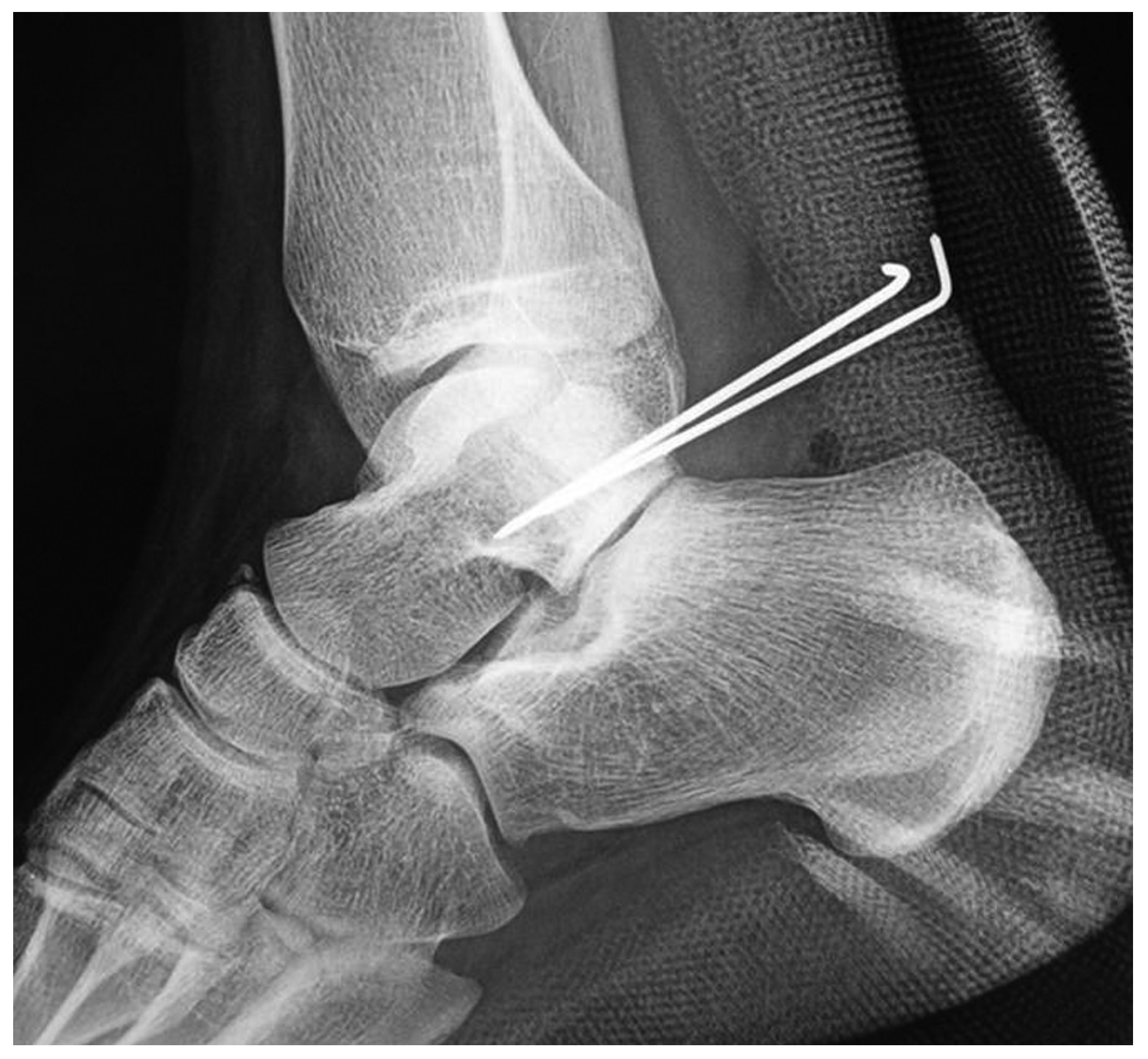
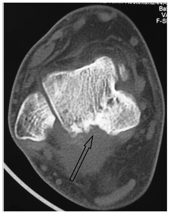
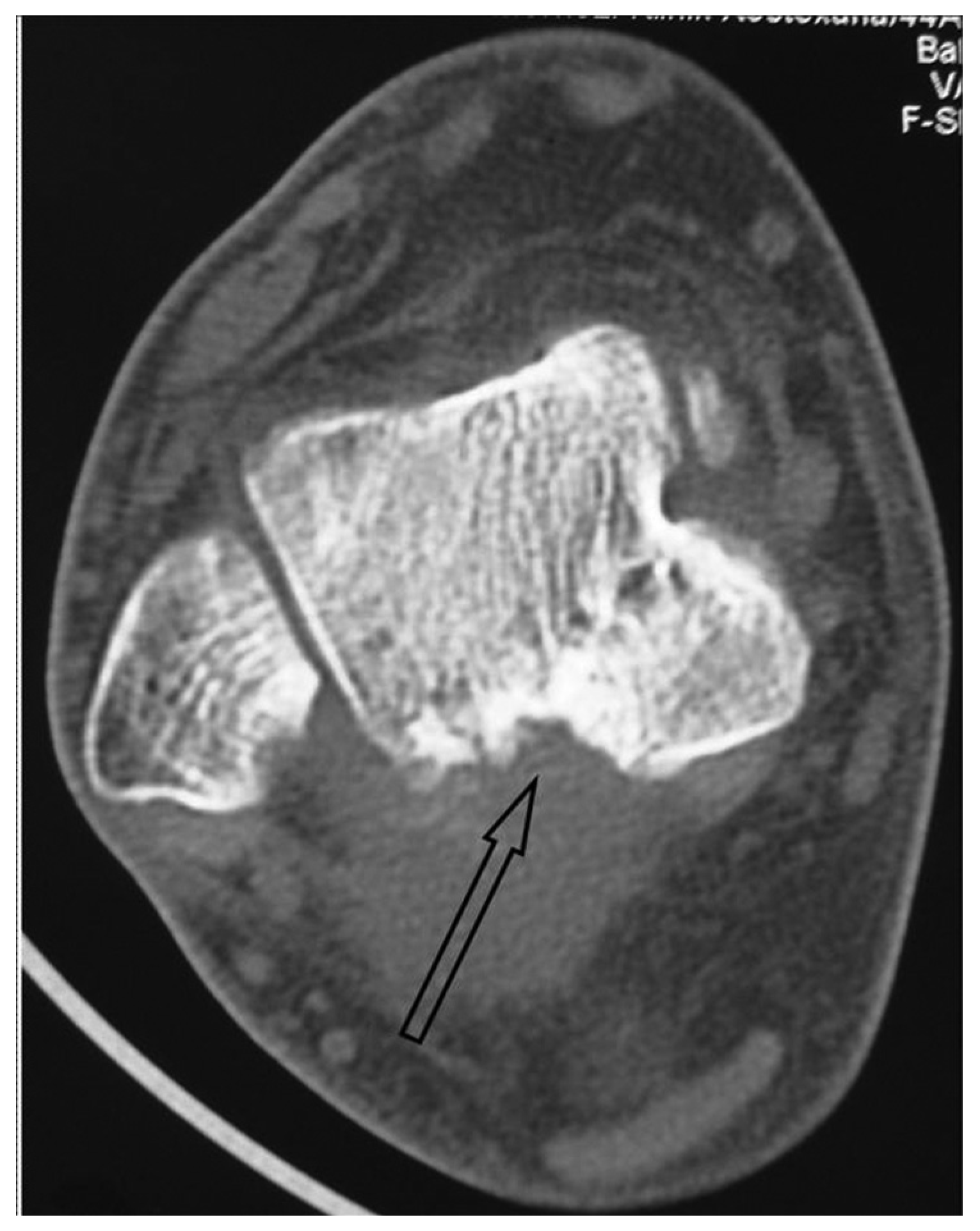
Figure 5.
Axial computed tomography scan 12 months after the operation. The posterolateral process was completely removed and the posteromedial fracture fragment healed with a solid union (arrow).
Discussion
The patient in this case had a comminuted talus fracture involving the posterior body and posterolateral process. This association was important in the decision for a posterior approach, which allowed us to debride the small fracture fragments of the posterolateral process as a part of the treatment, and thus to gain access to the main body fracture fragment to perform its anatomical osteosynthesis.
There is an inherent conflict in the management of displaced coronal plane fractures of the talar body. These fractures, often involving multiple joints, must be treated with immediate anatomical reduction and stable fixation to prevent long-term complications such as avascular necrosis, hindfoot malalignment, and peritalar arthritis.[5,8–10] However, this goal must be achieved with minimal disturbance of the soft tissue because of the tenuous nature of the talar body supply, which makes it again prone to avascular necrosis.[6,8,11]
All of these factors considered, surgeons dealing with displaced talus fractures have to balance the potential complications of suboptimal reduction against the risk of wound problems and avascular necrosis when deciding between a closed and open technique. Posterolateral process fracture adds to the problem with a potential risk of future posterior ankle pain. This can be caused by a symptomatic os trigonum or an FHL tenosynovitis attributable to irritation by the fracture fragments.[12–20]
Our patient was also prone to these problems by the comminuted posterolateral process fracture with small fragments adjacent to the tendon sheath of the FHL. The fracture also involved the subtalar joint, with osteochondral debris filling this space.
The open surgical approaches to the hindfoot are associated with some complications, mainly surgical wound-healing problems because of the poor blood supply to the posterior ankle region.[21–24] To avoid vascular damage and soft-tissue problems, fluoroscopy-assisted closed reduction and percutaneous fixation has been recommended in the treatment of less displaced fractures.[5,25]
The arthroscopically assisted approaches permit removal of free-floating, intra-articular osteochondral fragments, direct visual assessment of fracture reduction, and fixation stability as in the open technique, while causing minimal disruption of the remaining intact talar blood supply as in the closed fluoroscopic technique.[5–7] Although arthroscopy is widely used in the management of transchondral dome fractures, as far as we know, only three cases of arthroscopic treatment of talar body fractures have been reported. In all of these cases, classical anterior portals were used.[5–7]
The two-portal hindfoot endoscopy, first described by Van Dijk in 2000,[26] is a relatively new technique that is becoming increasingly popular.[13,20] Treatment of posteriorly located osteochondral defects of the ankle joint, removal of loose bodies, osteophytes, and ossicles in the posterior compartment of ankle and subtalar joints; posterior ankle impingment; deltoid ligament problems, flexor hallucis longus tendonitis, and tarsal tunnel syndrome are some of the indications of hindfoot endoscopy.[20]
The senior surgeon of this case (T.O.) learned the two-portal posterior approach from its author, Dr. Van Dijk, in Academic Medical Center, Amsterdam, and gained experience by performing more than 30 hindfoot endoscopies with Dr. Van Dijk’s technique.
We found this approach most appropriate for use with our patient with posterior process fracture. Debridement of the posterior ankle compartment eliminated the potential causes of future posterior ankle problems, such as posterior impingement syndrome, FHL tenosynovitis, and subtalar joint arthrosis. The approach also provided us a better exposure of the main fracture fragments and subtalar joint to assess the reduction.
We think that combined excision of the posterolateral process and fixation of the talar body fracture with the two-portal hindfoot endoscopy can be a reliable treatment option for the posterior part talar body fractures involving posterolateral process.
Financial Disclosure
None reported.
Conflicts of Interest
None reported.
References
- Frank A, Cohen P, Beaufils PH, et al: Arthroscopic treatment of osteochondral lesions of the talar dome. Arthroscopy5: 57, 1989.
- Pritsch M, Horoshowski H, Farine I: Arthroscopic treatment of osteochondral lesions of the talus. J Bone Joint Surg Am68: 862, 1986.
- Kelberine F, Frank A: Arthroscopic treatment of osteochondral lesions of the talar dome: a retrospective study of 48 cases. Arthroscopy15: 77, 1999.
- Van Buecken K, Barrack RL, Alexander AH, et al: Arthroscopic treatment of transchondral talar dome fractures. Am J Sports Med17: 350, 1989.
- Saltzman CL, Marsh JL, Tearse DS: Treatment of displaced talus fractures: an arthroscopically assisted approach. Foot Ankle Int15: 630, 1994.
- Monllau JC, Pelfort X, Hinarejos P, et al: Combined fracture of the talus: arthroscopic treatment. Arthroscopy17: 418, 2001.
- Subairy A, Subramanian K, Geary NP: Arthroscopically assisted internal fixation of a talus body fracture. Injury35: 86, 2004.
- Canale ST, Kelly FB Jr: Fractures of the neck of the talus. Long-term evaluation of seventy-one cases. J Bone Joint Surg Am60: 143, 1978.
- Hawkins LG: Fractures of the neck of the talus. J Bone Joint Surg Am52: 991, 1970.
- Kenwright J, Taylor RG: Major injuries of the talus. J Bone Joint Surg Br52: 36, 1970.
- Szyszkowitz R, Reschauer R, Seggl W: Eighty-five talus fractures treated by ORIF with five to eight years of follow-up study of 69 patients. Clin Orthop Relat Res199: 97, 1985.
- Blake RL, Lallas PJ, Ferguson H: The os trigonum syndrome: a literature review. JAPMA82: 154, 1992.
- Jerosch J, Fadel M: Endoscopic resection of a symptomatic os trigonum. Knee Surg Sports Traumatol Arthrosc14: 1188, 2006.
- Hedrick MR, McBryde AM: Posterior ankle impingement. Foot Ankle Int15: 2, 1994.
- Martin BF: Posterior triangle pain: the os trigonum. J Foot Surg8: 312, 1989.
- Bureau NJ, Cardinal E, Hobden R, et al: Posterior ankle impingement syndrome: MR imaging findings in seven patients. Radiol215: 497, 2000.
- Abramowitz Y, Wollstein R, Barzilay Y, et al: Outcome of resection of a symptomatic os trigonum. J Bone Joint Surg Am85-A: 1051, 2003.
- Karasick D, Schweitzer ME: The os trigonum syndrome: imaging features. AJR Am J Roentgenol166: 125, 1996.
- Wakeley CJ, Johnson DP, Watt I: The value of MR imaging in the diagnosis of the os trigonum syndrome. Skeletal Radiol25: 133, 1996.
- Van Dijk CN: Hindfoot endoscopy. Foot Ankle Clin11: 391, 2006.
- Morag G, Maman E, Arbel R: Endoscopic treatment of hindfoot pathology. Arthroscopy19: E13, 2003.
- McAllister JL, Thordarson DB: Complications of foot and ankle surgery. Clin Sports Med18: 927, 1999.
- Schepsis AA, Leach RE: Surgical management of Achilles tendinitis. Am J Sports Med15: 308, 1987.
- Nelen G, Martens M, Burssens A: Surgical treatment of chronic Achilles tendinitis. Am J Sports Med17: 754, 1989.
- Lemaire RG, Bustin W: Screw fixation of fractures of the neck of the talus using a posterior approach. J Trauma20: 669, 1980.
- Van Dijk CN, Scholten PE, Krips R: A 2-portal endoscopic approach for diagnosis and treatment of posterior ankle pathology. Arthroscopy16: 871, 2000.
© 2009 American Podiatric Medical Association