


Abstract
Lipoma arborescens is an uncommon pseudotumoral synovial lesion usually located in the suprapatellar pouch of the knee. Lipoma arborescens involving the synovial sheaths of the tendons is exceedingly rare. This diagnosis should be considered, particularly in patients with chronic joint effusion. We report a case with lipoma arborescens affecting the synovial sheaths of the peroneal tendons without involvement of the adjacent ankle joint. To our knowledge, this is the second reported case of lipoma arborescens involving tenosynovial sheaths of tendons arround the ankle joint without ankle joint involvment.
Lipoma arborescens (LA) is a rare benign condition of the synovial joints and bursae, characterized by villous proliferation of the synovium and diffuse hyperplasia of fat tissue in the subsynovial layer.[1] Lipoma arborescens is usually monoarticular and preferentially involves the suprapatellar pouch of the knee joint.[2,3,4,5,6] Bilateral involvement of the knees, wrists, and hips, and multiple affected joints has also been observed.[4,7,8] The condition sometimes occurs in the subacromial–subdeltoid and bicipital bursae.[9,10,11] Tenosynovial LA may occur in the hand.[12] Ankle joint involvement is very rare,[13,14] and isolated involvement of the synovial sheaths of the tendons around the ankle joint is extremely rare.[15]
To our knowledge only one case of LA involving the tendon sheaths around the ankle joint alone without involvement of the adjacent joint, has been reported in the English language literature.[15] We present the second case of LA with isolated involvement of the tendon sheath of the peroneus longus and peroneus brevis at the level of ankle in an adult patient.
Case Report
A 26-year-old female patient presented to the Mustafa Kemal University, Faculty of Medicine, Orthopaedic and Trauma Clinic, Antakya, Hatay, Turkey, with a 3-year history of pain behind the lateral malleolus of the right ankle. The pain began intermittently but gradually became continuous, especially upon standing and walking. In the previous 8 months, she had developed a painful, gradually enlarging soft mass behind the lateral malleolus. She had no history of trauma, arthritis, or any systemic disorder. She had been diagnosed with nonspecific tendinitis and received nonsteroidal analgesics, but her symptoms did not improve.
Physical examination revealed a tender palpable mass of approximately 3 × 2 cm in dimensions with no erythema or skin changes. Physical examination of the ankle joint revealed no effusion, erythema, or tenderness with free and full range of motion.
Routine laboratory tests including the erythrocyte sedimentation rate, white cell count, and C-reactive protein serum rate were within normal limits. Rheumatoid factor was negative.
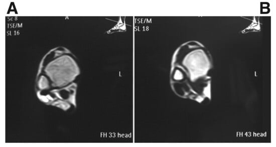
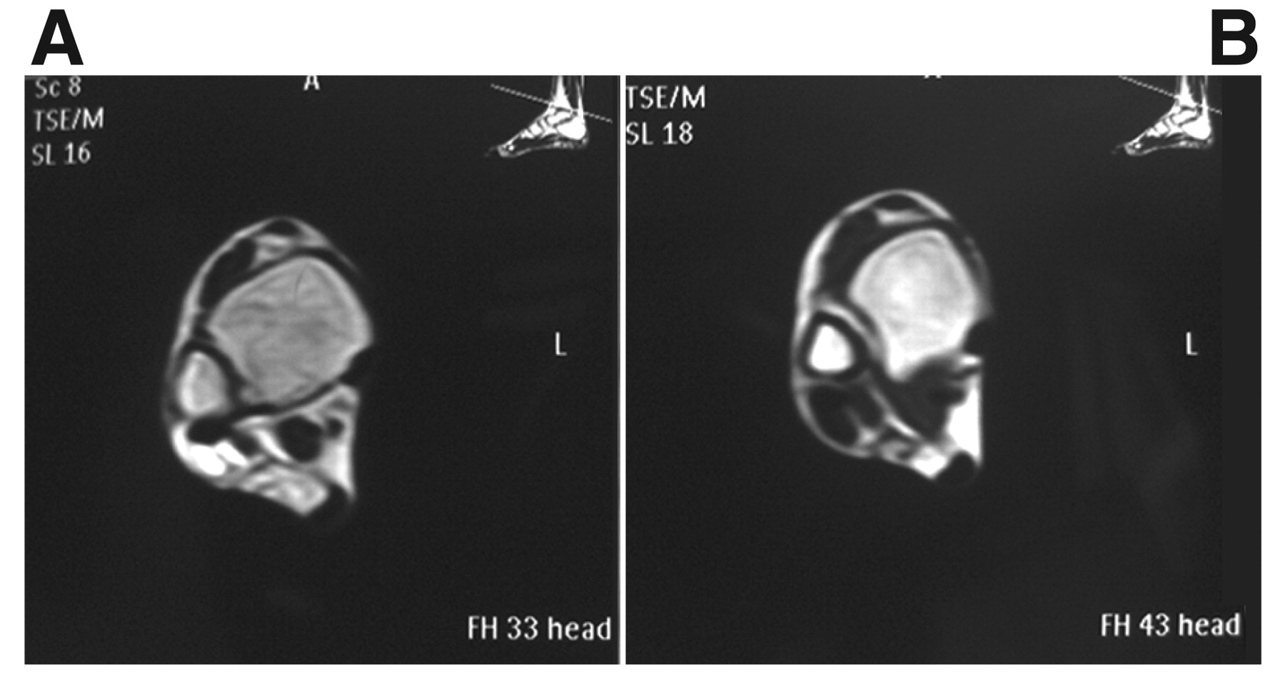
Standard radiographs of the ankle joint revealed normal findings with no underlying bone or joint abnormality. Magnetic resonance imaging (MRI) revealed a mass encasing the peroneal tendons with high-signal intensity on both T1- and T2-weighted sequences, with low-signal intensity on the fat-suppressed scan and enhancement on the post-contrast scans (Fig. 1). The high signal on the T1- and T2-weighted scans, with loss of signal on the fat-suppressed sequences, was compatible with the presence of fatty tissue. The underlying ankle joint was normal and had no joint effusion.
Figure 1.
Axial T1-weighted magnetic resonance image of the left ankle shows distension of the synovial sheaths of the peroneal tendons with fat tissues of high-signal intensity (A) that are suppressed with a fat suppression technique (B). The ankle joint was normal.
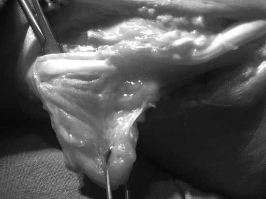
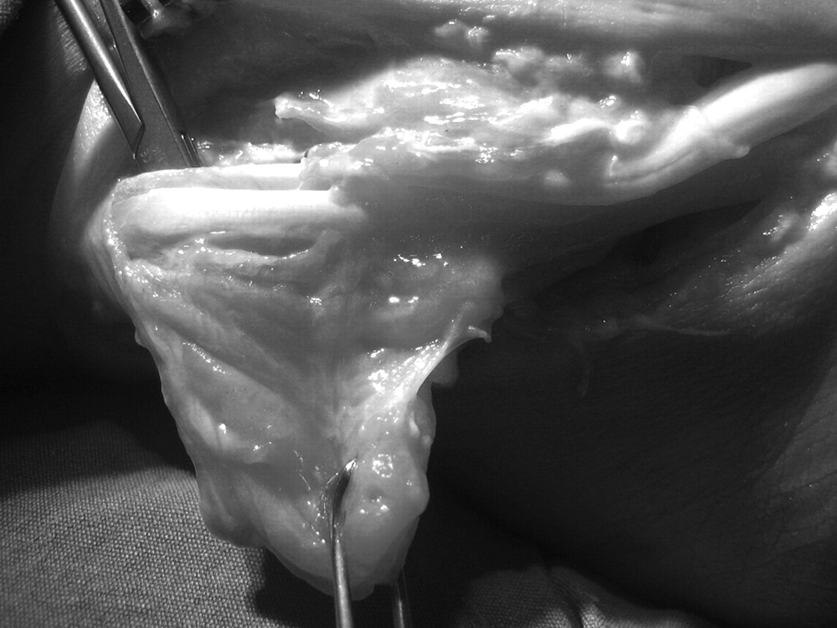
Under spinal anesthesia, a curvilinear lateral incision was used to approach the mass. A yellow mass, approximately 2 × 3 × 2 cm in dimensions was found arising from the peroneus longus and peroneus brevis tendon sheath (Fig. 2). The size and the compressive effects of the mass had led to flattening of the peroneus longus tendon, and the sural nerve had been under tension from the underlying mass. Total resection of the mass together with the synovial sheath of the peroneus longus and peroneus brevis tendon was conducted. The sural nerve was freed and decompressed.
Figure 2.
Surgical exposure of the lesion reveals macroscopic appearance of the lesion. Note the frond-like projection arising from the synovial sheath of peroneal tendons.
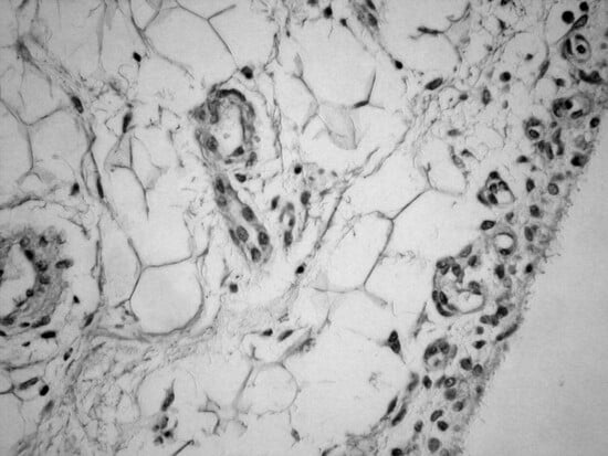
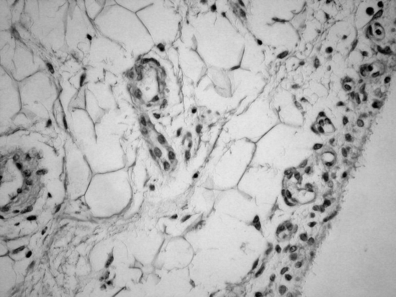
Histopathologic examination revealed synovial villous proliferations and diffuse replacement of the subsynovial tissue by mature fat cells with coexistence of congested hyperemic capillaries and chronic inflammatory cells, which confirmed the diagnosis of LA (Fig. 3).
Figure 3.
Photomicrograph of the specimens of the excised mass shows hyperplasia of the mature fat tissues of the subsynovial stroma. Focal areas of synovitis with inflammatory reaction were noted (H&E, × 100).
Discussion
Lipoma arborescens affecting the synovial sheaths of the tendons is extremely rare, and only a few cases have been reported in the English language literature. Siva et al[12] reported the first case of tenosynovial LA affecting the dorsal and volar tendon sheaths of the wrist.
Huang et al[13] reported a case of tenosynovial LA in a child, affecting synovial linings of the peroneal, flexor hallucis longus, flexor digitorum longus, and posterior tibialis tendon sheaths. Babar et al[14] reported a case of LA of the ankle joint in an adult patient with involvement of the intra-articular synovium as well as the synovial sheath of the tendons around the ankle. In all of the above mentioned cases, adjacent synovial joints were also involved. Senocak et al[15] reported a case of tenosynovial LA affecting synovial linings of the extensor digitorum longus tendon sheath at the ankle region; however, in that case the ankle joint appeared normal. To our knowledge, this is only the second reported case of LA involving the synovial lining of the extra-articular tendon sheaths of the ankle region without ankle joint involvement.
Lipoma arborescens may be a misleading term; Hallel et al[1] suggested that the lesion be called villous lipomatous proliferation of the synovial membrane, because LA implies a tumor. The exact etiology is unknown; it may be associated with degenerative joint disease, chronic rheumatoid arthritis, or prior trauma.[1,16,17,18,19] However, primary cases without underlying chronic intra-articular pathologic conditions have been reported. In our case the ankle joint was normal and there was no history of previous joint disease involving the ankle joint, and no history of prior trauma. Males are affected much more commonly than females. Patients range in age from 9 to 66 years.[16] When it affects the synovial joints, LA typically presents as an insidious onset of swelling over a period of months and years followed by pain, which can be debilitating. It can simulate oligoarthritis without specific signs and insidious symptoms, as was the case with our patient. In our case, pain was the presenting feature; the location of the lesion between the peroneus longus and brevis tendon may explain this painful presentation.
Laboratory tests in LA are not helpful. Radiographs of patients with LA are nonspecific and may reveal soft-tissue swelling, degenerative changes around the joints, and bone erosions. Ultrasound usually shows joint effusion and may also demonstrate villous frond-like projections, signifying underlying synovial hypertrophy.[15] Computed tomography (CT) may show a frond-like mass of fat attenuation, although the villous nature may be difficult to recognize on CT.[15] Magnetic resonance imaging alone is often sufficient to make the diagnosis. Indeed, in 21 of the 33 cases reported by Vilanova et al,[20] LA was diagnostically established on the MRI features alone.
Magnetic resonance imaging demonstrates large villous, frond-like masses with signal characteristics of fat.[20] The fat typically shows high-signal intensity on both T1- and T2-weighted sequences, mirroring the subcutaneous tissue signal, and suppresses on the fat saturation sequences. Enhancement following intravenous administration of contrast material may be seen in the overlying inflamed synovium. Lesions have been mistaken for liposarcoma, and the key to diagnosis is recognition of the diffuse synovial (joint or bursal) origin, as well as the multilobulated frond-like contour.
Huang et al[13] has demonstrated the fact that fat cells might have myxoid changes in regions of these lesions and thus cause difficulty in identifying the fat content by means of its signal intensity characteristics. These authors reported a case of LA around the ankle joint; the pattern of the LA was slightly different from that observed in its more classic location around the knee. The characteristic filiform frond-like appearance, as seen in the knee, was less evident and a more diffuse fatty but infiltrative pattern was observed in the ankle.
The differential diagnosis for LA may include pigmented villonodular synovitis, localized nodular synovitis or synovial giant cell tumor, synovial osteochondromatosis, synovial hemangioma, rheumatoid arthritis, and other types of inflammatory synovitis.[13,16] The suggested treatment for tenosynovial LA is open synovectomy. Synovectomy alleviates the synovial thickening and effusions but not the associated osteoarthritis.[1,4,6] Recurrence following synovectomy has been reported. In our case no recurrence was observed at 6 months follow-up.
Lipoma arborescens is a rare synovial disorder that must be considered in the differential diagnosis of chronic pain around the ankle joint. Careful planning and investigation for any suspected tenosynovial mass is essential to establish the diagnosis and evaluate the extent of involvement.
Financial Disclosure
None reported.
Conflicts of Interest
None reported.
References
- Hallel T, Lew S, Bansal M: Villous lipomatous proliferation of the synovial membrane (lipoma arborescens). .J Bone Joint Surg Am70::264. ,1988. .
- Martinez D, Millner PA, Coral A, et al: Case report: synovial lipoma arborescens. .Skeletal Radiol21::393. ,1992. .
- Narvaez J, Narvaez JA, Ortega R, et al: Lipoma arborescens of the knee. .Rev Rhum Engl Ed66::351. ,1999. .
- Martin S, Hernandez L, Romero J, et al: Diagnostic imaging of lipoma arborescens. .Skeletal Radiol27::325. ,1998. .
- Soler T, Rodriguez E, Bargiela A, et al: Lipoma arborescens of the knee: MR characteristics in 13 joints. .J Comput Assist Tomogr22::605. ,1998. .
- Grieten M, Buckwalter KA, Cardinal E, et al: Case report: lipoma arborescens (villous lipomatous proliferation of the synovial membrane). .Skeletal Radiol23::652. ,1994. .
- Nisolle JF, Blouard E, Baudrez V, et al: Subacromial-subdeltoid lipoma arborescens associated with a rotator cuff tear. .Skeletal Radiol28::283. ,1999. .
- Levadoux M, Gadea J, Flandrin P, et al: Lipoma arborescens of the elbow: a case report. .J Hand Surg [Am]25::580. ,2000. .
- Ryu KN, Jaovisidha S, Schweitzer M, et al: MR imaging of the lipoma arborescens of the knee joint. .Am J Roentgenol167::1229. ,1996. .
- Dinauer P, Bojescul JA, Kaplan KJ, et al: Bilateral lipoma arborescens of the bicipitoradial bursa. .Skelet Radiol31::661. ,2002. .
- Doyle AJ, Miller MV, French JG: Lipoma arborescens in the bicipital bursa of the elbow: MRI findings in two cases. .Skelet Radiol31::656. ,2002. .
- Siva C, Brasington R, Totty W, et al: Synovial lipomatosis (lipoma arborescens) affecting multiple joints in a patient with congenital short bowel syndrome. .J Rheumatol29::1088. ,2002. .
- Huang GS, Lee HS, Hsu YC, et al: Tenosynovial lipoma arborescens of the ankle in a child. .Skeletal Radiol35::244. ,2006. .
- Babar SA, Sandison A, Mitchell AW: Synovial and tenosynovial lipoma arborescens of the ankle in an adult: a case report. .Skeletal Radiol37::75. ,2008. .
- Senocak E, Gurel K, Gurel S, et al: Lipoma arborescens of the suprapatellar bursa and extensor digitorum longus tendon sheath. .J Ultrasound Med26::1427. ,2007. .
- Murphey MD, Carroll JF, Flemming DJ, et al: From the archives of the AFIP: benign musculoskeletal lipomatous lesions. .Radiographics24::1433. ,2004. .
- Marui T, Yamamoto T, Kimura T, et al: A true intra-articular lipoma of the knee in a girl. .Arthroscopy18::E24. ,2002. .
- Armstrong SJ, Watt I: Lipoma arborescens of the knee. .Br J Radiol62::178. ,1989. .
- Feller JF, Rishi M, Hughes EC: Lipoma arborescens of the knee: MR demonstration. .Am J Roentgenol163::162. ,1994. .
- Vilanova JC, Barcelo J, Villalon M, et al: MR imaging of lipoma arborescens and the associated lesions. .Skeletal Radiol32::504. ,2003. .
© 2009 American Podiatric Medical Association