


Abstract
Inclusion cysts are benign lesions that appear as a consequence of traumatic inclusion of epidermal cells into the dermis. They can be painful if they appear under pressure areas, especially the metatarsal heads. We report a case of a 36-year-old woman with an intractable plantar keratosis lesion under the third metatarsal head of 3 years’ duration. Ultrasonography revealed the presence of a subcutaneous mass with a growing epidermoid. It was surgically excised, and pathology confirmed the diagnosis of a plantar epidermoid cyst. All symptoms disappeared after the excision of the lesion. This case should alert the clinician about the existence of keratotic lesions in the metatarsal heads commonly diagnosed and treated as intractable plantar keratosis, although they are not directly derived from metatarsal overload. Some of the lesions could be directly derived from skin problems aggravated by pressure from the metatarsal head. This should be taken into consideration when addressing the management of these lesions.
Intractable plantar keratosis (IPK) is defined as a discrete lesion that appears as a corn within a callus usually under a metatarsal head. It is generally accepted that these lesions are attributable to pressure overload from a structural or rigidly adapted osseous deformity of the involved metatarsal.[1,2] The treatment of IPK is difficult as evidenced by the multiplicity of treatments, which usually include an osteotomy of the involved lesser metatarsal.[1,2,3,4,5] However, it is not clear what role the skin plays in these lesions and if skin disorders can be a causative factor for the development of IPK.
Inclusion cysts are benign soft-tissue lesions that result from the proliferation of epidermal cells within a circumscribed space of the dermis. They form a cavity, which is covered by polystratified epithelial tissue. These cavities can contain lipid and keratotic substances. Clinically they appear as firm, round, and mobile, and they have a white-yellowish color, with sizes that can vary from a few millimeters to several centimeters.[6,7,8] Occasionally they occur bilaterally.[6] These lesions are caused by deposition of epidermal cells into the dermis by a traumatic event or a foreign-body reaction.[6,7,8,9,10] Sometimes, a microtraumatic event such as shoe irritation can be the precipitating factor.[7]
We present here a clinical case of IPK caused by an epidermoid inclusion cyst in a plantar location and its surgical management and progress.
Case Report
A 36-year-old woman presented to Clínica Universitaria de Podología of Universidad Complutense de Madrid, Madrid, Spain, with a complaint of pain associated with a plantar hyperkeratotic lesion of her right foot. The patient reported a 3-year presence of the lesion; she stated that the pain associated with this lesion was exacerbated by weightbearing activities and she was limiting her overall activity. This was a recurrent problem that had received several episodes of conservative care, consisting of debridement and trimming of the callus and orthotic therapy with accommodative orthoses. The results of these treatments had been poor. The remainder of her medical history was unremarkable.
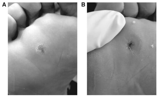
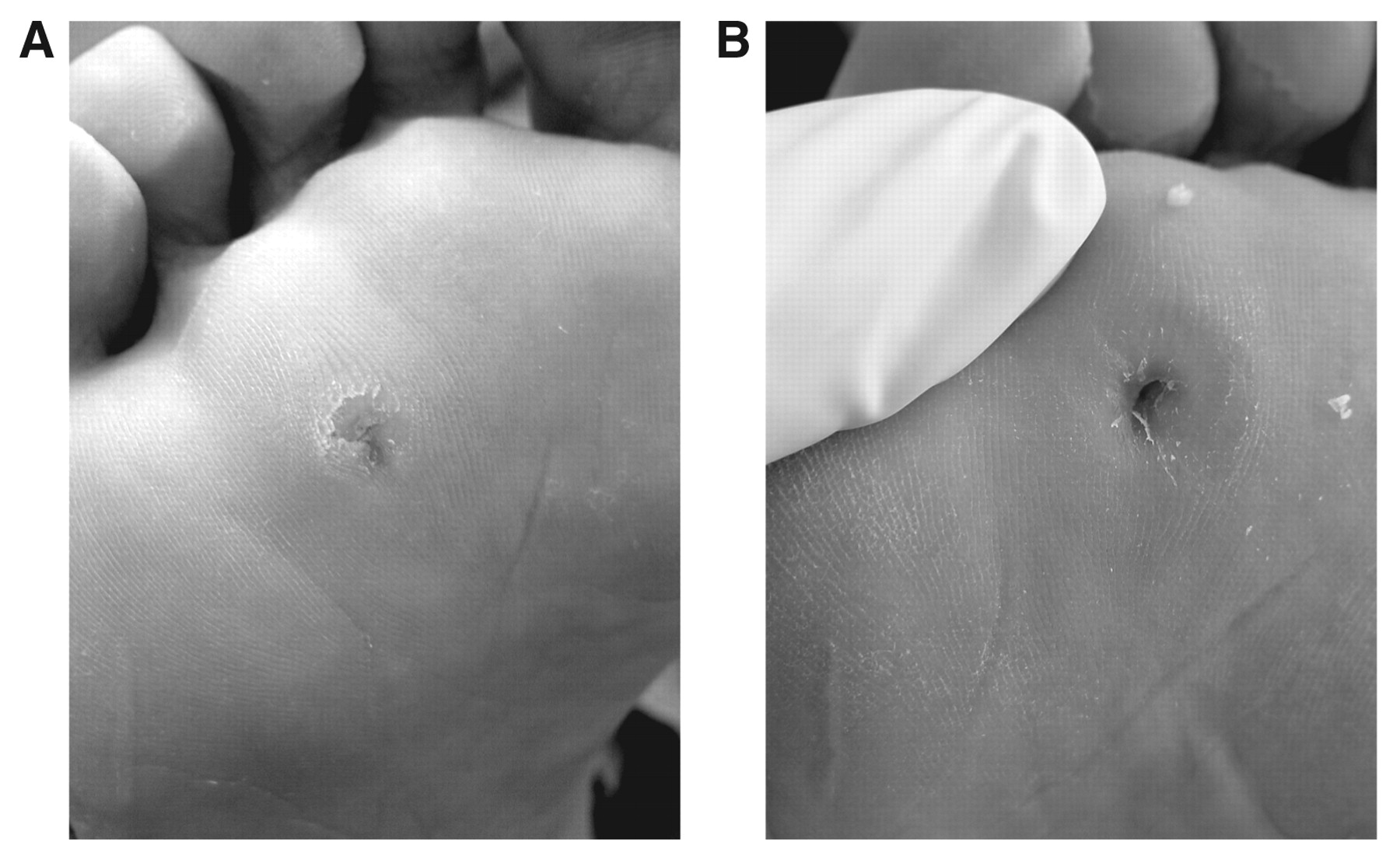
Physical examination revealed a discrete hyperkeratotic lesion located under the third and fourth metatarsal heads of the right foot. Palpation of the lesion elicited pain. Trimming of the callus revealed a discrete corn with a whitish soft hyperkeratotic ring (Fig. 1). The remainder of the physical examination findings were essentially unremarkable. Radiographic examination was performed on her right foot. Anteroposterior radiographs were taken of the right foot, with a marker under the lesion and with the beam oriented 90° to the supporting surface. The radiographs showed that the lesion was located between the third and fourth metatarsal heads. An ultrasound was ordered preoperatively in an attempt to explore the presence of soft-tissue masses. The ultrasound revealed a well-defined cystic image with fine and delineated borders and also confirmed that the location of the lesion was between the third and fourth metatarsal heads.
Figure 1.
External appearance of the plantar keratosis before (A) and after (B) debridement of the callus.
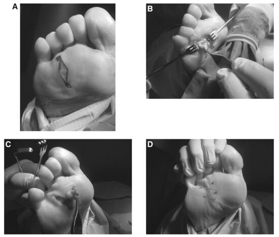
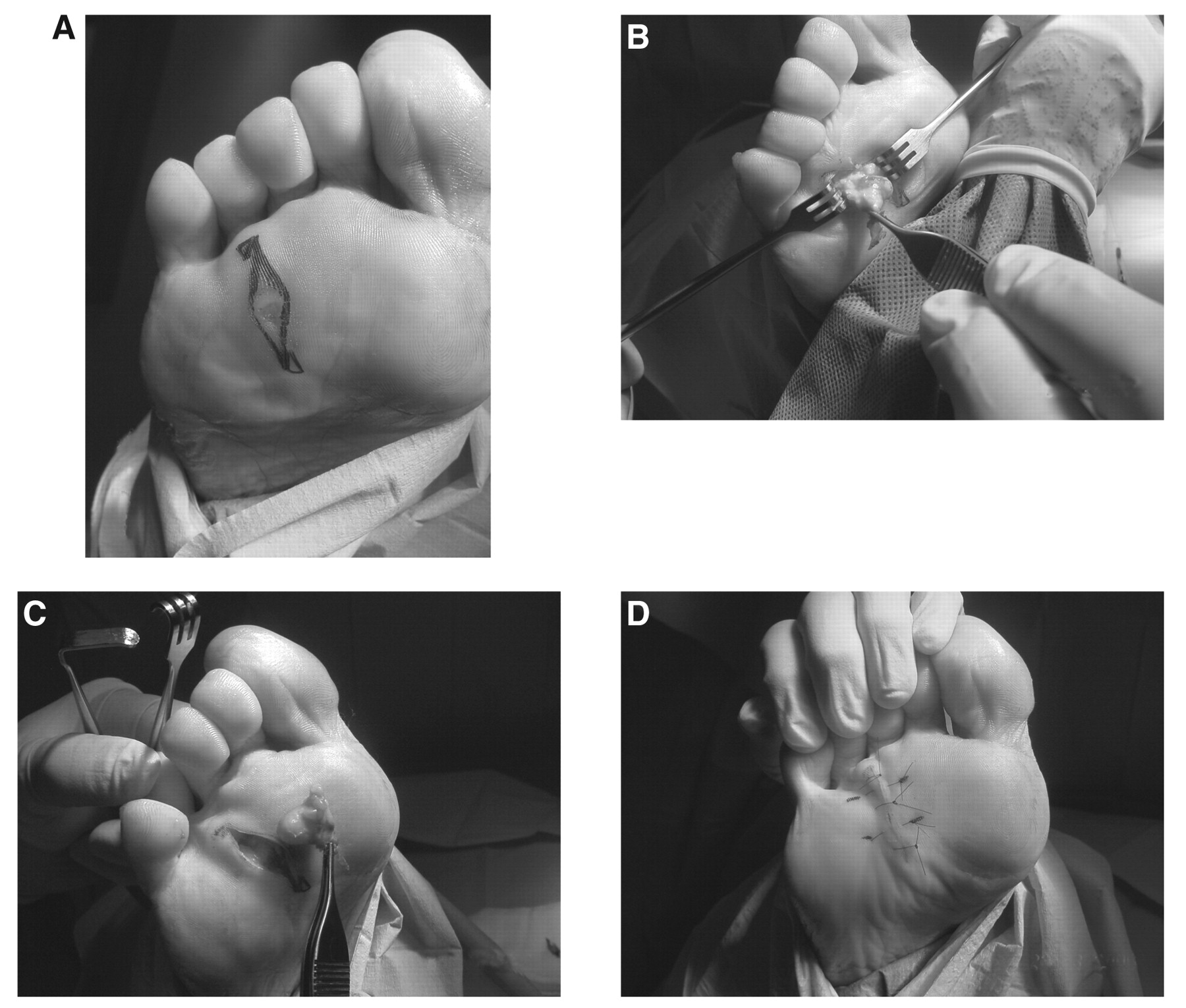
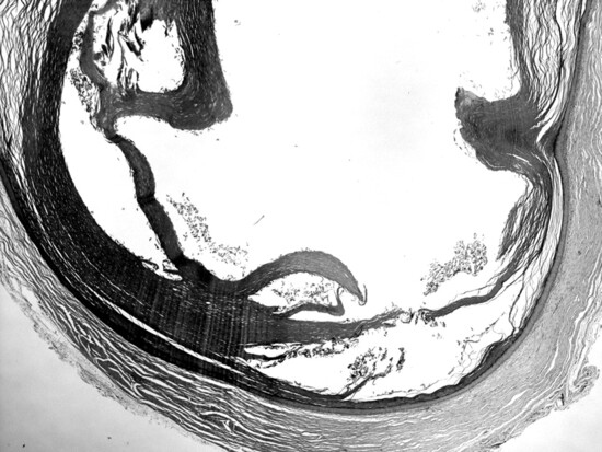
The patient was brought to the operating room for removal of the lesion. The procedure was performed under posterior tibial nerve-block anesthesia with an ankle tourniquet. A plantar semi-elliptical incision was planned for the excision of the whole lesion. The mass was isolated using sharp and blunt dissection until the complete excision of the subcutaneous mass was achieved (Fig. 2). Wound closure was performed with 4-0 nonabsorbable nylon sutures. Vertical mattress sutures were inserted with simple interrupted sutures. The excised mass was sent to the laboratory for pathologic study. The laboratory determined that the cutaneous piece had laminar content. Slides identified a cystic formation covered with polystratified and keratotic epithelium with content that came from the desquamation of the cover (Fig. 3). Some inflammatory changes surrounding the lesion were also noted. The laboratory findings allowed us to establish a definitive diagnosis of epidermoid inclusion cyst.
Figure 2.
Excision of the subcutaneous plantar lesion. A, Incision marked on the plantar surface. B, Subcutaneous mass dissected in the superficial fascia. C, Complete excision of the lesion with the subcutaneous mass. D, Final closure with vertical mattress and simple interrupted sutures.
Figure 3.
Histologic view of the lesion. A cystic formation covered with polystratified and keratotic epithelium was observed, consistent with an inclusion cyst.
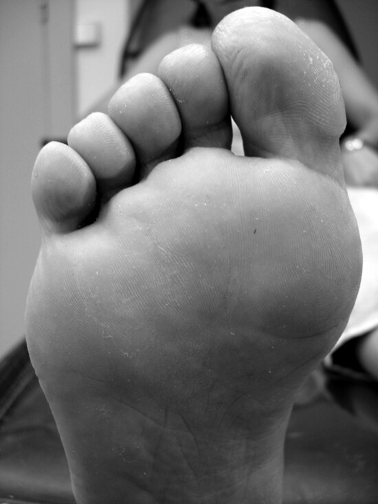
Postoperative visits were routinely performed until complete healing of the wound. Sutures were removed at 25 days, and the postoperative course developed without any incident. The patient was instructed to remain nonweightbearing for 3 weeks, followed by total weightbearing. The patient was followed clinically for 1 year after surgery without any recurrence of the symptoms or the lesion (Fig. 4).
Figure 4.
Appearance 3.5 months postoperatively.
Discussion
Intractable plantar keratoses are discrete hyperkeratotic lesions formed by a corn within a callus and usually develop on the sole under weightbearing prominences. They are a major cause of metatarsalgia and generally appear under metatarsal heads or sesamoids. Because of this location, it seems reasonable that overpressure from the metatarsal head is the etiology of these lesions. However, the present case provides a new explanation of some cases of IPK that may be created by an inclusion cyst. Inclusion cysts represent less than 10% of all soft-tissue masses of the lower extremities,[6,7,9,11] and the sole of the foot is the most common location probably because it is exposed to pressure and microtrauma.[6,12] They can also appear as a result of previous surgery in digits or nails or as the result of minimal incisional surgery.[6,8,13] Although they are generally painless, inclusion cysts can be very painful when located in a high-pressure area, such as the plantar foot. In these cases, they can generate intense pain on weightbearing, damage adjacent bone,[6,10,11,14,15,1617] and even calcify.[18]
In the case presented, the IPK lesion did not have any obvious structural or functional origin as its main etiology. It could be suggested, therefore, that skin processes such as inclusion cysts or foreign bodies could play a central role in the development of some cases of IPK. These primary dermatologic lesions would be aggravated from metatarsal overload instead of metatarsal overload forming an IPK lesion by itself. This case illustrates how some occurrences of IPK are attributable to primary skin conditions that are aggravated by pressure from the metatarsal heads. It is our opinion that under these circumstances, conservative measures and off-loading of the involved metatarsal result in poor outcomes. However, more studies are required to determine what the role of the skin is in the pathogenesis of IPK development.
Diagnostic accuracy is key to avoiding the general tendency to treat these lesions with metatarsal osteotomies. Determining if the plantar lesion is the result of a structural deformity from metatarsal overload or a primary skin condition aggravated from metatarsal pressure is a challenging process. The latter group of patients usually exhibit a firm fibrotic dermal nidus that can be palpated beneath the keratotic lesion. Imaging diagnosis techniques such as simple radiology, magnetic resonance imaging (MRI), or ultrasonography can help to establish a correct diagnosis. Inclusion cysts cannot be detected by simple radiology.[11,14] The use of a radiopaque mark under the lesion can help to establish the exact location of the lesion. If the mark is not directly located under a metatarsal head and appears between two metatarsal heads, suspicions of problems other than pressure overload can be raised. In these cases the clinician should rule out a primary skin condition aggravated from pressure as the origin of the lesion. Magnetic resonance imaging is a more specific test that allows visualization of soft-tissue masses and liquid or fatty collections (MRI on T1-weighted, T2-weighted, and short tau inversion recovery).[15,17,19] Ultrasonography is also an excellent imaging test to differentiate cystic lesions from other soft-tissue masses.[16] In the present case, ultrasonography was performed preoperatively, showing a good defined cystic image with fine and delimitated borders, which preoperatively confirmed the diagnosis of an inclusion cyst.
Treatment of inclusion cysts consists of surgical excision of the whole lesion, including capsule of the cyst, to avoid recurrences. It is important to avoid damaging the cyst during excision to avoid the formation of a keratotic granuloma of giant cells.[9] If there is bone implication, curettage of the affected zone of bone should be performed.[6,10,13,14] Although epidermoid cysts are usually benign lesions, cases have been described of malignization into basal cell carcinoma and squamous cell carcinoma.[6,7,14 ]The case presented here was treated by simple surgical excision without osteotomy procedures. Signs and symptoms were completely eradicated after surgery. At 1-year postoperative follow-up, the patient had a complete resolution of symptoms and an absence of hyperkeratosis.
Plantar incisional approaches on the feet have been a controversial subject for many years.[20] It is a common belief that plantar incisions predispose to painful hypertrophic scars. However, in a retrospective study, Richardson et al[21] reported good results (96%) using plantar incisional approaches for forefoot surgery. In our experience, plantar incisions heal satisfactorily if a strict postoperative regimen of absolute nonweightbearing during the first 3 weeks is followed. Therefore, this could be a good approach for removal of these lesions. In the case presented, the lesion was excised surgically by plantar semi-elliptical incision and healed without complications following an absolute nonweightbearing regimen for the first 3 weeks.
Conclusions
A clinical case of IPK under the third metatarsal head caused by an epidermoid inclusion cyst has been presented. Epidermoid inclusion cysts are cystic benign injuries of rare occurrence in the foot. These cysts are generally asymptomatic, except when they occur in high-pressure areas. This case should alert the clinician to the presence of keratotic lesions plantar to metatarsal heads that are commonly diagnosed in IPK and do not have a biomechanical etiology. Instead, they are the result of a primary dermatologic condition, which is aggravated from metatarsal pressure. Simple excision of the lesion is an optimum treatment for these cases.
Acknowledgments
Simon K. Spooner, PhD, for his support and assistance with manuscript preparation.
Financial Disclosure
None reported.
Conflicts of Interest
None reported.
References
- Pontious J, Lane GD, Moritz JC, et al: Lesser metatarsal V-osteotomy for chronic intractable plantar keratosis: retrospective analysis of 40 procedures. .JAPMA88::323. ,1998. .
- Kitaoka HB, Patzer GL: Chevron osteotomy of lesser metatarsals for intractable plantar callosities. .J Bone Joint Surg Br80-B::516. ,1998. .
- Idusuyi OB, Kitaoka HB, Patzer GL: Oblique metatarsal osteotomy for intractable plantar keratosis: 10-year follow-up. .Foot Ankle Int19::351. ,1998. .
- Mann RA, DuVries HL: Intractable plantar keratosis. .Orthop Clin North Am41::63. ,1973. .
- Leventen EO, Pearson SW: Distal metatarsal osteotomy for intractable plantar keratoses. .Foot Ankle Int10::247. ,1990. .
- Sheff J, Bregman P, Curran T: Bilaterally symmetrical epidermal inclusion cysts with foreign-body giant-cell reaction. .JAPMA88::410. ,1998. .
- Coulter PD, Bouche RT: Traumatically induced inclusion cyst secondary to shoe impingement: report of three cases. .J Foot Ankle Surg38::271. ,1999. .
- Hall JP, Sheffey RJ, Chagares WE, et al: Epidermal inclusion cyst in the foot of a Vietnam veteran. .JAPMA96::445. ,2006. .
- Harish S, Jan E, Ghert M, et al: Pseudotumoral appearance of a ruptured epidermal cyst in the foot. .Skeletal Radiol35::867. ,2006. .
- Patel K, Bhuiya T, Chen S, et al: Epidermal inclusion cyst of phalanx: a case report and review of the literature. .Skeletal Radiol35::861. ,2006. .
- Wang B, Eisler J, Springfield D, et al: Intraosseus epidermoid inclusion cyst in a great toe: a case report and review of the literature. .Arch Pathol Lab Med127::298. ,2003. .
- Lemont H: Traumatic inclusion cyst versus pilar cyst of the foot. .JAPMA82::218. ,1992. .
- Reinherz RP: Epidermal inclusion cysts. .J Foot Ankle Surg32::247. ,1993. .
- Radke S, Walther M, Ettl V, et al: Epidermoid cyst mimicking monoarticular arthritis of the great toe. .Rheumatol Int24::117. ,2004. .
- Chevrot A: Diagnóstico por Imagen de las Afecciones del Pie. Masson, Barcelona. ,2000. .
- Adler R, Sofka CM, Positano RG: Atlas of Foot and Ankle Sonography. Lippincott Williams & Wilkins, Philadelphia. ,2004. .
- Spouge AR, Pope TL: Practical MRI of the Foot and Ankle. CRC Press, Boca Raton, FL. ,2001. .
- Lemont H, Brady J: Infant heel nodules: calcification of epidermal cysts. .JAPMA92::112. ,2002. .
- Waldt S, Rechl H, Rummeny EJ, et al: Imaging of benign and malignant soft tissue masses of the foot. .Eur Radiol13::1125. ,2003. .
- Kuwada GT: Plantar incisional approaches: current perspectives. .JAPMA85::15. ,1995. .
- Richardson EG, Brotzman SB, Graves SC: The plantar incision for procedures involving the forefoot: an evaluation of one hundred and fifty incisions in one hundred and fifteen patients. .J Bone Joint Surg Am75::726. ,1993. .
© 2009 American Podiatric Medical Association