


Abstract
Background: Knee osteoarthritis, a common musculoskeletal disorder, can cause considerable pain and disability. This study investigates the effect of certain foot deformities on the functional status of women with knee osteoarthritis. Methods: The common foot deformities pes planus and hallux valgus were evaluated in 115 women with knee osteoarthritis and Kellgren-Lawrence grade 2 and 3 osteoarthritis. Anteroposterior and lateral foot-ankle standard radiographs were obtained bilaterally. A lateral talometatarsal angle greater than 4° was defined as pes planus. A hallux valgus angle greater than 21° was defined as hallux valgus. The Western Ontario and McMaster Universities Osteoarthritis Index (WOMAC) was used to objectively assess functional impairment. Knee pain intensity was measured with a visual analog scale. Results: Fifty-nine patients had bilateral pes planus, hallux valgus, or both. The mean visual analog scale value was higher for the deformity group, but the difference was not statistically significant. There was a significant difference in WOMAC scores between the group with foot deformities versus the group without (P = .000). Visual analog scale scores were positively correlated with WOMAC scores (r = 0.499, P = .000). Also, there was a significant correlation between WOMAC scores and lateral talometatarsal angle (r = 0.266, P = .004) and hallux valgus angle (r = 0.362, P = .000) values. Conclusions: There is a significant correlation between indicators of pain and disability (visual analog scale and WOMAC scores). Also, the presence of foot deformities increased disability levels in women with knee osteoarthritis.
Knee osteoarthritis is one of the most common musculoskeletal disorders, and it can cause considerable pain and disability.[1,2] The prevalence of symptomatic knee osteoarthritis is higher in women than in men.[3] Although factors responsible for sex differences in knee osteoarthritis are unclear, there is strong evidence that women have a greater risk of prevalent and incident knee osteoarthritis, and women also tend to have more severe knee osteoarthritis than men.[4] Anatomical differences between the sexes also exist regarding knee osteoarthritis. Women have a thinner patella, a slightly larger quadriceps angle, and a smaller lateral tibial condyle relative to the medial tibial condyle.[5] It is also established that isolated patellofemoral arthritis is more common in women.[6]
To evaluate disease severity and response to treatment, functional assesment in knee osteoarthritis has gained importance. The Western Ontario and McMaster Universities Osteoarthritis Index (WOMAC) questionnaire is commonly used for functional assessment of patients with knee osteoarthritis. Previously, studies[7–9] have considered the effect of foot characteristics on lower-limb osteoarthritis. Some studies[10,11] have shown that there is a relationship between foot type and athletic problems, such as patellar pain and lower-extremity overuse injuries. Moreover, it has been reported that foot characteristics affect the mechanical alignment of the lower limb.[12]
Pes planus and hallux valgus are common foot deformities.[13,14] These are also possible causes of disability and pain. In clinical practice, these foot deformities are correctable and partly preventable. Therefore, we investigated the effect of certain foot deformities on the functional status of women with knee osteoarthritis.
Materials and Methods
Female patients admitted to the physical medicine and rehabilitation outpatient clinics of Mustafa Kemal University, Hatay, Turkey, between January 1, 2005, and December 31, 2007, were included in this study. The patients fulfilled the American College of Rheumatology criteria for knee osteoarthritis and had Kellgren-Lawrence grade 2 and 3 osteoarthritis.[15,16] Knee osteoarthritis of the medial tibiofemoral joint has been extensively studied. This localization is usually the one selected for clinical trials. However, lateral tibiofemoral involvement occurs preferentially in women. Therefore, we aimed to evaluate disability in patients with medial and lateral (bilateral) compartment involvement. Patients with only medial or lateral or patellofemoral involvement and tricompartmental involvement were not included. The patients were examined for genu varum deformity. We divided the patients into two groups according to the presence of foot deformity (hallux valgus and pes planus). Patients with at least one deformity were classified as the deformity group; patients who had nondeformed normal feet were defined as the control group. Patients gave their informed consent, and the Ethical Committee of Mustafa Kemal University School of Medicine approved the protocol.
Age, sex, body mass index, and duration of knee pain were recorded for each patient. All of the patients underwent neuromuscular and radiographic examinations. Patients with a history of previous trauma or surgery on the lower limb or neurologic problems were excluded from the study. Also, patients with other conditions associated with knee pain, such as hip joint disorders, fibromyalgia, and radiculopathies, were exluded.
Pes planus and hallux valgus were evaluated in this study. Anteroposterior and lateral foot-ankle standard radiographs were obtained for the bilateral feet. For assessment of pes planus, the lateral talometatarsal angle was used. The lateral talometatarsal angle was defined as the angle between the longitudinal axis of the talus drawn through a point midway between the upper and lower margins of the neck of the talus and a point midway between the lower and upper margins of the body of the talus. Similarly, the longitudinal axis of the first metatarsal was drawn through points equidistant from the upper and lower margins of the first metatarsal at the proximal and distal metaepiphyseal junction. The continuity of the two lines or the presence of an angle less than 4° between them was a criterion for a normal longitudinal arch of the foot. If the distal end of the talar line fell below the metatarsal line and an angle greater than 4° was formed between the two lines, a pes planus configuration of the foot was defined.[17] The hallux valgus angle is formed by the intersection of the longitudinal axes of the proximal phalanx and the first metatarsal. The axis of the first metatarsal was drawn through points proximally and distally bisecting the shaft of the bone and the axis of the proximal phalanx through the midpoints of the proximal and distal articular surfaces. A hallux valgus angle greater than 21° was defined as hallux valgus.[18]
The WOMAC questionnaire was used to objectively assess functional impairment. It is extensively validated, widely recommended, and is used in studies of individuals with knee osteoarthritis.[19,20] The WOMAC questionnaire consists of three items: pain, stiffness, and physical function.[20,21] The WOMAC scores were recorded on a Likert scale from 0 to 4 (0 = no pain/limitation, 1 = mild pain/limitation, 2 = moderate pain/limitation, 3 = severe pain/limitation, and 4 = very severe pain/limitation). Maximum scores for pain, stiffness, and physical function were 20, 8, and 68, respectively, with a possible total score of 96.
Knee pain intensity was measured by visual analog scale (0 = no pain and 100 = most severe pain) during walking. Demographic data, levels of disability, and pain scores were computed by descriptive statistical analysis. For comparison between groups, Mann-Whitney U and χ2 tests were performed. The associations among WOMAC score, lateral talometatarsal angle, hallux valgus angle, and visual analog scale score were tested by Pearson correlation after evaluation for normal distribution. Differences of P < .05 were considered significant.
Results
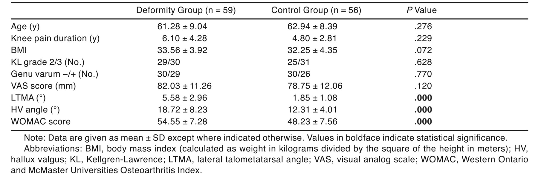
The study group comprised 115 women with knee osteoarthritis. Fifty-nine of these patients (51.3%) had pes planus, hallux valgus, or both in their bilateral feet. Forty-four patients (38.3%) had pes planus and 26 (22.6%) had hallux valgus with clinical and radiographic evidence. Eleven patients (9.6%) had both deformities. Mean values for age, duration of knee pain, grade of knee osteoarthritis, genu varum deformity, body mass index, hallux valgus angle, lateral talometatarsal angle, visual analog scale score, and total WOMAC score for both groups are listed in Table 1. The mean visual analog scale score was higher for the deformity group, but the difference was not statistically significant. There was a statistically significant difference between the WOMAC scores of the two groups. For further assessment, we grouped patients according to the number of deformities: patients with no deformity, with one deformity, and with two deformities. The group with one deformity was composed of patients with either hallux valgus or pes planus. The group with two deformities was composed of patients with both hallux valgus and pes planus. There was a significant difference between the WOMAC scores of the groups with no deformity and one deformity (P = .000) or two deformities (P = .000). Also, the difference between the groups with one and two deformities was significant (P = .049).

Table 1.
Clinical Features of the Deformity and Control Groups
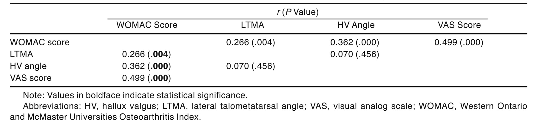
This analysis showed a significant correlation between indicators of pain and disability (visual analog scale and WOMAC scores). Correlation coefficients between pain and disability measures are given in Table 2. Both the lateral talometatarsal angle and hallux valgus angle, defined as objective measures of deformities, were correlated with WOMAC scores (Table 2). We found that as the hallux valgus and lateral talometatarsal angles increased, disability scores also increased in accordance with subjective measures.
Table 2.
Correlation Between Objective HV and Pes Planus Measurements and Disability and Pain Scores
Discussion
Knee osteoarthritis is one of the most common chronic musculoskeletal disorders associated with aging, affecting approximately 20% to 40% of people older than 65 years.[1] The prevalence of symptomatic knee osteoarthritis is higher in women than in men (11.4% versus 6.8%).[3] Pain and physical disability are major symptoms in knee osteoarthritis, profoundly affecting the everyday life of patients.[1,2] Foot pain has previously been shown to be related to disability in older women. Factors such as obesity and improper footwear affect foot malalignment. These factors are reversible and correctable.[22] In previous studies,[7–9] it has been reported that there is a relationship between lower-limb osteoarthritis (hip and knee) and foot characteristics. Furthermore, the effects of foot structure on lower-limb musculoskeletal injuries have been frequently reported.[23–25] Recently, it has been shown that orthoses that alter rearfoot characteristics may prove to be useful in the conservative treatment of knee osteoarthritis.[26] To our knowledge, no previous studies have evaluated the effect of certain foot deformities on the functional status of women with knee osteoarthritis. Therefore, we aimed to investigate the impact of certain foot pain syndromes on the disability of women with knee osteoarthritis.
We found that visual analog scale scores were higher in the deformity group than in the control group. Also, we observed that measurements of pain (visual analog scale) significantly correlated with measurements of disability in women with knee osteoarthritis. Kaufman et al[27] found that flat or pronated feet were associated with shin splints or knee pain. Several studies[10,11,27,28] have demonstrated a relationship between foot type and knee pain in athletes. Rubin and Menz[29] suggested that laterally wedged foot orthoses may reduce the pain of mild-to-moderate osteoarthritis in the medial compartment of the knee. Furthermore, it has been reported that the biomechanics of the foot and ankle may play a role in the biomechanics of the knee and hip at the heel strike and midstance phases of gait.[9] Many studies[7–9,26] have considered that there was a relationship between foot characteristics and lower-limb osteoarthritis (hip and knee).
Mean ± SD scores on the WOMAC questionnaire for the deformity and control groups were 54.55 ± 7.28 and 48.23 ± 7.56, respectively. We found that WOMAC scores were significantly higher in the group with foot deformities than in the control group. Moreover, WOMAC scores were significantly higher in patients with two deformities than in those with one deformity. These data show that coexisting foot deformities in women with knee osteoarthritis increased disability levels. We examined whether there was a correlation between WOMAC scores and the lateral talometatarsal and hallux valgus angles. The lateral talometatarsal and hallux valgus angles were correlated with disability scores, displaying a direct proportional increase. These angles are objective indicators of deformity. Therefore, these results confirm that the existence and severity of foot deformities correlate with more disability in women with knee osteoarthritis compared with no deformity or a less deformed foot.
Variables such as sex and obesity may affect pain perception and disability. Therefore, only women were recruited in this study. It has been reported that as body mass index increases, loading on the forefoot results in flattening of the arch and incongruency of the first phalanx.[30] Mean body mass index was higher for the deformity group; however, there was no statistically significant difference in body mass index between the deformity and control groups.
Pain duration is another variable in pain assessment. In some studies,[31,32] it has been reported that depressive symptoms increase with pain duration. In the present study, the mean ± SD duration of knee pain was 6.10 ± 4.28 years for the deformity group and 4.80 ± 2.81 years for the control group (P = .229).
Knee osteoarthritis, pes planus, and hallux valgus are common health problems. Whether these coexisting conditions (knee osteoarthritis and foot deformities) are comorbid diseases or have a cause-outcome relation is unclear. Women with knee osteoarthritis may have different foot deformities. However, there is a question regarding whether the foot influences the site at which knee osteoarthritis develops or whether these changes are secondary to osteoarthritis and develop as compensatory mechanisms. The patients in the study could not state whether they had low-arched feet currently or whether they had changed recently. Acquired flatfoot is a recognized condition in the adult population; however, if the varus deformity typical of medial compartment knee osteoarthritis influenced rearfoot characteristics, it would favor the development of a supinated foot position rather than a pronated foot position.[33]
In this study, we did not assess the cause-outcome relationship of these possibly related conditions. We aimed to display the effect of the presence of foot deformities on pain intensity and disability. We evaluated two somewhat preventable or correctable deformities in this study: pes planus and hallux valgus. The goal of nonoperative care for pes planus was to support the medial longitudinal arch and valgus deformity of the calcaneus. Treatment was augmented with the use of shoe modifications with medial sole and heel wedges with longitudinal arch support; also, a permanent foot orthosis should be used.[34] We found that the presence of foot deformities increased disability in women with knee osteoarthritis. Therefore, we suggest that simple correction may reduce disability in women with knee osteoarthritis.
Financial Disclosure
None reported.
Conflict of Interest
None reported.
References
- Dekker J, Boot B, van der Woude LH, et al: Pain and disability in osteoarthritis: a review of biobehavioral mechanisms. J Behav Med 15: 189, 1992.
- McAlindon TE, Cooper C, Kirwan JR, et al: Determinants of disability in osteoarthritis of the knee. Ann Rheum Dis 52: 258, 1993.
- Hooper MM, Holderbaum D, Moskowitz RW: “Clinical and Laboratory Findings in Osteoarthritis,” in Arthritis and Allied Conditions: A Textbook of Rheumatology, Vol 2, ed by WJ Koopman, LW Moreland, p 2227, Lippincott Williams & Wilkins, Philadelphia, 2005.
- Srikanth VK, Fryer JL, Zhai G, et al: A meta-analysis of sex differences prevalence, incidence and severity of osteoarthritis. Osteoarthritis Cartilage 13: 769, 2005.
- Hitt K, Shurman JR II, Greene K, et al: Anthropometric measurements of the human knee: correlation to the sizing of current knee arthroplasty systems. J Bone Joint Surg Am 85 (suppl 4):115, 2003.
- McAlindon TE, Snow S, Cooper C, et al: Radiographic patterns of osteoarthritis of the knee joint in the community: the importance of the patellofemoral joint. Ann Rheum Dis 51: 844, 1992.
- Keating EM, Faris PM, Ritter MA, et al: The use of lateral heel and sole wedges in the treatment of medial osteoarthritis of the knee. Orthop Rev 22: 921, 1993.
- Felson DT, Lawrence RC, Hochberg MC, et al: Osteoarthritis: new insights: part 2. Treatment approaches. Ann Intern Med 133: 726, 2000.
- Reilly KA, Barker KL, Shamley D, et al: Influence of foot characteristics on the site of lower limb osteoarthritis. Foot Ankle Int 27: 206, 2006.
- Powers CM, Chen PY, Reischl SF, et al: Comparison of foot pronation and lower extremity rotation in persons with and without patellofemoral pain. Foot Ankle Int 23: 634, 2002.
- Stergiou N, Bates NT: The relationship between subtalar and knee joint function as a possible mechanism for running injuries. Gait Posture 6: 177, 1997.
- Guichet JM, Javed A, Russell J, et al: Effect of the foot on the mechanical alignment of the lower limbs. Clin Orthop 415: 193, 2003.
- Dunn JE, Link CL, Felson DT, et al: Prevalence of foot and ankle conditions in a multiethnic community sample of older adults. Am J Epidemiol 159: 491, 2004.
- Lazarides SP, Hildreth A, Prassanna V, et al: Association amongst angular deformities in hallux valgus and impact of the deformity in health-related quality of life. Foot Ankle Surg 11: 193, 2005.
- Altman R, Asch E, Bloch D, et al: Diagnostic and Therapeutic Criteria Committee of the American Rheumatism Association: development of criteria for the classification and reporting of osteoarthritis: classification of osteoarthritis of the knee. Arthritis Rheum 29: 1039, 1986.
- Ravaud P, Auleley GR, Amor B, et al: Radiographic assessment of progression in knee osteoarthritis. Rheumatol Europe 24: (suppl 2):129, 1995.
- Ceceli E, Ergun S, Yorgancioglu ZR: Pes planus and lumbar lordosis in adolescents. Fiziksel Tip 2: 5, 1998.
- Budiman-Mak EB, Roach KE, Stuck R, et al: Radiographic measurement of hallux valgus in the rheumatoid arthritic foot. J Rheumatol 21: 623, 1994.
- Bellamy N, Buchanan WW, Goldsmith CH, et al: Validation study of WOMAC: a health status instrument for measuring clinically important patient relevant outcomes following total hip and knee arthroplasty in osteoarthritis. J Orthop Rheum 1: 95, 1988.
- Bellamy N, Buchanan WW, Goldsmith CH, et al: Validation study of WOMAC: a health status instrument for measuring clinically important patient relevant outcomes to antirheumatic drug therapy in patients with osteoarthritis of the hip or knee. J Rheumatol 15: 1833, 1988.
- McConnell S, Kolopack P, Davis AM: The Western Ontario and McMaster Universities Osteoarthritis Index (WOMAC): a review of its utility and measurement properties. Arthritis Care Res 45: 453, 2001.
- Leveille SG, Guralnik JM, Ferrucci L, et al: Foot pain and disability in older women. Am J Epidemiol 148: 657, 1998.
- Evans P: Clinical biomechanics of the subtalar joint. Physiotherapy 76: 47, 1990.
- Fu FH, Feldman A: The biomechanics of running: practical considerations. Tech Orthop 5: 8, 1990.
- Tiberio D: The effect of excessive subtalar joint pronation on patellofemoral mechanics: a theoretical model. J Orthop Sports Phys Ther 9: 160, 1987.
- Toda Y, Tsukimura N, Kato A: The effects of different elevations of laterally wedged insoles with subtalar strapping on medial compartment osteoarthritis of the knee. Arch Phys Med Rehabil 85: 673, 2004.
- Kaufman KR, Brodine SK, Shaffer RA, et al: The effect of foot structure and range of motion on musculoskeletal overuse injuries. Am J Sports Med 27: 585, 1999.
- Dahle LK, Mueller M, Delitto A, et al: Visual assessment of foot type and relationship of foot type to lower limb extremity injury. J Orthop Sports Phys Ther 14: 70, 1991.
- Rubin R, Menz HB: Use of laterally wedged custom foot orthoses to reduce pain associated with medial knee osteoarthritis: a preliminary investigation. JAPMA 95: 347, 2005.
- Tanaka Y, Takakura Y, Takaoka T, et al: Radiographic analysis of hallux valgus in women on weightbearing and nonweightbearing. Clin Orthop Res 336: 186, 1997.
- Hopman-Rock M, Odding E, Hofman A, et al: Physical and psychological disability in elderly subjects in relation to pain in the hip and/or knee. J Rheumatol 23: 1037, 1996.
- Klinger L, Spaulding SJ, Polatajko HJ, et al: Chronic pain in the elderly: occupational adaptation as a means of coping with osteoarthritis of the hip and/or knee. Clin J Pain 15: 275, 1999.
- Van Boerum DH, Sangeorzan BJ: Biomechanics and pathophysiology of flat foot. Foot Ankle Clin 8: 419, 2003.
- Dipreta JA, Chao W, Wapner KL: “Conservative Management of Acquired Flatfoot,” in Physical Medicine and Rehabilitation: State of the Art Reviews, ed by DDJ Kim, SF Wainapel, p 625, Hanley & Belfus Inc, Philadelphia, 2001.
© 2009 American Podiatric Medical Association