


Abstract
The modified Brostrom procedure has been a proven procedure with excellent utility in the treatment of lateral ankle instability within limitation. Multiple variations of the original technique have been described in the literature to date. Included in these variations are differences in anchor placement, suture technique, or both. In this research study, we propose placing a bone screw anchor into the lateral shoulder of the talus rather than the typical placement at the lateral malleolus for anatomic reconstruction of the lateral ankle ligaments. (J Am Podiatr Med Assoc 98(6): 473–476, 2008)
Lateral ankle sprains are among the most common injuries, particularly with sports-related activities. Katcherian[1] estimated that one such injury occurs per day in a population of 10,000. The classical mechanism of injury involves forced adduction, supination, and internal rotation of the foot, thereby making this injury commonplace. Zwipp et al[2] identified predisposing factors for ankle sprains. Extrinsic factors include unsuitable footwear, namely high heels. Intrinsic factors such as general ligamentous laxity, calcaneus varus, muscular exhaustion of the peroneal musculature, and neurologic deficits, including peroneal palsy or proprioceptive deficit after previous trauma also contribute to lateral ankle instability. Zwipp et al[2] and Dehne[3] noted factors for the pathomechanics of acute ankle instability, which is associated with anterior talofibular ligament (ATFL) and calcaneofibular ligament (CFL) rupture, which are the usual suspects when the injury progresses to chronic lateral instability. Dehne[3] also noted that the talus dislocates in three directions: 1) varus tilt in the frontal plane, 2) anterior dislocation in the sagittal plane (identified on clinical examination as the “anterior drawer” sign), and 3) internal rotation in the transverse plane. This in turn results in three-dimensional instability.
Anatomically, the lateral ligamentous complex consists of three parts: the ATFL, the CFL, and the posterior talofibular ligament (PTFL). The ATFL is the weakest of the lateral ligaments, is the first to rupture, and is usually torn in up to 75% of cases surgically explored. The ATFL originates at the anterior aspect of the distal fibula and runs to the lateral aspect of the talar head.[2],[4] The CFL originates at the fibular tip and inserts at the lateral wall of the calcaneus; the CFL is ruptured in about 60% of cases and insufficient or disrupted in another 25% of cases.[2],[4] The PTFL is the strongest of the lateral ankle ligaments, originates at a small groove at the posterior aspect of the dorsomedial aspect of the distal fibular, and runs almost horizontally to the lateral tubercle of the posterior process of the talus. The PTFL is rarely involved, with injury reports of only 5% of cases of lateral ankle instability.[2],[4]
There have been a myriad of surgical procedures noted for ATFL and CFL repair for chronic lateral ankle instability. These can be categorized into anatomic repairs and nonanatomic repairs. Much discussion has occurred in the literature regarding which procedure is most beneficial for a particular type of patient, yet ultimately, both groups of procedures tend to provide generally satisfying results in the right surgical circumstance. One procedure often utilized is the modified Brostrom procedure, which consists of direct suture with shortening and reinsertion of the torn ligaments and reefing of the ATFL and CFL.[2],[4],[5] There are several advantages to the modified Brostrom procedure. It is relatively easy with a small cosmetic incision; there is little danger to the sural nerve and does not sacrifice other anatomical structures such as the peroneal tendons. The procedure is anatomic in its reconstruction, and it maintains full range of motion of the ankle and subtalar joints. Finally, unlike various weaving procedures that are nonanatomic reconstructions, the Brostrom procedure makes it difficult to make the repair over-tight such that the subtalar joint is locked in eversion.[4]
The modified Brostrom is indicated for chronic, symptomatic lateral ankle instability that has not responded to physical therapy and rehabilitation efforts. It is not a recommended procedure for patients with a fixed heel varus without correction of deformity, or for patients with increased body weight, such as athletes weighing more than 250 pounds.[4]
The present report is intended to demonstrate the success of a modification of the modified Brostrom technique. The modification consists of an alternative site for anchor placement, namely the lateral talar shoulder, during the anatomic reconstruction of the ATFL and CFL.
Technique
Prior to introducing the alternative method of fixation, it is necessary to understand the standard technique typically used for the modified Brostom procedure. Typically, a curvilinear incision is made over the distal anterior border of the lateral malleolus. Dissection then proceeds through the subcutaneous tissue where the inferior extensor retinaculum is identified and preserved. The retinaculum can later be used as further reinforcement with attachment to the anterior edge of the fibula if necessary. The inferior extensor retinaculum runs parallel to the CFL deep to the peroneal tendons. The ATFL is identified, which usually appears as a thickening in the anterior joint capsule. Personal experience of both authors seems to indicate that when the ATFL is torn, it is usually from the fibula with a cuff intact. An anterior capsular incision is made along the anterior margin of the fibula down to its distal tip, leaving a small cuff of tissue attached to the fibular to facilitate later repair. The CFL is then identified at the inferior tip of the fibula as the ankle is placed in dorsiflexion and valgus to assess the redundancy of the ligaments. One method of anchoring is fixation with an anchor placed in the distal fibula, such as a Mitek (DuPuy Mitek; Johnson and Johnson, Raynham, Massachusetts) or Stryker (Stryker Orthopedics, Kalamazoo, Michigan) anchor. The suture lines are passed through the proximal aspects, then through the distal aspects of the ATFL and CFL, and are then incorporated with the capsule, with reduction in redundancy, and then tied with imbrication of the capsule. Alternatively, drill holes are made in the distal fibula with sutures passed through the proximal edges of the ATFL and CFL then passed distally through the ligaments and tied.
Our procedure is a modification of the above. A curvilinear incision measuring 3.5 to 4 cm is made centered over the anterior lateral ankle and dissected deep, taking care to avoid all neurovascular structures. The peroneal retinaculum is identified and incised. The capsule is identified, and a linear capsulotomy is created down to the level of the lateral shoulder of the talus. When the ATFL is ruptured, it is completely breeched off the talus with a cuff intact at the level of the fibula. The condition of the CFL is also noted, and if ruptured or disrupted, it too is addressed. A 3.5-mm twin fix anchor with #2 Ethibond (Ethicon; Johnson and Johnson, Somerville, New Jersey) is then inserted into the lateral shoulder of the anterior talus instead of the normal insertion into the distal aspect of the fibula. The ATFL and CFL is then repaired with imbrication and tightening of the ligaments in conjunction with the periosteum of the fibula holding the foot in dorsiflexion and eversion. The placement of the anchor into the shoulder of the talus is unique in performing the modified Brostrom procedure and allows excellent intra-operative stability noted with an anterior drawer test.
On the basis of anatomical observations, we propose that it is preferable to place a bone anchor into the lateral talar shoulder. The fibula, unlike the talus, contains a thick envelope of periosteum with which to secure the primary repair. The talus is devoid of a soft-tissue envelope. Therefore, from a functionally corrective approach, it is preferable to insert the anchor into the lateral talar shoulder of the ATFL “pocket” and suture into the fibular periosteum at the corresponding ATFL origin (Fig. 1).
We have performed 32 modified Brostrom procedures with anchoring into the lateral talar shoulder instead of the distal tip of the fibula over the past 8 years. All patients were noted to have insufficient ATFL, CFL, or both as identified by magnetic resonance imaging, and clinical instability symptoms were specifically present. All patients exhibited a positive anterior drawer test of +2 or +3 and positive varus tilt of greater than 5° compared to contralateral limb or 10° total. Clinically, patients displayed distinct pain along the lateral ligament area. Dissection was carried out in each as described above, with the only exception of possible ankle joint debridement via ankle arthroscopy performed as necessary in some patients. After surgery, all 32 patients exhibited a negative anterior drawer test on clinical examination. At 1 year, all patients appeared to be free of pain with palpation or with range of motion, and continued to have a negative anterior drawer test clinically.
Complications noted were transient numbness at distal lesser digits and transient stiffness. All such cases resolved over the immediate postoperative course. Two patients experienced wound dehiscence with subsequent complete healing with local wound care without sequelae. No complications to the structure of the talus or fibula have been identified to date.
Discussion
The strength and success of the modified Brostrom procedure is well documented in the current literature. Messer et al[6] reported on 22 patients with chronic lateral ankle instability who underwent surgical repair of their lateral ankle ligaments using suture anchors. At a mean follow-up of 34.5 months, 20 patients, (91%), reported good or excellent functional outcome assessments, and 14 of 16 patients had no evidence of instability on examination or stress radiographs.[6] Junis et al[7] and Schnirring-Judge et al[5] conducted a retrospective analysis of 26 lateral ankle stabilizations with primary repair and found that in 23 patients, primary repair performed with the remnant ligament was sufficient.
However, not all cases of chronic lateral ankle instability are amenable to primary repair. In a study by Peters et al[8], generalized hypermobility and a delay of 10 years between injury and diagosis was reported to be a relative contraindication to antomic repair. Compared to tenodesis procedures, Visser et al[9] believed primary repair to be inferior, although more recent studies have disputed this and shown the modified Brostom procedure to equal the stabilization strength when compared to the Chrisman-Snook and Evans procedures. Lastly, Zwipp[10] and Schnirring-Judge et al[5] suggested that patients with a long history of instability, namely greater than 10 years, in conjunction with a talar tilt of greater than 15° and anterior drawer of 10 mm, are more suitable to the tenodesis reconstruction.
Conclusion
The modified Brostom procedure is an excellent procedure for anatomic stabilization of the lateral ankle ligaments, given the proper patient selection following careful physical examination. We have tried to illustrate an alternative for anchor placement in the talus, which provides very favorable results in our limited study.
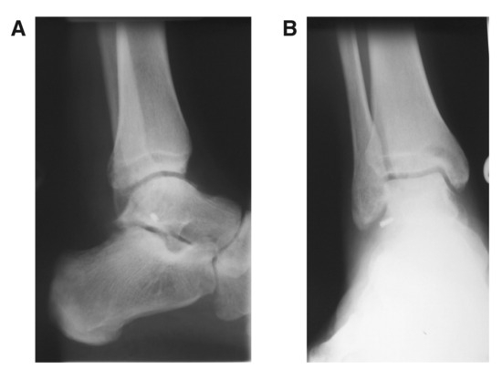
Figure 1.
Radiograph of bone anchor into the lateral shoulder of the talus. A, Lateral view of the right ankle. B, Anteroposterior view of the right ankle.
Financial Disclosure: None reported.
Conflict of Interest: None reported.
References
- Katcherian D: “Soft tissue injuries of the ankle,” in Foot and Ankle, ed by LD Luter, MS Mizel, GB Pfeffer, p 241, American Academy of Orthopaedic Surgeons, Rosemont. ,1994. .
- Zwipp H, Rammelt S, Gruns R: Ligamentous injuries about the ankle and subtalar joints. .Clin Podiatr Med Surg19::195. ,2002. .
- Dehne E: Die Klinik der frischen und habituellen Adduktionssupinationsdistorsion des Fuβes. .Dtsch Z Chir40::242. ,1933. .
- Hamilton HG: “Ankle Instability Repair: The Brostrom-Gould Procedure,” in Master Techniques in Orthopaedic Surgery: The Foot and Ankle, ed by HB Kitaoka, p 487, Lippincott Williams & Wilkins, Philadelphia. ,2002. .
- Schnirring-Judge M, Perlman MD: “Chronic Ankle Conditions,” in McGlamery’s Comprehensive Textbook of Foot and Ankle Surgery, Vol 2, ed by AS Banks, MS Downey, DE Martin, et al, p 1595, Lippincott Williams & Wilkins, Philadelphia. ,1994. .
- Messer TM, Cummins CA, Kelikian AS: Outcome of the modified Brostrom procedure for chronic lateral ankle instability using suture anchors. .Foot Ankle Int21::996. ,2000. .
- Junis LR, Kittleson RS, Cox DG: Chronic lateral ankle instability: assessment of subjective outcomes following delayed primary repair and a new secondary reconstruction. .J Foot Ankle Surg37::369. ,1998. .
- Peters JW, Trevino SG, Renstrom PA: Chronic lateral ankle instability. .Foot Ankle12::182. ,1991. .
- Visser HJ, Oloff LM, Jacobs AM: Lateral ankle stabilization procedures: criteria and classification. .J Foot Surg19::74. ,1980. .
- Zwipp H: Treatment of chronic, two-plane instability of the ankle joint: syndesmoplasty versus periosteal flap versus tenodesis. .J Foot Surg29::3. ,1990. .
© 2008 American Podiatric Medical Association