


Abstract
Pure open dislocation of the ankle, or dislocation not accompanied by rupture of the tibiofibular syndesmosis ligaments or fractures of the malleoli or of the posterior border of the tibia, is an extremely rare injury. A 62-year-old man injured his right ankle in a motor vehicle accident. Besides posterolateral ankle dislocation, there was a 7-cm transverse skin cut on the medial malleolus, and the distal end of the tibia was exposed. After reduction, we made a 2- to 2.5-cm longitudinal incision on the lateral malleolus; the distal fibular fracture was exposed. Two Kirschner wires were placed intramedullary in a retrograde manner, and the fracture was stabilized. The deltoid ligament and the medial capsule were repaired. The tibiofibular syndesmosis ligaments were intact. At the end of postoperative year 1, right ankle joint range of motion had a limit of approximately 5° in dorsiflexion, 10° in plantarflexion, 5° in inversion, and 0° in eversion. The joint appeared normal on radiographs, with no signs of osteoarthritis or calcification. The best result can be obtained with early reduction, debridement, medial capsule and deltoid ligament restoration, and early rehabilitation. Clinical and radiographic features at long-term follow-up also confirm good mobility of the ankle without degenerative change or mechanical instability. (J Am Podiatr Med Assoc 98(6): 469–472, 2008)
Pure open dislocation of the ankle, or dislocation not accompanied by rupture of the tibiofibular syndesmosis ligaments or fractures of the malleoli or of the posterior border of the tibia, is an extremely rare injury.[1] D’Anca[2] explained that this rarity was attributable to the mechanical efficiency of the mortise and the resistance of the ankle ligaments being greater than that of bone, thus causing malleolar fracture in the case of injury.
The mechanism of the injury generally consists of high-energy trauma, which produces a combination of plantarflexion and forced inversion or eversion of the foot. In most cases, the dislocation is open by the laceration of soft tissues, which make up the only covering of the malleoli.[3] We report the results of treatment of a patient who had open posterolateral dislocation of the ankle without tearing of the tibiofibular syndesmosis ligaments.
Case Report
A 62-year-old man injured his right ankle in a motor vehicle accident. Immediately after the accident, he was taken to the Gülhane Military Medical Academy, Orthopedics and Traumatology Department, Ankara, Turkey. His medical history was normal. Immediately after the injury, he noticed that he could not move his right ankle. Physical examination revealed loss of movement in all directions. The vital findings of the patient were stable, and a deformation was observed on the right ankle. There was a 7-cm transverse skin cut on the medial malleolus, and the distal end of the tibia was exposed (Fig. 1). The wound was dirty, with sand, pieces of soil, and torn sock present in it. Although the dorsalis pedis artery was palpable, the tibialis posterior artery was not. Capillary filling at the fingertips was normal. Radiography showed that the tibiotalar joint was posterolaterally dislocated, and there was a transverse fracture in the distal fibula. No pathologic abnormalities were detected in other bony structures (Fig. 2).
The patient was taken to surgery approximately 180 min after injury. General anesthesia was used, and swabs of the wound were taken for culture and sensitivity. After thorough wound irrigation and debridement, the reduction was accomplished relatively easily. After reduction, the tibialis posterior artery became palpable. By applying a 2- to 2.5-cm longitudinal incision on the lateral malleolus, the distal fibular fracture was exposed. Two Kirschner wires were placed intramedullary in a retrograde manner, and the fracture was stabilized. After stabilization, the deltoid ligament and the medial capsule were repaired. It was observed that the tibiofibular syndesmosis ligaments were intact. One plastic suction drainage tube was inserted, and the wound was closed primarily (Fig. 3).
The patient received tetanus immunization, a third-generation cephalosporin, and an aminoglycoside to prevent infection. At the end of the operation, a short-leg cast was applied in the neutral position, to be kept in place for 45 days. Walking assisted with crutches with no weightbearing was begun after 3 days. The cast was removed at the end of week 6. Then the rehabilitation program was started. Low-molecular-weight heparin was given subcutaneously daily until the patient was mobilized. Several days after the operation, the patient was able to walk nonweightbearing with crutches. At the end of the following 10 days, he was allowed to bear weight gradually as much as he could tolerate. The pins were retained for 4 weeks and then were removed under local anesthesia.
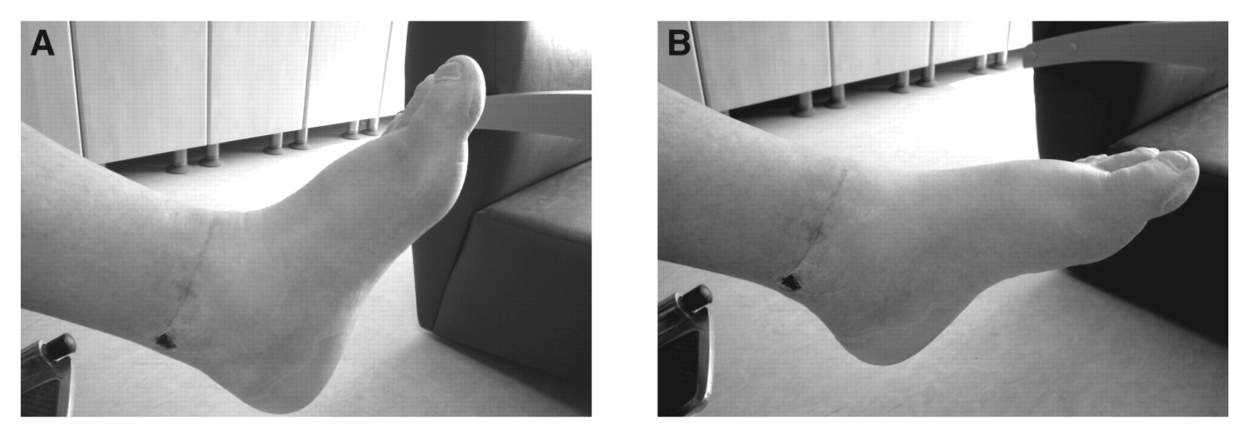
At the end of month 6, he regained comfortable movement of the ankle. Clinical and radiographic follow-up continued for 1 year after the accident (Fig. 4). The patient reported complete disappearance of pain and was able to return to work. At the end of postoperative year 1, the range of motion of the right ankle joint had a limit of approximately 5° in dorsiflexion, 10° in plantarflexion, 5° in inversion, and 0° in eversion. The joint appeared normal on radiographs, with no signs of osteoarthritis or calcification.
Discussion
The ankle does not have discrete stabilization structures ventrally and dorsally. Only medially and laterally does the ankle have strong stabilization structures: the tibial and fibular malleoli and the two collateral ligamentous complexes that reinforce the thin capsule. The unstable position of the tibiotalar joint is plantarflexion because the narrow part of the talar body lies within the ankle mortise. In this position, all of the ligamentous and capsular attachments to the talus are stretched, except for the posterior talofibular ligament.
Sufficient force in inversion results in posteromedial displacement of the foot on the fixed tibia, with tears in the anterolateral capsule attachments and in the anterior talofibular and calcaneofibular ligaments, allowing for talar ascent and tilt.[3]–[6] Likewise, an eversion force results in lateral displacement of the foot, with tearing of the medial capsule attachments and the talotibial ligament.[7] In the case of an applied posteroanterior force on the foot in plantarflexion, the ankle is displaced anteriorly.[7],[8] Posterior dislocation is the result of an anteroposterior force on the foot in plantarflexion and inversion.
Posterolateral dislocation of the ankle without disruption of the tibiofibular ligaments or fracture is rare. The mechanism of injury is forced eversion, plantarflexion, and axial loading.[4] To our knowledge, there have been only a few reports in the English-language literature.[3],[4],[9]–[13] In most cases, soft-tissue lacerations are observed on the malleolus.[10],[14] Medial dislocation of the tibiotalar joint without associated fractures or disruption of the tibiotalar syndesmosis is common compared with lateral dislocation. In the present case, besides the posterolateral dislocation of the ankle there was a fracture at the distal fibula; on the other hand, the distal tibiofibular ligament was intact.
A review of the literature[14] indicates that pure dislocation of the ankle is caused prevalently by road accidents (40%), particularly motorcycle accidents (33%). The second most common cause is sports trauma (35%), particularly sports in which jumping is a fundamental component, such as volleyball (13%) and basketball (8%).
The diagnosis is made clinically. The deviation of the foot with respect to the leg determines an evident alteration of the normal articular profile. Often, laceration of the soft tissue on the opposite side of the dislocation is present. Radiographic examinations are needed to exclude the presence of fractures.[3]
Because this is an uncommon injury, there is no standard treatment protocol. The length of rehabilitation and the time before returning to work or play will be determined on an individual basis. The short-term goal is to obtain a congruent reduction, which allows most patients to make a full recovery after a period of splinting, weightbearing, and rehabilitation.[14],[15] An anatomical reduction should be performed immediately by a physician to prevent further compromise of the neurologic and vascular structures, which could lead to serious consequences, including life-long neurovascular complications and loss of the foot.[14],[16] There are no data on long-term effects, such as instability or degenerative changes.
The present patient was treated with debridement and primary repair of the deltoid ligament and capsular repair after reduction. Kaneko et al[10] reported that they performed medial capsule and deltoid ligament restoration. However, Colville et al[4] and Moehring et al[11] reported a large series of this condition, but because all of the cases were posteromedial dislocations, they did not require repair of the deltoid ligament. In our case, it was necessary to repair the deltoid ligament and the capsule.
In the literature, it is reported that cast immobilization is enough in the treatment of closed ankle dislocations, but in cases with open dislocations, pin fixation is recommended.[3],[9] It is well known that the best results are obtained by using cast stabilization for 6 weeks after fixation and immediate rehabilitation after removing the cast. In our present case, after immediate reduction, deltoid ligament and medial capsule restoration, and fixation of the lateral malleolus fracture, 6 weeks of short-leg cast immobilization and an early rehabilitation program were applied. We obtained an acceptable result.
As reported by Elisé et al,[17] negative prognostic factors may include advanced age of a patient, injury to the inferior tibiofibular ligaments, the presence of vascular injuries, and delayed reduction. Complications in elderly patients are a concern because moderate swelling can cause vascular deficit. Rarely are vascular injuries of the anterior tibialis artery[3],[13] and dorsalis pedis artery[4],[13] present. In addition, neurologic injury of the tibial nerve, superficial peroneal nerve, and sural nerve are seen infrequently.[3] In severe cases, damage to the neurovascular structures may necessitate amputation.
As a result, open posterolateral dislocation of the ankle without tearing of the syndesmosis ligaments is observed rarely. The best result can be obtained with early reduction, debridement, medial capsule and deltoid ligament restoration, and early rehabilitation. Evaluation of clinical and radiographic features at long-term follow-up also confirms good mobility of the ankle without degenerative change or mechanical instability.
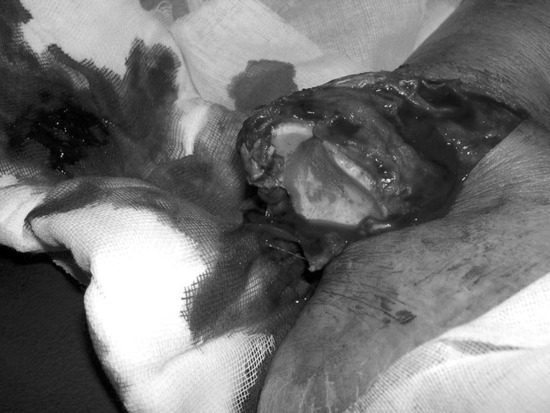
Figure 1.
A 62-year-old man injured his right ankle in a motor vehicle accident. There was a 7-cm transverse skin cut on the medial malleolus, and the distal end of the tibia was exposed.
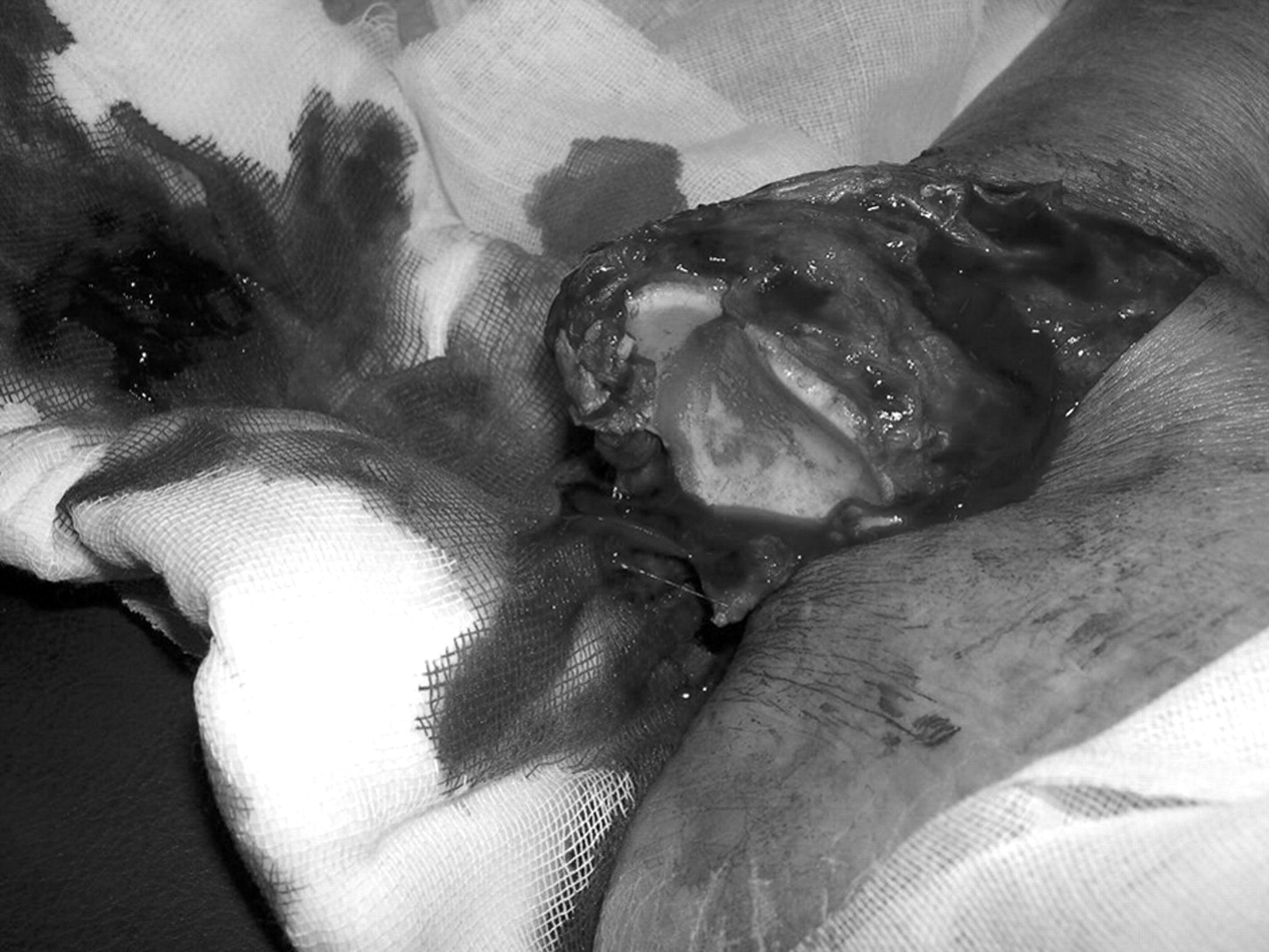
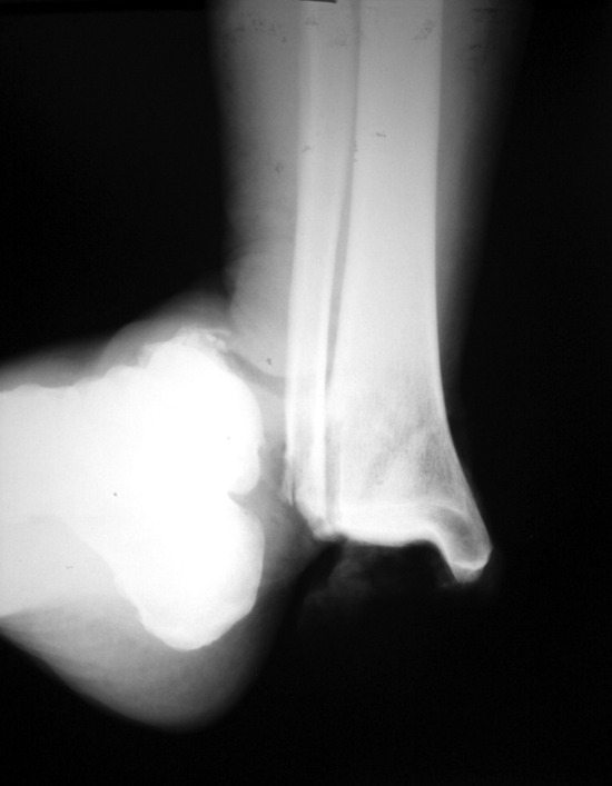
Figure 2.
Radiograph showing that the tibiotalar joint was posterolaterally dislocated and that there was a transverse fracture in the distal fibula. No pathologic abnormalities were detected in other bony structures.
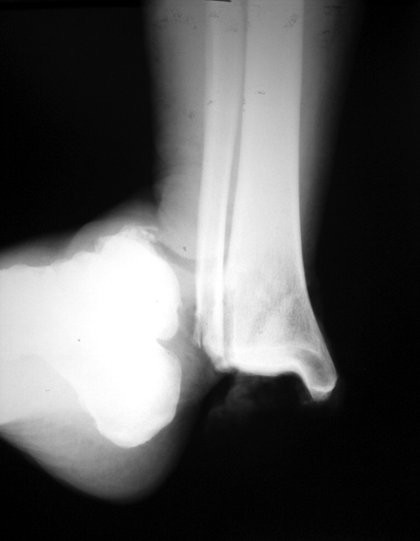
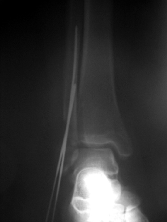
Figure 3.
Postoperative radiograph of the ankle.
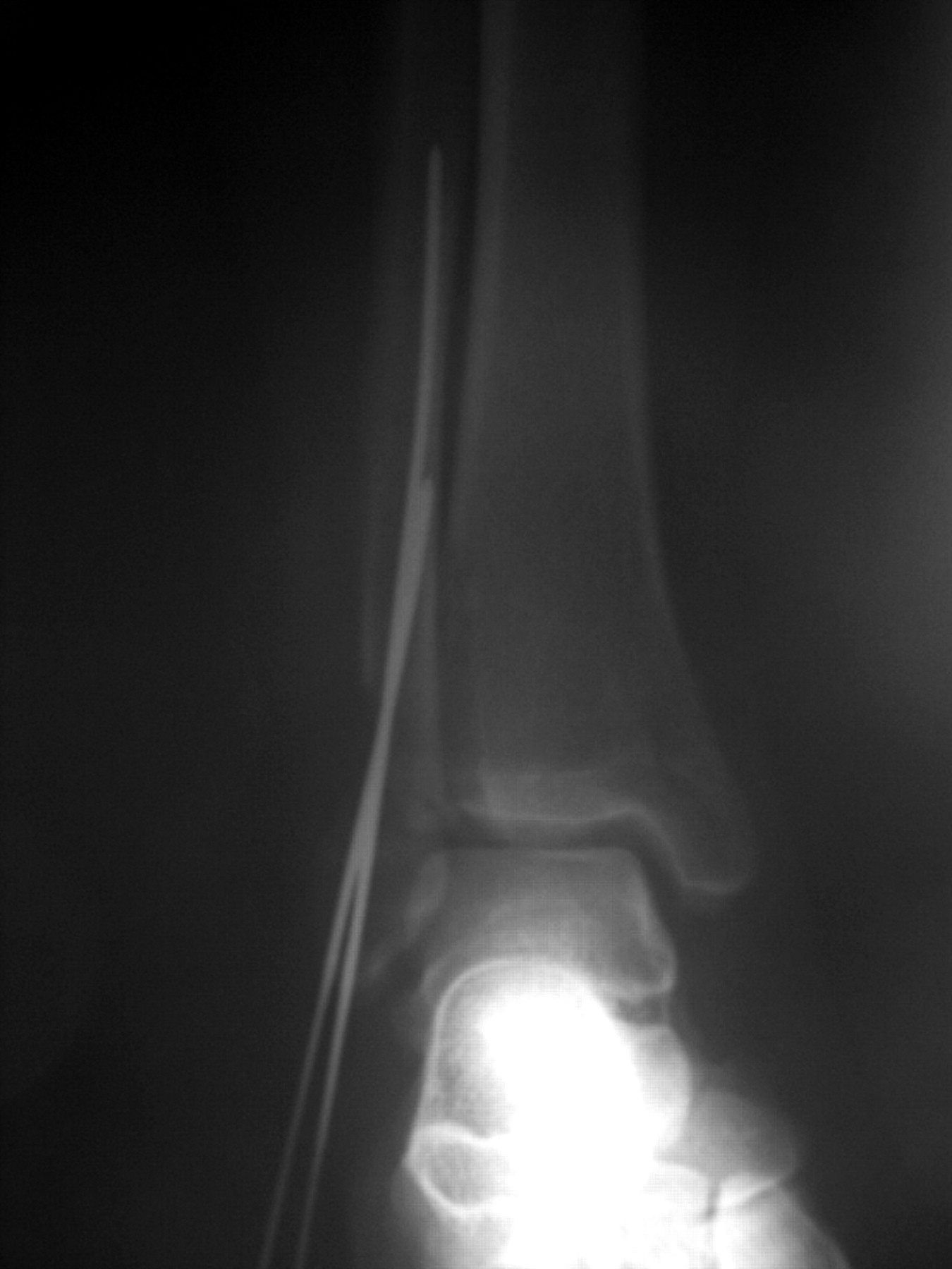
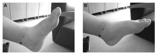
Figure 4.
At the end of postoperative year 1, the range of motion of the right ankle joint had a limit of approximately 5° in dorsiflexion (A) and 10° in plantarflexion (B).
Financial Disclosure: None reported.
Conflict of Interest: None reported.
References
- Leitner B: The mechanism of total dislocation of the talus. .J Bone Joint Surg Am37::89. ,1955. .
- D’Anca AF: Lateral rotatory dislocation of the ankle without fracture. .J Bone Joint Surg Am52::1643. ,1970. .
- Garbuio P, Gerard F, Gagneux E: Pure dislocations of the tibiotalar joint: apropos of 9 cases. .Rev Chir Orthop81::601. ,1995. .
- Colville MR, Colville JM, Manoli A: Posteromedial dislocation of the ankle without fracture. .J Bone Joint Surg Am69::706. ,1987. .
- Krishnamurthy S, Schultz RJ: Pure posteromedial dislocation of the ankle joint. .Clin Orthop201::68. ,1985. .
- Wilson M, Michele A, Jacobson E: Ankle dislocation without fracture. .J Bone Joint Surg Am21::198. ,1939. .
- Siuni E, Cabiddu M, Cara PP, et al: Pure ankle dislocation. .Chir Piede16::139. ,1992. .
- Segal D, Wasilewski S: Total dislocation of the talus. .J Bone Joint Surg Am62::1370. ,1980. .
- Finkemeier C, Engebretsen L, Gannon J: Tibiotalar dislocation without fracture: treatment principles and outcome. .Knee Surg Sports Traumatol Arthrosc3::47. ,1995. .
- Kaneko K, Mogami A, Maruyama Y, et al: Posterolateral dislocation of the ankle without fracture. .Injury31::740. ,2000. .
- Moehring HD, Tan RT, Marder RA, et al: Ankle dislocation. .J Orthop Trauma8::167. ,1994. .
- Mooney JF, Naylor PT, Poehling GG: Anterolateral ankle dislocation without fractures. .South Med J84::244. ,1991. .
- Toohey JS, Worsing RA Jr: A long-term follow-up study of tibiotalar dislocations without associated fractures. .Clin Orthop239::207. ,1989. .
- Rivera F, Bertone C, De Martino M, et al: Pure dislocation of the ankle: three case reports and literature review. .Clin Orthop382::179. ,2001. .
- Tranovich M: Ankle dislocation without fracture. .Physician Sports Med31::42. ,2003. .
- Kiefer EA, Wikstrom EA, Douglas McDonald J: Ankle dislocation without fracture: an on-field perspective. .Clin J Sport Med16::269. ,2006. .
- Elise S, Maynou C, Mestdagh H, et al: Tibiotalar dislocations without associated fracture: report on 16 cases. .Acta Orthop Belg64::25. ,1998. .
© 2008 American Podiatric Medical Association