


Abstract
Brown tumors represent the terminal stage of the remodeling process during primary or secondary hyperparathyroidism. We report the first case of brown tumor on the third metatarsal associated with secondary hyperparathyroidism caused by vitamin D deficiency. Radiography showed an expansile tissue mass in the third metatarsal bone. The diagnosis was suggested by the clinical history and was confirmed by biochemical, radiologic, and histopathologic determinations. After intravenous therapy with calcidiol, 1g/d, her symptoms were relieved. The brown tumor showed regression and ossification during the 3 months after therapy.
Brown tumors that are not true neoplasms occur in association with hyperparathyroidism and can arise in any bone.[1] Brown tumors appear as single or multiple well-defined lesions of axial or appendicular skeleton, are frequently eccentric or cortical in location, and are locally destructive bone lesions caused by rapid osteoclastic bone resorption due to hyperparathyroidism.[2,3] The incidence of brown tumors is 3% in primary hyperparathyroidism, and they are caused by adenoma or hyperplasia of the parathyroid gland. In secondary hyperparathyroidism, the incidence of brown tumors is 1.5% to 1.7%, and they are caused by chronic renal failure.[4] Brown tumors commonly affect the mandible, facial bones, clavicle, ribs, pelvis, and femur.[5] Involvement of the foot bones is rare. Previously, in primary hyperparathyroidism, calcaneal brown tumor due to parathyroid carcinoma has been reported in one case.[6] To our knowledge, only one case has been reported in the literature with brown tumor and aseptic bone necrosis as a cause for sesamoid bone disease of the first metatarsal bone.[7] We present, for the first time in the literature, a case of brown tumor of the third metatarsal bone in a patient who developed secondary hyperparathyroidism attributable to vitamin D deficiency.
Case Report
A 46-year-old woman with a 4 × 3-cm painful mass in the region of the third metatarsal of the left foot without redness or warmth was admitted to Harran University (Sanliurfa, Turkey). Her lower-extremity movements were extremely restricted and painful. Laboratory evaluation showed a serum calcium level of 8.1 mg/dL (reference range, 8.4–10.6 mg/dL), a phosphate level of 2.5 mg/dL (reference range, 2.3–4.5 mg/dL), an alkaline phosphatase level of 635 pg/mL (reference range, 53–128 pg/mL), a urinary calcium level of 0.6 mg/d (reference range, 5.2–30.7 mg/d), and a urinary phosphate level of 276 mg/d (reference range, 250– 1,000 mg/d). Her serum intact parathyroid hormone level was 1,205 pg/mL (reference range, 15–65 pg/mL), and her 25-hydroxy vitamin D level was 10 pg/mL (reference range, 30–65 pg/mL). The results of liver and kidney function tests were normal.
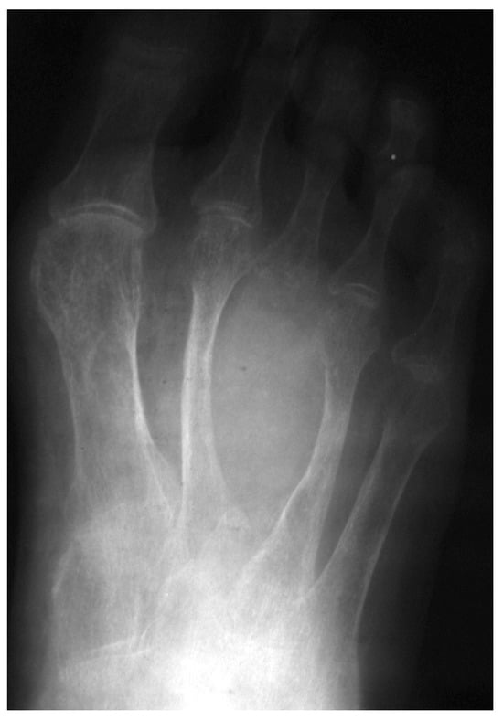
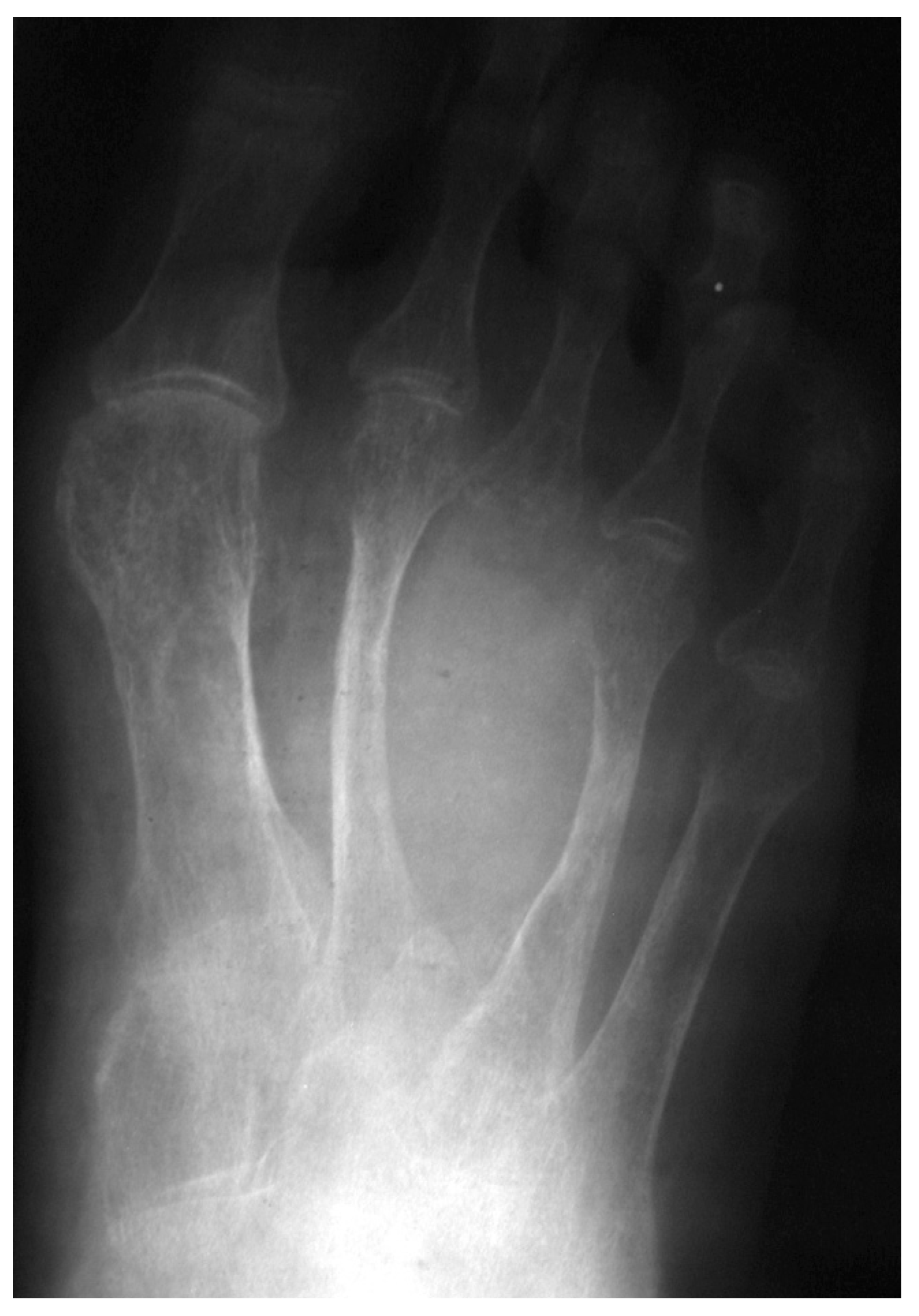
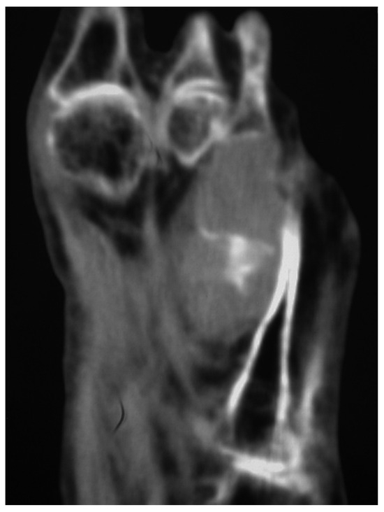
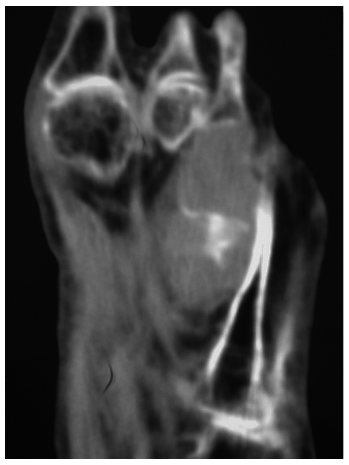
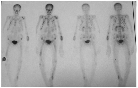
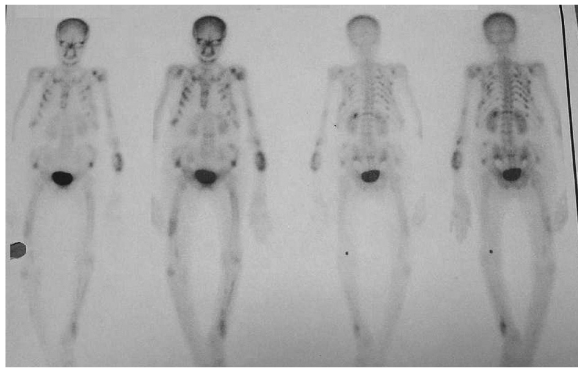
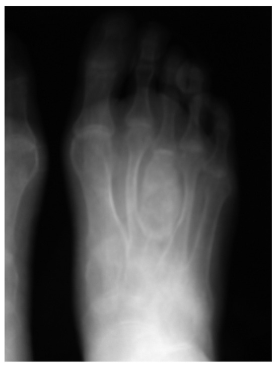
Radiographs revealed no bone structure in the third metatarsal area, but an increase in soft-tissue density was observed in this area (Figure 1). Computed tomography (CT) revealed a fusiform-shaped, well-defined mass of soft tissue localized to the third metatarsal and bony isodense structures in this mass (Figure 2). Bone mineral density measurements using dual-energy x-ray absorptiometry were as follows: left femur neck, 0.134 g/cm2; total femur, 0.218 g/cm2; and lumbar total (L2 –L4), 0.222 g/cm2. A technetium bone scan revealed increased focal uptake in the third metatarsal, costas, sternum, both scapulas, left humerus, left radius and ulna proximal, pelvis, both femurs, both knees, and left tibia (Figure 3). Scintigraphic and ultrasonographic evaluations of the parathyroid gland did not show any adenomas, nodules, or carcinomas.
Figure 1.
Anteroposterior radiograph showing increasing soft-tissue density in the third metatarsal area.
Figure 2.
Computed tomography of the foot reveals a fusiform-shaped, well-defined mass of soft tissue localized to the third metatarsal and bony isodense structures in this mass.
Figure 3.
Anterior and posterior whole-body bone scan showing increased activity in areas of brown tumors.
She was not taking any medications, and she had no history of steatorrhea. Her nutritional status was estimated from the self-administered food frequency questionnaire. She ingested very little food that was rich in vitamin D, and because she wore a religious veil that covered her face, she had little exposure to sunlight. The patient had weakness and a lack of energy on walking, especially on climbing stairs, for the past year. Her menstrual periods were normal. She had three healthy children. Her family history was normal. She did not smoke, drink alcohol, or use illicit drugs. On physical examination, she was 160 cm tall and weighed 45 kg. Her blood pressure was 106/70 mm Hg.
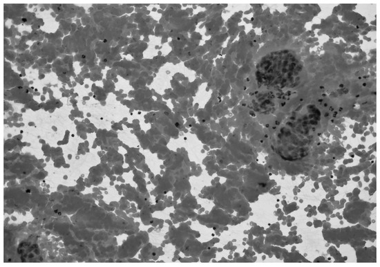
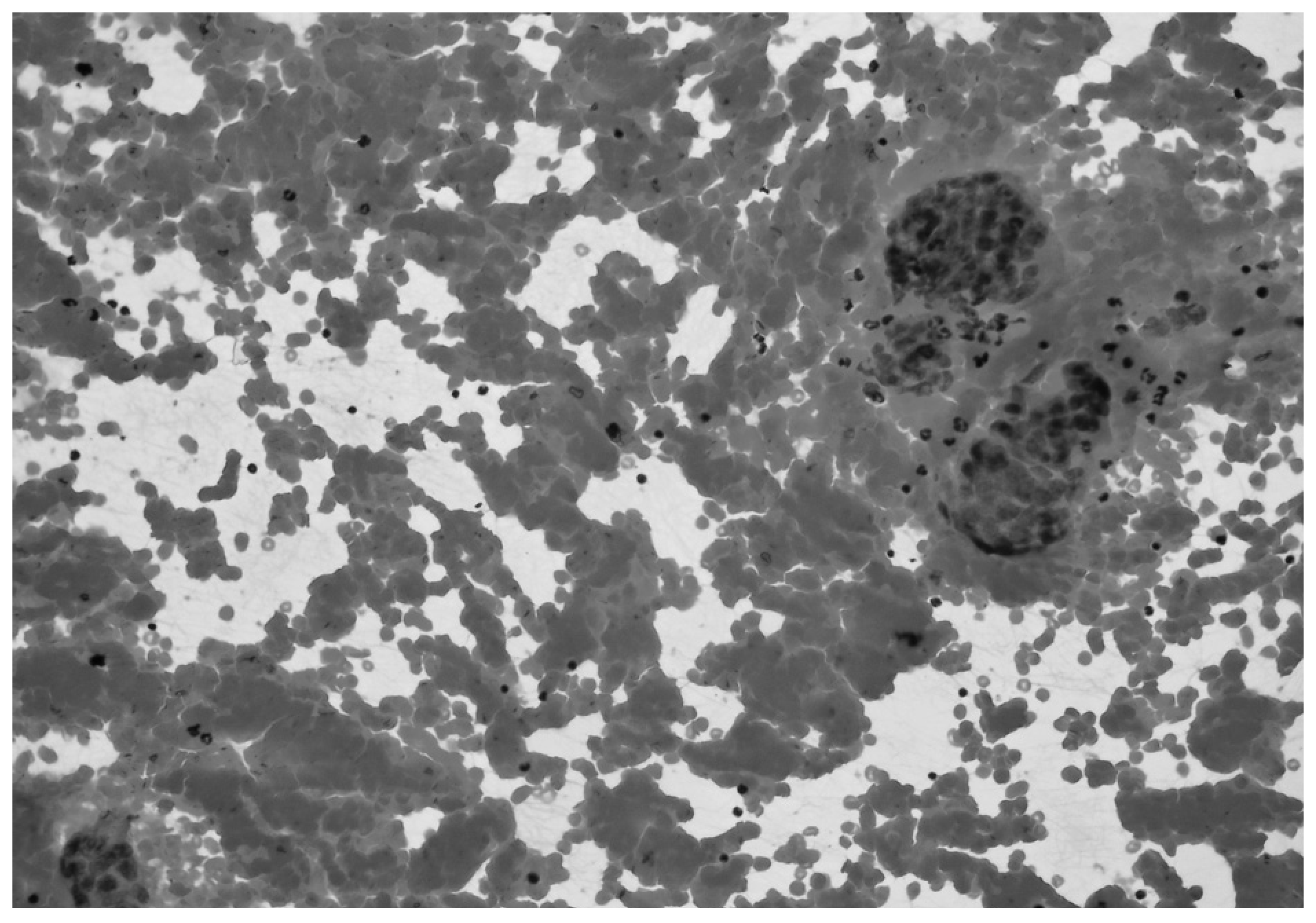
The lesion of the third metatarsal underwent fineneedle biopsy, and histologic examination showed osteoclast-type multinuclear giant cells along with erythrocytes, polymorphonuclear leukocytes, lymphocytes, and rare hemosiderin-laden macrophages (Figure 4). After the patient was diagnosed as having secondary hyperparathyroidism associated with hypovitaminosis D and multiple brown tumors, she was administered intravenous calcidiol, 1 μg/d, and oral elemental calcium, 3 g/d, until her blood calcium level recovered. The metatarsal lesion was treated with immobilization of the foot.
Figure 4.
Photomicrograph of the aspiration specimen from a third metatarsal brown tumor showing osteoclast-type multinuclear giant cells along with erythrocytes, polymorphonuclear leukocytes, lymphocytes, and rare hemosiderin-laden macrophages (H&E, ×200).
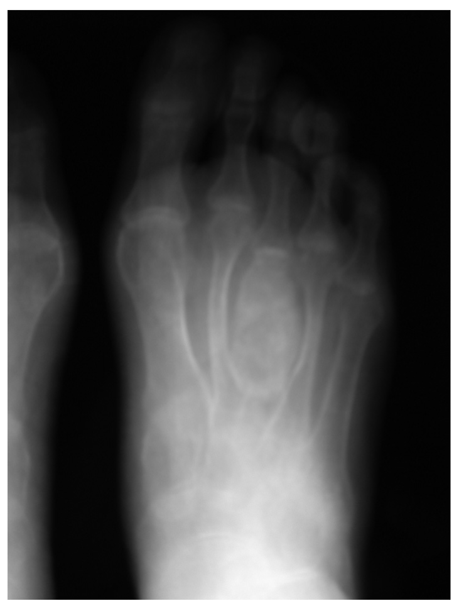
After 1 month, her widespread bone pain diminished, and the swelling and pain in her foot receded. Biochemical analysis and radiography performed 3 months later revealed reossification and an expansile lytic lesion with calcification involving the third metatarsal region (Figure 5). Serum calcium, alkaline phosphatase, and serum intact parathyroid levels were found to be 9.1 mg/dL (reference range, 8.4–10.6 mg/dL), 245 IU/L (reference range, 53–128 IU/L), and 312 pg/mL (reference range, 15–65 pg/mL), respectively. The patient’s medical treatment continues.
Figure 5.
Radiograph of the left foot after 3 months of vitamin D treatment.
Discussion
Bone involvement has similarly changed from the well-known, but now rarely seen, osteitis fibrosa cystica (brown tumor) to various degrees of bone loss in hyperparathyroidism.[8] Brown tumors occur as a result of development of a vascularized fibrotic tissue in the bone stemming from parathyroid hormone-stimulated osteoclastic activity. Brown tumor of the bone has long been recognized as a complication of primary hyperparathyroidism. Not until 1963, however, was a brown tumor noted in association with secondary hyperparathyroidism.
Brown tumors are rarely seen on the axial skeleton. Previously, involvements of the foot with primary hyperparathyroidism due to parathyroid adenoma or carcinoma have been reported in the calcaneal bone and in the first metatarsal bone.[6,7] The clinical course of the present patient with secondary hyperparathyroidism is different from previous cases because of hypocalcemia, multiple brown tumor, and vitamin D deficiency. Radiographically, brown tumors usually appear as well-defined lytic lesions, sometimes containing a few trabeculations, with thinning, expansion, or violation of the cortex. They may produce osseous expansion through endosteal indentation. Computed tomography is useful for evaluating brown tumors in the spine, skull, or lower limbs. Particularly, contrast enhancement of the lesion on CT rules out cystic transformation of brown tumor.[9] When CT shows a fluid-filled cyst, local surgery must be performed to prevent a pathologic fracture.[10] The present patient had an atypical location for a brown tumor. And, because the tumor was dilated and, thus, resulted in expanding of the cortex, it looked like a soft-tissue tumor. Therefore, differential diagnosis was made with the following, which may cause primary lesions in the third metatarsal: pigmented villonodular synovitis, digital papillary adenoma, intraosseous ganglion, intermetatarsal ganglion, elastofibroma, primary lymphoma of bone, chondromyxoid fibroma, telangietatic osteogenic sarcoma, osteochondroma, atypical neuroma, osteosarcoma, and plantar ganglion cyst; also, metastatic involvement of the third metatarsal bone was suspected as the initial differential diagnosis.[2]
Histologically, brown tumors are hard to distinguish from giant cell tumors, giant cell reparative granuloma, osteosarcoma, aneurysmal bone cysts, and telangietatic osteosarcoma.[6] Brown tumor and giant cell tumor of bone are histologically indistinguishable but are differentiated on the basis of clinical presentation and laboratory findings.[11] We diagnosed the brown tumor located in the third metatarsal bone, which is a first in the literature, by using radiography, CT, and laboratory and histologic examinations.
After the exclusion of other secondary hyperparathyroidism causes, such as chronic renal failure, high-dose phosphate treatment, malabsorption, pseudohypoparathroidism, and renal tubular phosphate-wasting disorders, the present patient was diagnosed, as having secondary hyperparathyroidism due to insufficient exposure to sunlight and lack of nutritional vitamin D.[12] Brown tumor development associated with secondary hyperparathyroidism is rare. Multiple brown tumor development is more common in patients who develop hyperparathyroidism due to chronic renal failure.[9] However, in the literature, multiple brown tumors were previously observed in nutritional secondary hyperparathyroidism, developing after gastric resection, as in the present patient.[13] Patients who develop secondary hyperparathyroidism together with osteomalacia, like the present patient, differ from patients with primary hyperparathyroidism in terms of serum calcium level, which is low or normal in secondary hyperparathyroidism. Serum calcium values were less than normal values in the present patient.[14] Parathyroid surgery is not indicated for secondary hyperparathyroidism as it is for primary hyperparathyroidism. It can generally be performed on patients with severe disease if the parathyroid hormone level is greater than 1,000 pg/mL.[14] Although the parathyroid hormone level was above the reference range in the present patient, because she totally rejected surgical treatment, initially we administered intravenous calcidiol and calcium. After 3 months of treatment, her clinical condition improved, the brown tumor became smaller, and these areas reossified, hence, showing that the treatment was successful.
In conclusion, the first case of a brown tumor involving the third metatarsal bone in a patient with secondary hyperparathyroidism has been presented. The presence of a mass or swelling in the metatarsal bone of the patient, and other findings, such as a low level of calcium, a high level of parathyroid hormone, difficulty walking, and weakness, should suggest, besides primary tumors and metastasis, development of brown tumors due to hyperparathyroidism.
Financial Disclosure
None reported.
Conflict of Interest
None reported.
References
- EL-MOFTY, S.K.; KYRIAKOS, M. “Soft Tissue and Bone Lesions,” in Diagnosis Pathology of the Head and Neck,; Gnepp, D.R., Ed.; WB Saunders: Philadelphia, 2000; Vol 21, p. 505. [Google Scholar]
- RESNICK, D.; NIWAYAMA, G. “Parathyroid Disorders and Renal Osteodistrophy,” in Diagnosis of Bone and Joint Disorders, In p 2012; WB Saunders: Philadelphia, 1995; Vol 52. [Google Scholar]
- CHEW, F.S. HUANG-HELLINGER F: Brown tumor. AJR Am J Roentgenol 1993, 160, 752. [Google Scholar] [CrossRef] [Scilit] [PubMed]
- KANAAN, I.; AHMED, M.; RIFAI, A. ET AL: Sphenoid sinus brown tumor of secondary hyperparathyroidism: case report. Neurosurgery 1998, 42, 1374. [Google Scholar] [CrossRef] [Scilit] [PubMed]
- TAKESHITA, T.; TANAKA, H.; HARASAWA, A. ET AL: Brown tumor of the sphenoid sinus in a patient with secondary hyperparathyroidism: CT and MR imaging findings. Radiat Med 2004, 22, 265. [Google Scholar] [PubMed]
- DOGAN, A.; ALGUN, E.; KISLI, E.; et al. Calcaneal brown tumor with primary hyperparathyroidism caused by parathyroid carcinoma: an atypical localization. J Foot Ankle Surg 2004, 43, 248. [Google Scholar] [CrossRef] [Scilit] [PubMed]
- RESKE, W. Osteitis fibrosa localisata (brown tumor or osteoclastoma) and aseptic bone necrosis as a cause for sesamoid bone disease of the first metatarsal: case report [in German]. Z Orthop Ihre Grenzgeb 1954, 85, 409. [Google Scholar] [PubMed]
- CHAROPOULOS, I.; TOURNIS, S.; TROVAS, G.; et al. Effect of primary hyperparathyroidism on volumetric bone mineral density and bone geometry assessed by peripheral quantitative computed tomography in postmenopausal women. J Clin Endocrinol Metab 2006, 91, 1748. [Google Scholar] [CrossRef] [Scilit] [PubMed]
- BROWN, T.W.; GENANT, H.K.; HATTNER, R.S. ET AL: Multiple brown tumors in a patient with chronic renal failure and secondary hyperparathyroidism. AJR Am J Roentgenol 1977, 128, 131. [Google Scholar] [CrossRef] [Scilit] [PubMed]
- DOPPMAN, J.L.; MARX, S.; SPIEGEL, A.; et al. Differential diagnosis of brown tumor vs. cystic osteitis by arteriography and computed tomography. Radiology 1979, 131, 339. [Google Scholar] [CrossRef] [Scilit] [PubMed]
- ZWICK, O.M.; VAGEFI, M.R.; COCKERHAM, K.P. ET AL: Brown tumor of secondary hyperparathyroidism involving the superior orbit and frontal calvarium. Ophthal Plast Reconstr Surg 2006, 22, 304. [Google Scholar] [CrossRef] [Scilit] [PubMed]
- MIHAI, R.; FARNDON, J.R. Parathyroid disease and calcium metabolism. Br J Anaesth 2000, 85, 29. [Google Scholar] [CrossRef] [Scilit] [PubMed]
- MERGENTHALER, H.G.; FINK, M.; SAUER, H. ET AL: Multiple brown tumors in a patient with nutritional secondary hyperparathyroidism. Klin Wochenschr 1989, 67, 42. [Google Scholar] [CrossRef] [Scilit] [PubMed]
- AHMAD R, HAMMOND JM: Primary, secondary, and tertiary hyperparathyroidism. Otolaryngol Clin North Am 2004, 37, 701. [CrossRef] [Scilit] [PubMed]
© 2008 American Podiatric Medical Association