


Abstract
We report a case of an unusual and unsuspected chronic infection creating a soft-tissue mass in the foot of a 35-year-old woman. The causative agent, Mycobacterium gordonae, is usually encountered as a laboratory contaminant. Only rarely does it manifest as a clinical infection. The patient’s presumed predisposing risk factor was a history of barefoot gardening. An iatrogenic source, corticosteroid injections, was also considered.
Soft-tissue infections of the foot caused by nontuberculous mycobacteria are distinctly uncommon. Nonetheless, the incidence of these infections is well documented in case reports and occasional small series.[1,2,3,4,5,6] In immunocompetent individuals, the disease is generally localized and self-limited, although deep-seated infections can induce osteomyelitis.[7] Severe immunocompromise is a predisposing factor to life-threatening systemic involvement and necessitates aggressive therapeutic intervention.[8,9] These organisms are often resistant to first-line antituberculous medications but are typically susceptible to a variety of routine antibiotics.[10,11]
Two species-specific infections exist: swimming-pool or fish-tank granuloma caused by Mycobacterium marinum, and Buruli ulcer caused by Mycobacterium ulcerans.[12,13] Infections caused by other species are more sporadic, with nonspecific clinical presentations and pathologic findings. Tissue biopsies often demonstrate only vague granulomatous features that are often disregarded, resulting in the erroneous diagnosis of an unspecified infectious and inflammatory or benign neoplastic process. The causal agent in this case, Mycobacterium gordonae, has only rarely been acknowledged as a pathogen.[14,15] Presumed injury to the patient’s foot incurred while gardening provided a provocative explanation for the source of the infection.
Case Report
A 35-year-old woman presented in August 2006 with the complaint of numbness and tingling in her toes and a painful mass in her left foot. She had no memory of recent trauma or injury to the left foot but did admit to gardening barefoot, often with exposure of her feet to dirty groundwater. Her past medical history was noncontributory; in particular, there was no evidence of immunocompromise, human immunodeficiency virus infection, or intravenous drug use.
Physical examination revealed a mobile, nodular lesion in the dorsal-proximal aspect of the second interspace of the left foot, which was painful with manipulation. Radiographic studies were negative for osteomyelitis and skeletal abnormality. A magnetic resonance imaging, performed without contrast, revealed minimal edema and inflammatory changes in the involved area of the left foot. Prominent fatty tissue was noted in this region as well, leading the radiologist to suggest a traumatized lipoma as the most likely diagnosis.
Corticosteroid injections to the area on follow-up office visits were unsuccessful in relief of pain. As a result of failed conservative therapy, the patient underwent incision and drainage of the site several months later. Based on the pathologic findings of the initial excision specimen, she underwent excision again, to obtain tissue for microbiological cultures. At that time, a routine chest radiograph and tuberculin skin test were performed, with normal and nonreactive results, respectively. Her postoperative course was unremarkable. An infectious disease consultation was obtained, but treatment with antibiotics was not initiated.
Pathologic Findings
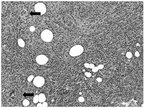
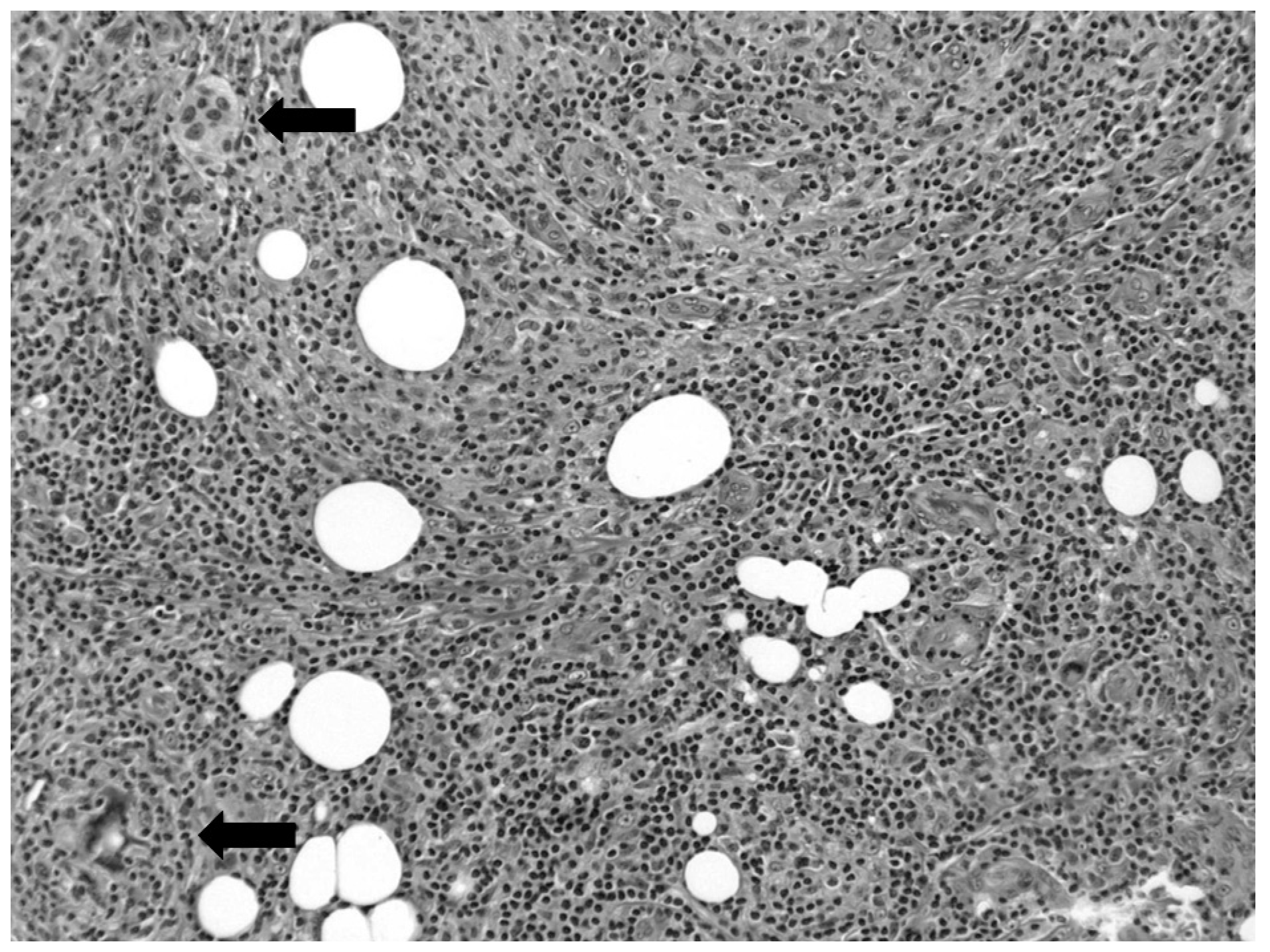
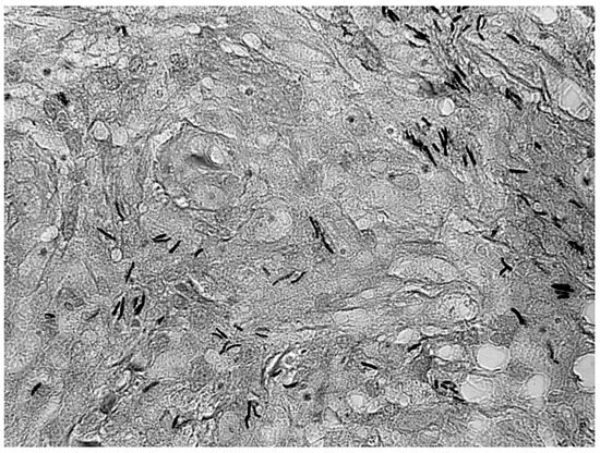
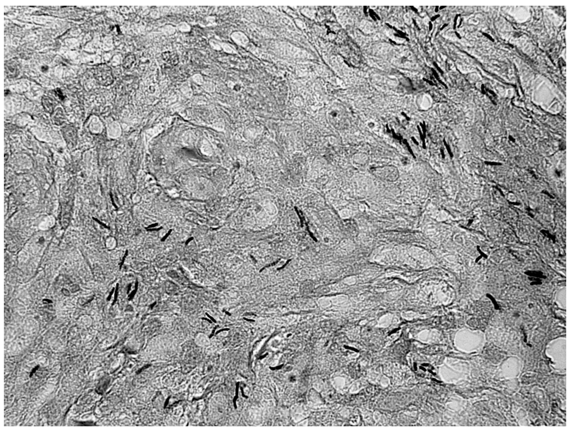
The specimen was received in formalin and consisted of a 1.5-cm fragment of firm, tan-gray tissue, grossly inconsistent with a typical lipoma. Histologic examination revealed a chronic lymphocytic-histiocytic inflammatory process with subtle granulomatous features (Figure 1). Caseous necrosis was not present. Acid-fast stain revealed numerous acid-fast bacilli in the lesion, morphologically consistent with non-tuberculous mycobacteria (Figure 2). Special stains for fungal elements and routine bacteria were negative. Microbiological cultures from the second excision and debridement of the lesion were positive for M gordonae.
Figure 1.
Fibroadipose tissue demonstrating a chronic lymphocytic-histiocytic inflammatory reaction with subtle granulomatous features (×200). Giant cells are designated with arrows.
Figure 2.
Acid-fast stain demonstrating acid fast bacilli in the tissue (oil immersion ×1,000).
Discussion
Mycobacterium gordonae is a ubiquitous environmental organism, existing predominantly in soil and groundwater. As such, it is identified in the laboratory most frequently as a nosocomial water contaminant,[16,17,18] which must be distinguished from other, more typical, pathogenic mycobacteria. Documented infections have been reported only sporadically.[8,12,13] In the majority of these reported cases, a confirmed or presumptive exposure to environmental soil or water was present. At least one article alluded to a probable gardening-related acquisition of the infection.[13]
Histologic features suggesting the granulomatous nature of these lesions (e.g., multinucleated giant cells, palisading histiocytes, or focal necrosis) may be extremely subtle and easily overlooked. Of four pathologists who independently reviewed the histologic slides in this case, only one suspected an infectious etiology; the other three favored a benign fibrohistiocytic lesion or a pseudopyogenic granuloma. Therefore, these lesions are almost certainly under-recognized pathologically as well as clinically.
Differential diagnostic considerations include other chronic cutaneous and soft-tissue infections, benign tumor and tumor-like conditions (e.g., tenosynovitis, traumatic neuroma, lipoma, and giant cell tumor of the tendon sheath), and, rarely, malignant tumors that may mimic granulomatous processes (e.g., epithelioid sarcoma and necrotizing metastases). Directed microbiological cultures in conjunction with the histologic findings will permit the correct diagnosis in most instances. However, routine aerobic/anaerobic cultures are generally insufficient for the detection of these “atypical” mycobacteria. A high index of clinical suspicion is necessary to prompt the appropriate microbiological evaluation. In cases with atypical clinical presentations, a small representative portion of the specimen can be submitted to the laboratory in a sterile container for retention until the correct microbiological appraisal is determined by the histologic examination.
The possibility that the etiology of the patient’s infection was iatrogenic, secondary to the corticosteroid injections,[19] cannot be entirely excluded. The vial and syringe used for the injections were not available for microbiological cultures. However, the appearance of the lesion during the patient’s gardening season and the lack of clinical change following the injections favors an environmental acquisition of the infection. Since the presentation of the initial lesion, she has developed a similar, smaller lesion in the right foot, which is currently being followed clinically. If this lesion persists, excision with culture could provide additional support for this supposition.
Conclusion
Mycobacterium gordonae is an uncommon cause of a painful foot mass. In this case, exposure to ground-water while gardening barefoot was the presumptive etiology of the soft-tissue infection. Conservative surgical excision is generally curative, except in individuals with severely compromised immune systems, for whom aggressive systemic antibiotic therapy is indicated.
Acknowledgment
Jill Woods for typing the manuscript.
Financial Disclosure
None reported.
Conflict of Interest
None reported.
References
- CAREY, M.J.; MACLAREN, H.M.C.; MILLER, M.V. A cutaneous infection with Mycobacterium fortuitum. Ann Emerg Med 1994, 23, 347. [Google Scholar] [CrossRef] [Scilit] [PubMed]
- ESTEBAN J, FERNANDEZ ROBLAS R, GARCIA CIA JI, ET AL: Clinical significance and epidemiology of non-pigmented rapidly growing mycobacteria in a university hospital. J Infect 2007, 54, 135. [CrossRef] [Scilit] [PubMed]
- MATTILA, J.O.; KATILA, M.L.; VORNANEN, M. Slowly growing mycobacteria and chronic skin disorders. Clin Infect Dis 1996, 23, 1043. [Google Scholar] [CrossRef] [Scilit] [PubMed]
- ROSENMEIER, G.; KEELING, J.; GRABSKI, W. ET AL: Latent cutaneous Mycobacterium fortuitum infection in a healthy man. J Am Acad Dermatol 1991, 25, 898. [Google Scholar] [CrossRef] [Scilit] [PubMed]
- SPIEGL P, FEINER C: Mycobacterium phlei infection of the foot: a case report. Foot Ankle Int 1994, 15, 680. [CrossRef] [Scilit] [PubMed]
- SUBBARAO, E.; TARPA, M.; MARKS, M. Soft-tissue infections caused by Mycobacterium fortuitum complex following penetrating injury. Am J Dis Child 1987, 141, 1018. [Google Scholar] [CrossRef] [Scilit] [PubMed]
- BARTON A, BERNSTEIN RM, STRUTHERS JK, ET AL: Mycobacterium marinum infection causing septic arthritis and osteomyelitis. Br J Rheumatol 1997, 36, 1207. [CrossRef] [Scilit] [PubMed]
- GAROFALO, R.; CHADWICK, E.G.; YOGEV, R. Mycobacterium gordonae bacteremia in an asymptomatic adolescent with acquired immunodeficiency syndrome. Pediatr Infect Dis J 2003, 22, 569. [Google Scholar] [CrossRef] [Scilit] [PubMed]
- STREIT, M.; BOHLEN, L.M.; HUNZIKER, T.; ET, A.L. Disseminated Mycobacterium marinum infection with extensive cutaneous eruption and bacteremia in an immunocompromised patient. Eur J Dermatol 2006, 16, 79. [Google Scholar] [PubMed]
- BROWN-ELLIOTT, B.A.; CRIST, C.J.; MANN, L.B.; ET, A.L. In vitro activity of linezolid against slowly growing nontuberculous mycobacteria. Antimicrob Agents Chemother 2003, 47, 1736. [Google Scholar] [CrossRef] [Scilit] [PubMed]
- RODRIGUEZ DIAZ, J.C.; LOPEZ, M.; RUIZ, M.; ET, A.L. In vitro activity of new fluoroquinolones and linezolid against nontuberculous mycobacteria. Int J Antimicrob Agents 2003, 21, 585. [Google Scholar] [CrossRef] [Scilit] [PubMed]
- BARTRALOT, R.; GARCIA-PATOS, V.; SITJAS, D.; et al. Clinical patterns of cutaneous nontuberculous mycobacterial infections. Br J Dermatol 2005, 152, 727. [Google Scholar] [CrossRef] [Scilit] [PubMed]
- KERN, W.; VANEK, E.; JUNGBLUTH, H. Fish breeder granuloma: infection caused by Mycobacterium marinum and other atypical mycobacteria in the human. Analysis of 8 cases and review of the literature [in German]. Med Klin (Munich) 1989, 84, 578. [Google Scholar] [PubMed]
- MCINTYRE, P.; BLACKLOCK, Z.; MCCORMACK, J.G. Cutaneous infection with Mycobacterium gordonae. J Infect 1987, 14, 71. [Google Scholar] [CrossRef] [Scilit] [PubMed]
- ZALA, L.; JUNZIKER, T.; BRAATHEN, L.R. Chronic cutaneous infection caused by Mycobacterium gordonae. Dermatology 1993, 187, 301. [Google Scholar] [CrossRef] [Scilit] [PubMed]
- ESTEBAN, J.; FERNANDEZ-ROBIAS, R.; ORTIZ, A.; ET, A.L. Pseudooutbreak of Mycobacterium gordonae: usefulness of randomly amplified polymorphic DNA analysis to assess the clonality of the isolates. Clin Microbiol Infect 2006, 12, 677. [Google Scholar] [CrossRef] [Scilit] [PubMed]
- FUJITA, J.; NANK, N.; NEGAYAMA, K. Nosocomial contamination by Mycobacterium gordonae in hospital water supply and super-oxidized water. J Hosp Infect 2002, 51, 65. [Google Scholar] [CrossRef] [Scilit] [PubMed]
- LABOMBARDI, V.J.; O’BRIEN, A.M.; KISLAK, J.W. Pseudo-outbreak of Mycobacterium fortuitum due to contaminated ice machines. Am J Infect Control 2002, 30, 184. [Google Scholar] [CrossRef] [Scilit] [PubMed]
- BAACK, B.R.; BROWN, R.E. Atypical mycobacterium soft-tissue infection of the dorsal radial wrist: a possible complication of steroid injection for de Quervain’s disease. Ann Plast Surg 1991, 27, 73. [Google Scholar] [PubMed]
© 2008 American Podiatric Medical Association