


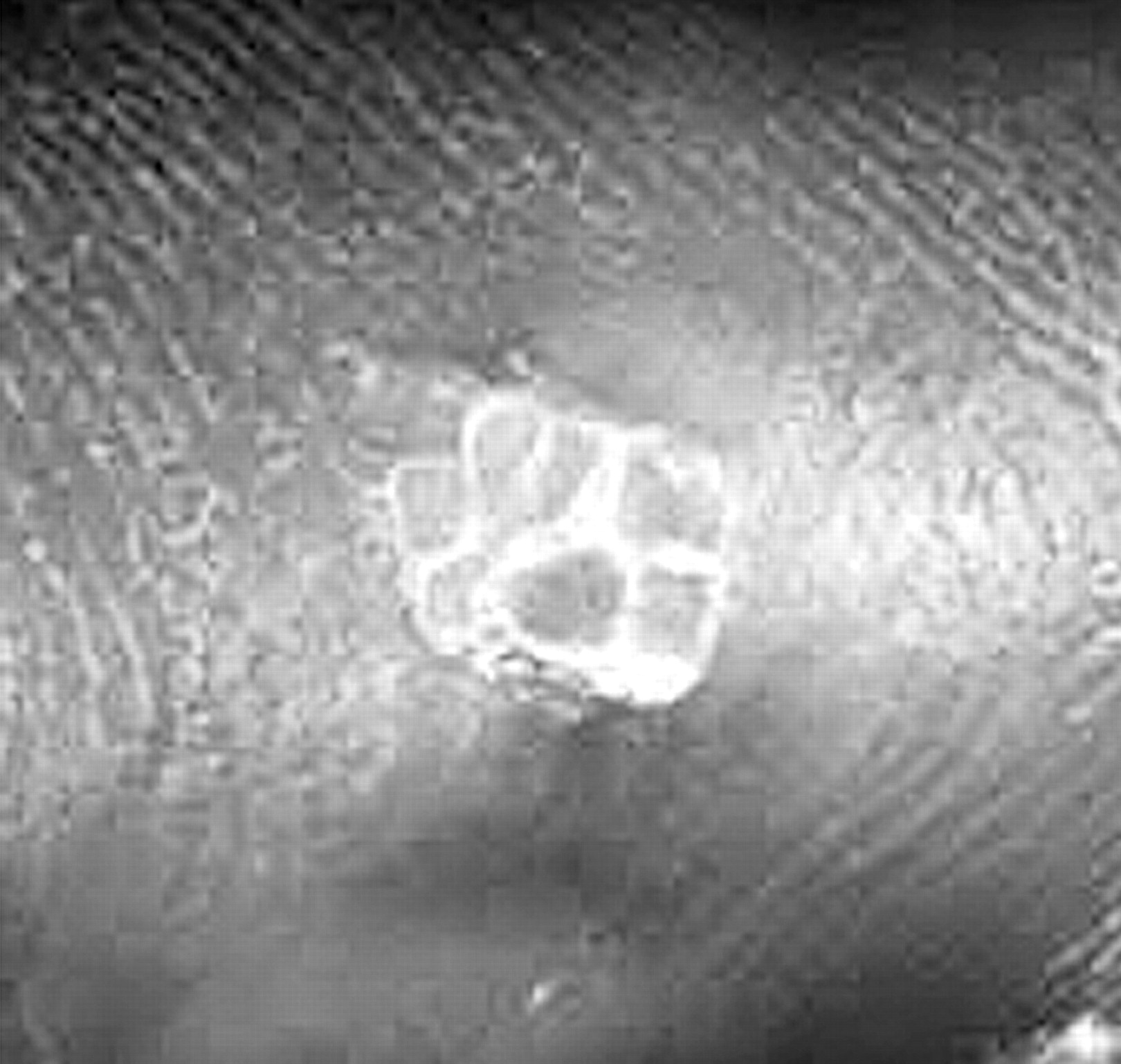
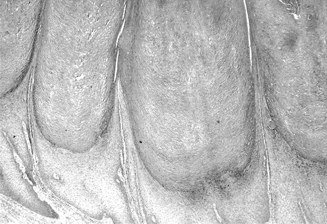
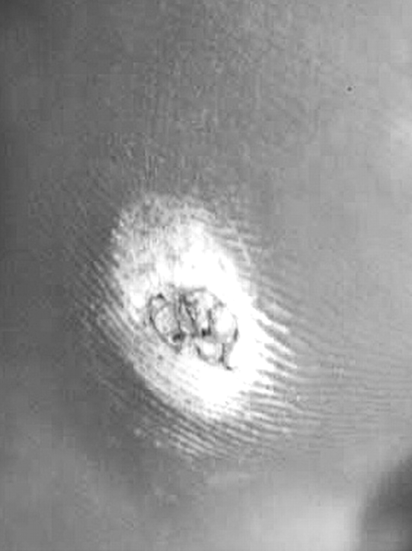
A 63-year-old female with a 25-year history of an exquisitely painful callus located at the lateral margin of the right fifth metatarsal is seen by you in your office. The patient states that she must have the lesion debrided every two months to avoid pain when wearing shoes. She warns you in advance to be careful when you debride the lesion because the lesion also hurts when it is debrided. After you remove the overlying callus, you notice a mosaic-looking lesion with multiple small keratotic plugs seated next to each other, forming a plaque (Fig. 1A and 1B). No pinpoint bleeding is noted. You decide to bluntly remove the lesion, thinking it is not a simple callus. Histologically, multiple rectangular-shaped plugs of keratin are seen with rounded bases. Beneath these keratinous plugs, the underlying epithelium is hyperplastic. Each plug is separated by air spaces so they do not appear to be attached to each other (Fig. 1C).
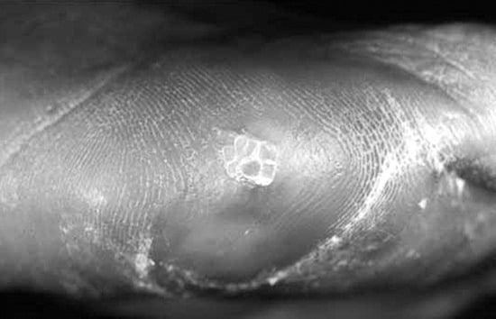
Figure 1A.
Lesion located at lateral aspect of the fifth metatarsal.
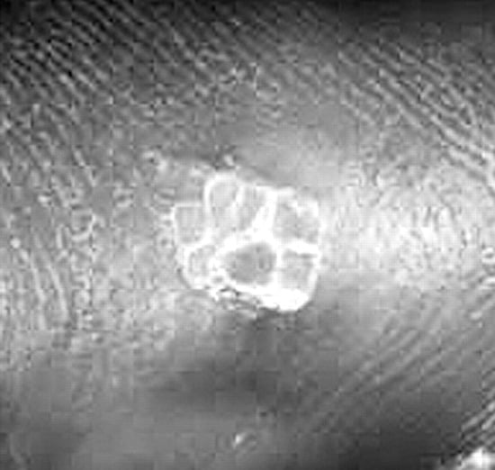
Figure 1B.
Close-up view of polygonal keratinous stalks.
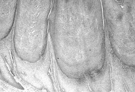
Figure 1C.
Multiple stalks of keratin with rounded bases located on hyperplastic epithelium (H&E, ×2.5).
Answer: Heloma neurofibrosum
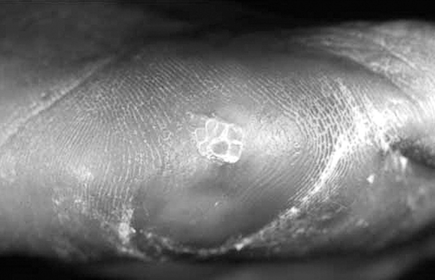
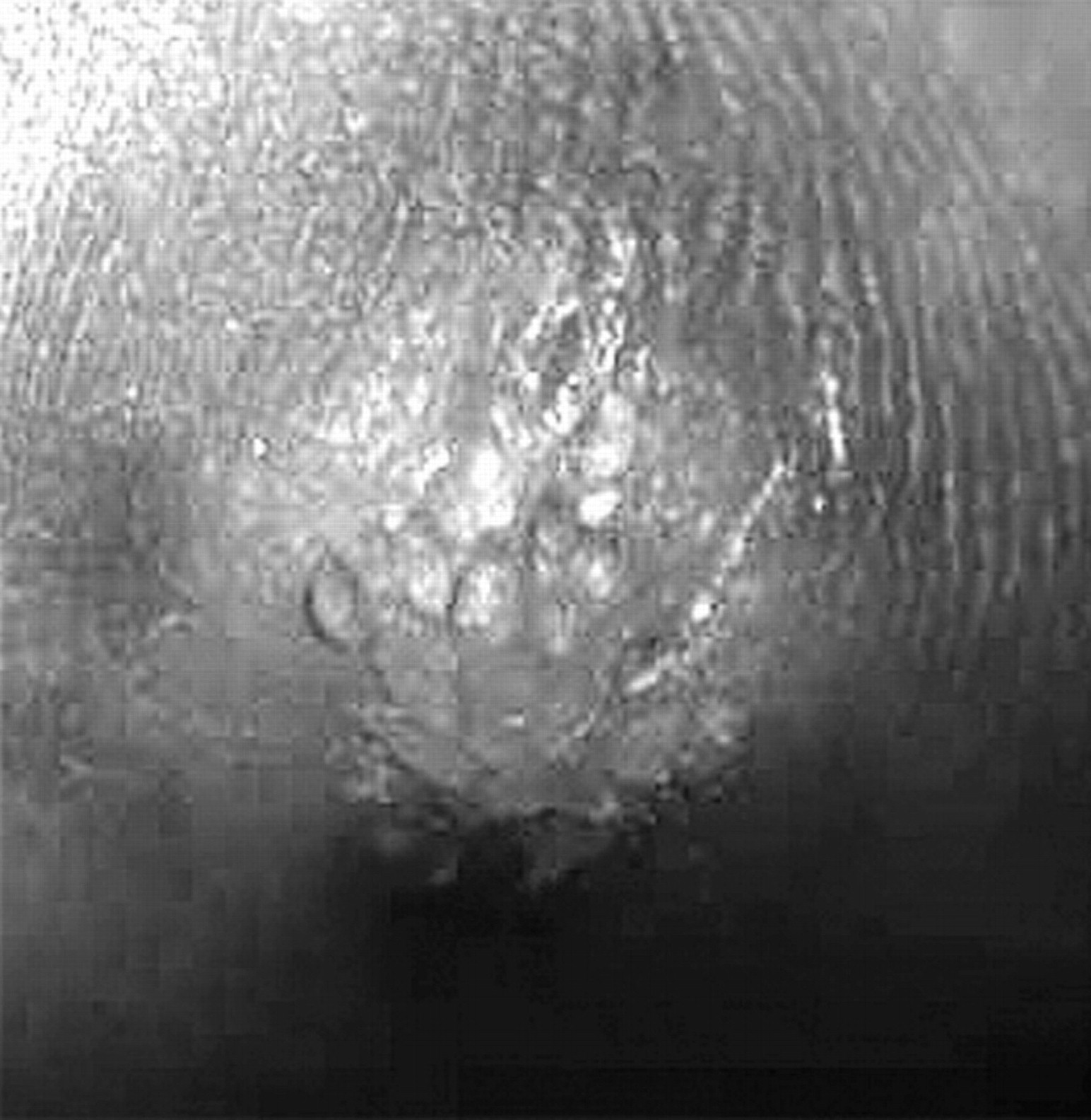
Heloma neurofibrosum, known also as a neurofibrous corn, is a lesion that has had scant attention in modern literature. Described by Durlacher in 1845 [1] and by Gross and Burnett in 1933, [2] these extremely painful lesions were thought to represent entrapped nerve tissue with the stalks representing nerve filament. These lesions are often seen in certain fixed locations, particularly along the margins of the heel and forefoot. These lesions are often mistaken for mosaic warts (Fig. 2A and 2B). After reviewing the histology of many such lesions, our laboratory began to notice material within the granular layer, which suggests that these lesions might be viral. It is our feeling that these lesions are probably best described as “burnt-out warts,” which are warts that have resolved in most cases but are maintained under weightbearing areas for years by mechanical pressure.
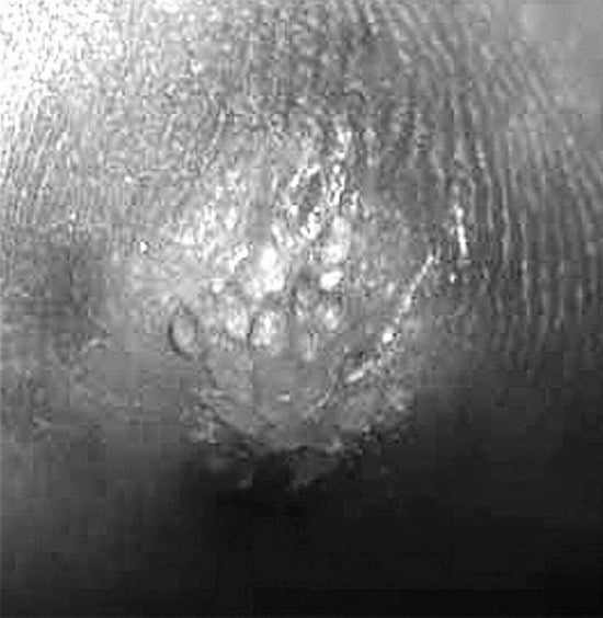
Figure 2A.
Mosaic-like plaque.
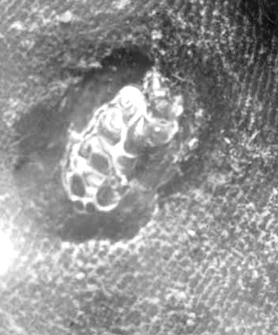
Figure 2B.
Multiple polygonal keratinous plugs resembling a mosaic wart.
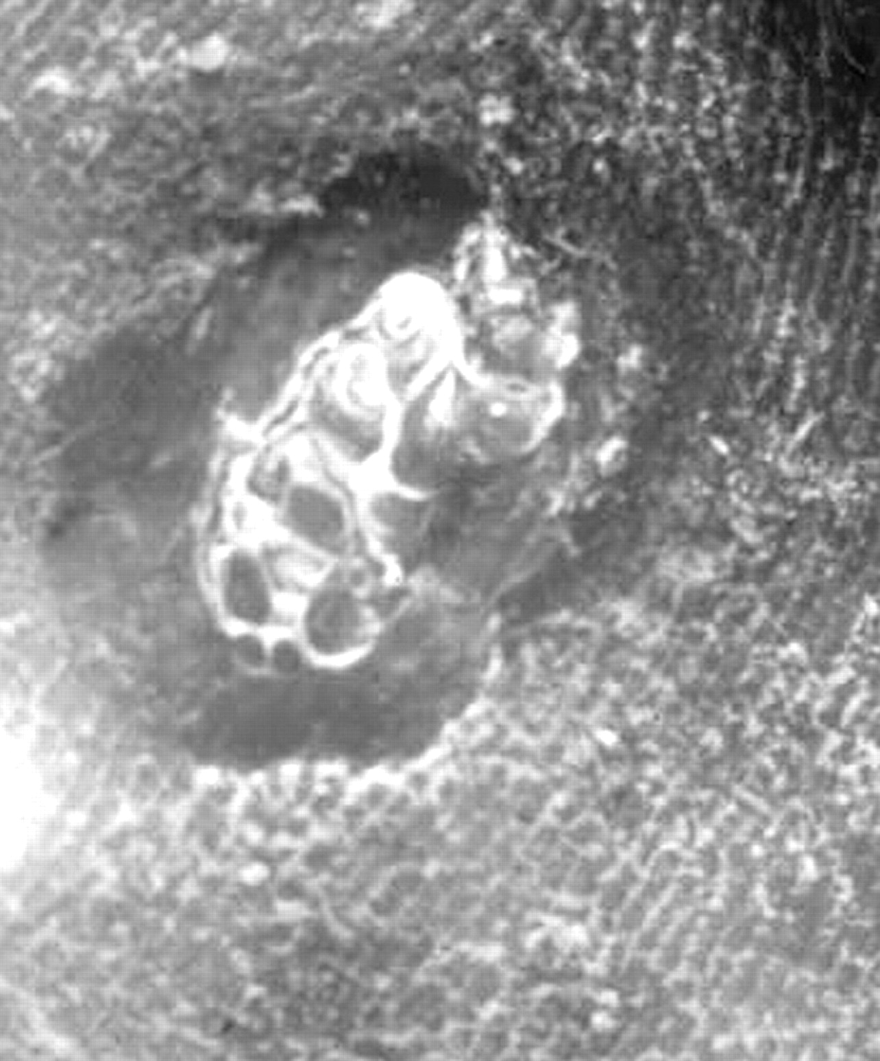
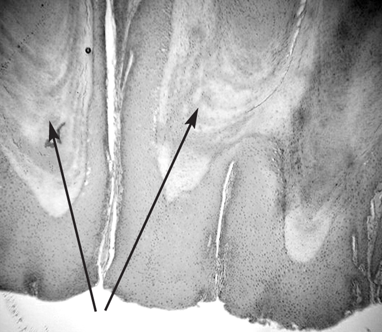
When treated with salicylic acid, the stalks or projections become characteristically prominent and clinically diagnostic of heloma neurofibrosum (Fig. 2C). The separate keratinous blocks were thought by clinicians to represent superficially located cutaneous nerve filaments, which were thought to explain the extreme pain upon debridement. Histologically, however, these cutaneous projections represent keratin and not nerve tissue (Fig. 3).
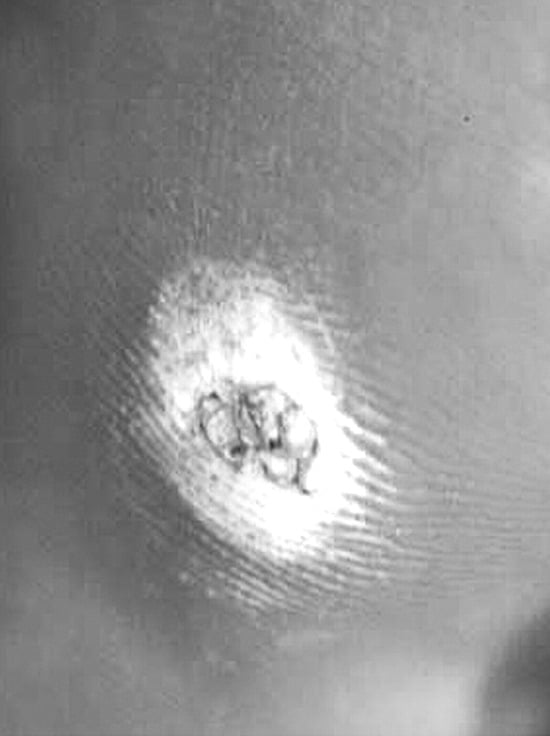
Figure 2C.
Stalks become prominent with acid therapy.
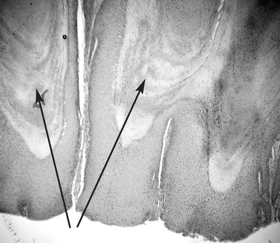
Figure 3.
Keratinous projections (arrows) (H&E, ×2.5).
References
- Durlacher L: “Neurovascular corns,” in A Treatise on Corns, Bunions, the Diseases of Nails, and the General Management of the Feet, p 65, Simpkin, Marshall and Co., London. ,1845. .
- Gross RH and Burnett EK: “Heloma neurofibrosum,” in The Practice of Podiatry, ed by MJ Lewi, p 154, Harriman Printing Co., New York. ,1933. .
© 2008 American Podiatric Medical Association