Muscle flaps for soft-tissue defects have been widely used in reconstructive surgery. Soft-tissue coverage over bony defects can be difficult to treat. Exposed bone in a wound can lead to increased infection and a delay in healing. Osteomyelitis compounds this problem, and extensive debridement of all infected, devitalized bone and soft tissue is necessary to aid healing before wound closure can be attempted.[
1–
6]
The abductor digiti minimi muscle belly can be used to cover defects that encompass the lateral ankle joint, calcaneus, or lateral heel.[
7–
11] Meticulous dissection and soft-tissue handling is key to the success of this procedure. Knowledge of the major arterial supply and perforators that supply the muscle is necessary, and loupe magnification is important in this task.[
7,
9] The abductor digiti minimi muscle is supplied by the lateral plantar artery.[
7,
9] The abductor digiti minimi muscle is reflected proximally and stops at the fifth metatarsal tuberosity.[
7] At this level, terminal branches of the lateral calcaneal branch of the peroneal artery perforate the abductor digiti minimi muscle and provide the vascular supply to the muscle.[
7–
14] The abductor digiti mini muscle flap requires use of a split-thickness skin graft on its superficial surface to achieve complete closure. This is a straightforward procedure that does not involve significant morbidity, and the split-thickness skin graft can be easily harvested from the ipsilateral medial arch,[
15] calf, or thigh.[
16]
We present a case report of a patient who had successful reconstruction of a chronic nonhealing traumatic wound associated with calcaneal osteomyelitis after a failed open reduction internal fixation procedure that was successfully closed using an abductor digiti minimi muscle flap and a split-thickness skin graft.
Case Report
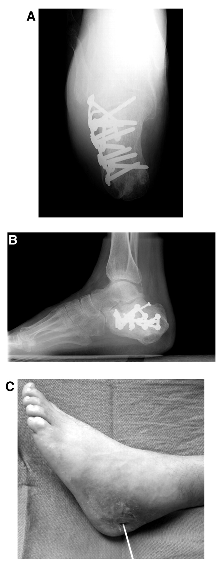
A 32-year-old healthy man was admitted from the emergency department at The University of Texas Health Science Center at San Antonio with a chronic open wound on the lateral aspect of the left heel with pain and cellulitis. His medical history revealed a previous intra-articular comminuted calcaneal fracture (after falling off a roof in 2004). The fracture required open reduction and internal fixation with a plate and multiple screws. Since the surgery, an open wound has been present on the lateral aspect of the left heel. At the initial visit 15 months after surgical repair, the wound measured 3.0 cm long, and mild purulence was expressed (
Fig. 1). The patient had several courses of antibiotic therapy for almost a year before admission to the hospital, and his initial radiographs of the left foot revealed signs of osteomyelitis of the calcaneus.
Figure 1.
Calcaneal axial (A) and lateral (B) radiographs showing signs of osteomyelitis, along with the initial clinical presentation of the chronic open wound at the lateral aspect of the calcaneus (C).
Figure 1.
Calcaneal axial (A) and lateral (B) radiographs showing signs of osteomyelitis, along with the initial clinical presentation of the chronic open wound at the lateral aspect of the calcaneus (C).
The initial surgery consisted of excision of the ulcer and incision and drainage of the lateral calcaneal wound of the left foot. The calcaneal plate and screws were also removed during this initial procedure. The distal inferior portion of the calcaneus was noted to be soft and brittle, and partial resection and decortication of the lateral calcaneal wall in addition to aggressive curettage of the remaining large calcaneal holes after the removal of screws was also performed. Intraoperative cultures of soft tissue and bone were also performed. Negative-pressure wound therapy was performed in the central aspect of the wound to help stimulate granulation tissue. Intravenous antibiotic drug therapy was started after this procedure. The patient was given intravenous cefazolin, 1 g every 8 hours. Bone culture results revealed light growth of oxacillin-sensitive Staphylococcus aureus.
The negative-pressure wound therapy was in place for 4 days before the second procedure was performed. The wound base exhibited healthy granulation tissue. In the central aspect of the wound, the lateral aspect of the calcaneus was visible in the base of the wound. Surgical planning consisted of bone grafting the calcaneal deficit after removal of the multiple large cortical screws, with wound closure via an abductor digiti minimi muscle flap.
Attention was directed to the lateral aspect of the fifth metatarsal of the left foot. A linear incision measuring 7 cm in length was made along the fifth metatarsal to expose the insertion of the abductor digiti minimi muscle. The abductor digiti minimi muscle was transected at its insertion at the base of the proximal phalanx of the fifth digit. Loupe magnification was necessary to ensure identification and maintenance of all neurovascular structures to the abductor digiti minimi muscle belly.
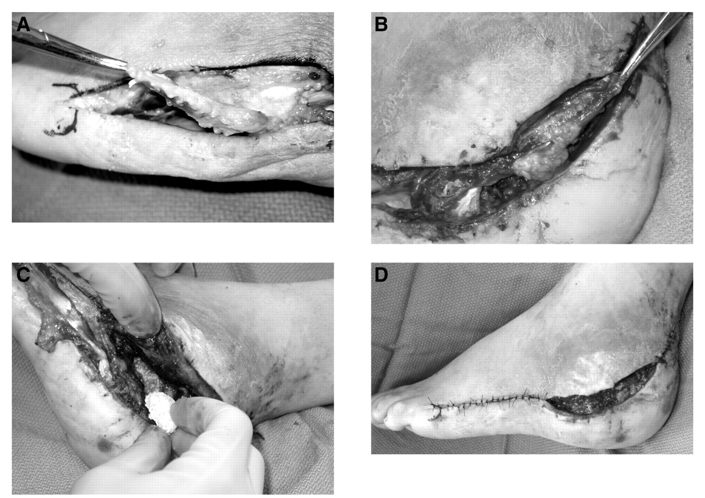
After transection of the muscle tendon, the abductor digiti minimi muscle was advanced proximally. Advancement was stopped approximately 2 cm proximal to the styloid process of the fifth metatarsal to preserve the perforating artery of the lateral calcaneal branch of the peroneal artery to the muscle belly. An allogeneic corticocancellous bone graft was used to fill the calcaneal defect from the previous bone removal due to osteomyelitis. The abductor digiti minimi muscle belly was placed over the defect to provide bone coverage. Sutures of 2-0 polypropylene (Prolene; Ethicon, Somerville, New Jersey) were used to hold the muscle belly in place as it was sutured to the posterior aspect of the Achilles tendon. Sutures of 3-0 and 4-0 nylon were used to close the distal and proximal aspects of the wound site (
Fig. 2).
Figure 2.
Transection of the muscle tendon from its insertion (A), followed by proximal advancement (B), application of corticocancellous bone grafting (C), and muscle laying over the major calcaneal bone and soft-tissue defect (D).
Figure 2.
Transection of the muscle tendon from its insertion (A), followed by proximal advancement (B), application of corticocancellous bone grafting (C), and muscle laying over the major calcaneal bone and soft-tissue defect (D).
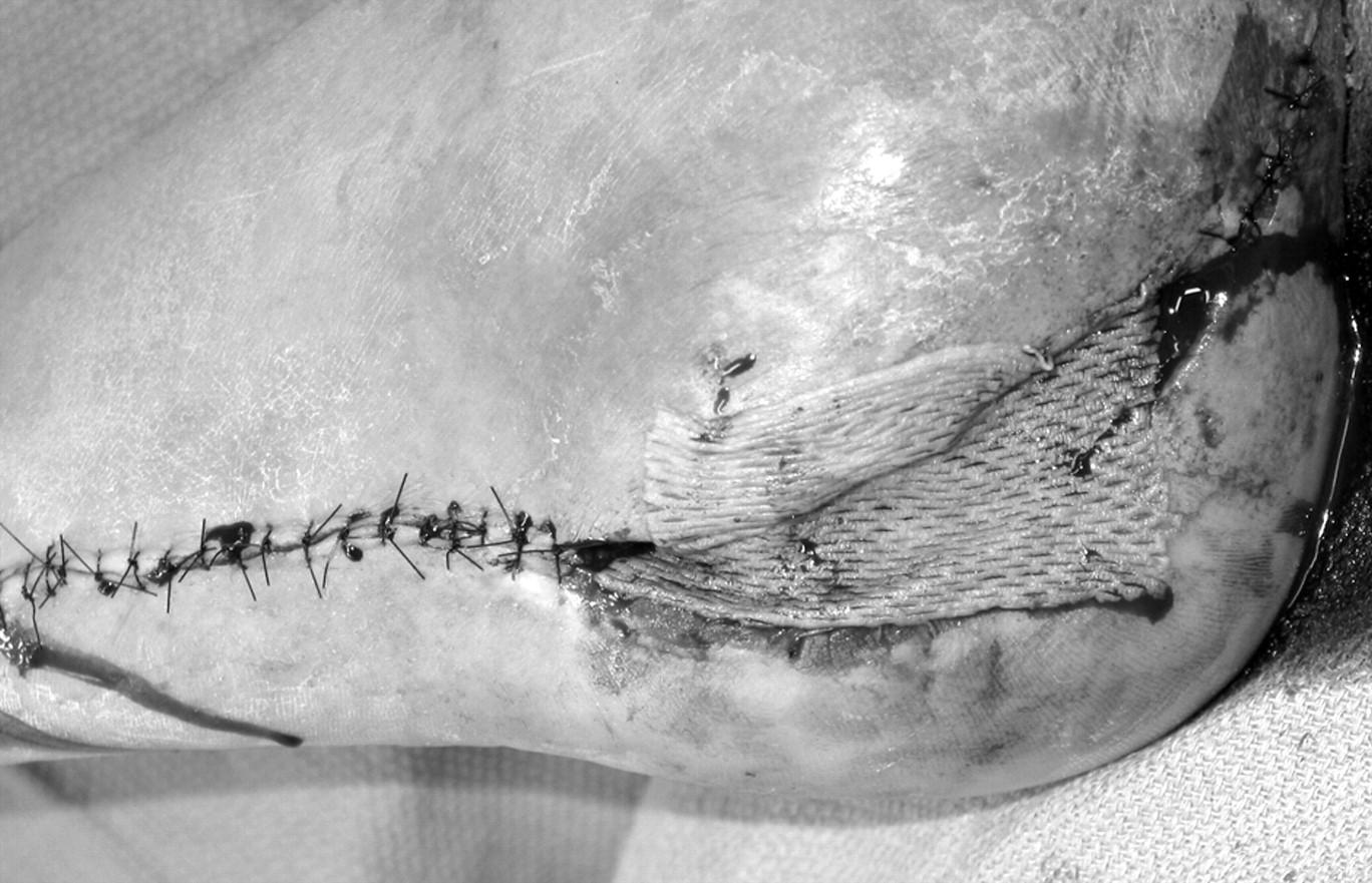
To provide closure over the muscle flap, a split-thickness skin graft was taken from the lateral aspect of the patient’s left leg (
Fig. 3). The graft was held in place with skin staples and a bolster dressing. The patient was placed in a posterior splint. After surgery, the patient was monitored for pain management and continued the same intravenous antibiotic drug regimen verified by the intraoperative cultures before closure. The patient was discharged 2 days after surgery with oral antibiotic drugs that were taken for 6 weeks after wound closure.
Figure 3.
Application of an ipsilateral split-thickness skin graft over the muscle to provide complete wound coverage.
Figure 3.
Application of an ipsilateral split-thickness skin graft over the muscle to provide complete wound coverage.
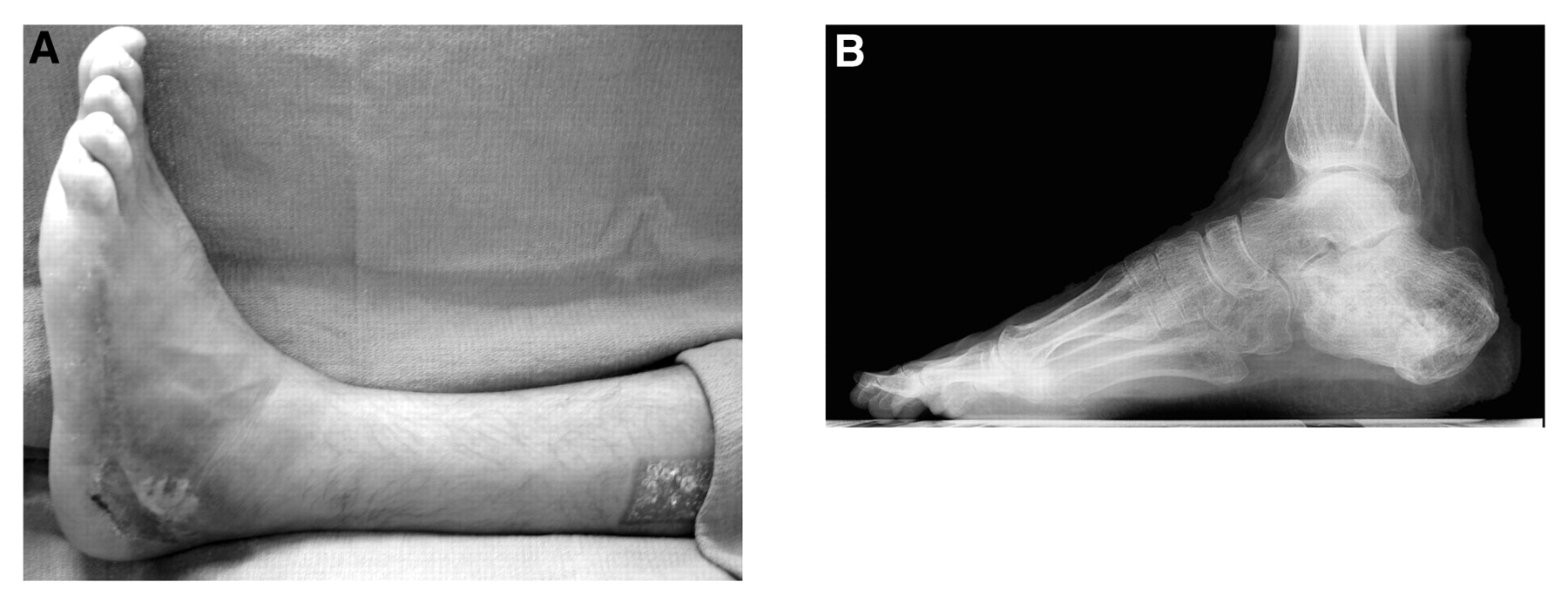
His postoperative course included a nonweightbearing status for 6 weeks. His bolster dressing was removed 3 weeks after surgery. The patient was allowed to bear weight 6 weeks after surgery while wearing a surgical shoe and using crutches for assistance. The final postoperative visit 6 months after surgery revealed a completely healed wound and bone, and the patient was wearing his normal shoes (
Fig. 4).
Figure 4.
Postoperative clinical (A) and radiographic (B) images showing healed soft-tissue and bone defects.
Figure 4.
Postoperative clinical (A) and radiographic (B) images showing healed soft-tissue and bone defects.
Discussion
The abductor digiti minimi muscle flap is an effective means of closure for lateral heel defects complicated by osteomyelitis after traumatic injury.[
7,
9,
17] Dissection of this muscle is relatively straightforward, but care must be taken to preserve the main vascular pedicle during dissection and transposition of the muscle flap to avoid ischemic compromise and venous congestion. The abductor digiti minimi muscle flap for reconstructive purposes provides patients with a shorter hospitalization, faster healing, and quicker progression to normal daily activities.[
7–
15]
The patient in this case report progressed well, and complete healing was achieved. This case report demonstrates a quick and reliable method for osseous and soft-tissue reconstruction after severe complication of a calcaneal osteomyelitis and wound infection from an open reduction and internal fixation of a comminuted calcaneal fracture.