


Abstract
Traditional methods of diagnosing onychomycosis, such as microscopy, histologic staining, and cultures, may not provide the clinician with documentation before initiating antifungal drug therapy. DNA technology now supplies the tools for increased sensitivity, speed, and accuracy in the diagnostic arena by allowing for the amplification, qualification, and quantitation of DNA. These techniques, already being used to identify many infectious agents, may soon be commonly applied to onychomycosis. This report reviews some of the DNA-based techniques that are currently being used to identify dermatophytes and their possible diagnostic use.
The prevalence of onychomycosis, a general term for fungal nail infections, has been reported to range from 2% to almost 9% in the general US population[1] and may be increasing in North America.[2] When the ensuing nail infection is exclusively dermatophytic, it is known as “tinea unguium.” This nail infection is predominantly caused by the dermatophyte species Trichophyton, principally Trichophyton rubrum and Trichophyton mentagrophytes var interdigitale,[3] which are capable of parasitizing keratinized human tissues.[4] These dermatophytes secrete metalloproteases and serine proteases, with T mentagrophytes reported to have higher proteolytic activity than T rubrum.[5] The seven-member gene family encoding the serine protease activity in T rubrum includes two proteases with high specificity for keratin, the main protein in nails, and others that may be involved in immediate- and delayed-type hypersensitivity skin reactions.[6]
Trichophyton rubrum and T mentagrophytes account for 80% of all dermatophytic nail infections and approximately 50% of all nail infections,[7] with T rubrum being the most significant agent.[8] Though considered non–life threatening, the presence of onychomycosis could cause more serious dermatophytic infections and complications in certain populations and should be given careful clinical treatment. Onychomycosis is much more frequent in patients with human immunodeficiency virus disease and may be linked to the level of immunosuppression,[9] and, although the condition is rarely invasive, reports have confirmed the presence of deep dermatophytic infections in an immunocompromised host.[10]
A report[11] on elderly patients found diabetes mellitus to be the most prevalent disease associated with onychomycosis. Recent epidemiologic data[12] indicate that the total prevalence of diabetes in the United States is 20.8 million people, 10.3 million of whom are older than 60 years. In general, onychomycosis is thought to affect one-third of the diabetic population (a rate 2.77 times higher than in healthy individuals[13]), representing approximately 7 million people.
The established diagnostic techniques of microscopy, staining, and cultures for diagnosing onychomycosis often cannot identify the specific infectious agent or can cause a 3- to 4-week lapse between the initial patient visit and the final diagnosis. Negative culture results can frustrate the clinician when there is a strong suggestion of onychomycosis, which can delay treatment. The advent of molecular biology techniques and specifically the application of polymerase chain reaction (PCR)[14] and related nucleic acid techniques provide the laboratory with a powerful but relatively simple method for detection and diagnosis of human infections by microorganisms.[15]
The purposes of this article are to briefly review the traditional diagnostic techniques, to explain the basics of the most common DNA techniques currently used in dermatophyte studies, and to compare these techniques as diagnostic tools for identifying dermatophytes and diagnosing onychomycosis.
Traditional Diagnostic Procedures for Onychomycosis
The traditional methods of dermatophyte morphotyping are hyphal microscopy of potassium hydroxide (KOH) wet-mount preparations (which identifies hyphae under low microscopic lighting), Calcofluor white stain, histologic staining using periodic acid (ρ-aminosalicylic acid)–Schiff, and cultures. Although a KOH preparation, Calcofluor white stain, or periodic acid–Schiff stain may be used to confirm the presence of fungi,[16] a fungal culture is required to identify the specific genus and species of the infectious agent.[17] Periodic acid–Schiff, although not as widely performed, is becoming a more important clinical tool in diagnosing onychomycosis. This stain, performed by a dermatopathologist, has been used in the past to study glycoproteins, histochemistry, and ultrastructure,[18–20] and the detection of glycoproteins allows for the visualization of hyphae. Calcofluor white, a fluorescent brightener, stains cellulose and chitin, fluoresces when exposed to ultraviolet light, and has been reported to have high sensitivity in identifying the presence of dermatophytes.[4] When diluted with KOH, this stain will dissolve mammalian cells and other nonfungal debris, although the tougher cell walls of the fungi remain intact. It requires a fluorescent microscope with an excitation wavelength less than 400 nm and a barrier filter at 420 nm.[21]
The periodic acid–Schiff histochemical stain is thought to increase diagnostic yield,[22] with some authors concluding that it is the most sensitive method for the diagnosis of onychomycosis that they have reviewed.[23] Another study[24] reported 85% sensitivity with periodic acid–Schiff, compared with 32% with Mycosel agar (Sabouraud’s + chloramphenicol and cycloheximide) (BBL, Coffeyville, Maryland) and 57% with KOH with centrifugation (see the discussion of culture medium later in this section). The greatest sensitivity in this study was reported for a combination of periodic acid–Schiff plus cultures, whereas other studies have shown that a combined KOH and periodic acid–Schiff stain is a simple and accurate tool for diagnosing onychomycosis,[25] although periodic acid–Schiff staining may miss nonviable fungal elements.[22] A similar comparative study[26] used Calcofluor white as the gold standard and found that a combination of nail plate biopsy and periodic acid–Schiff was slightly more positive than Calcofluor white (78 and 76 of 93 cases, respectively) in identifying onychomycosis in a group of patients.
The traditional methods of hyphae visualization through KOH analysis, even when coupled with periodic acid–Schiff, do not provide specific identification of the etiologic agent and thus may not provide a good scientific basis for selecting the most appropriate antimycotic agent.[4] A recent study[24] on standard methods used for the diagnosis of onychomycosis reported that 25% of patients suspected of having onychomycotic nails on the basis of clinical examination in fact had negative results on all of the traditional diagnostic tests used in this study. Although nononychomycotic agents may explain these results, more sensitive diagnostic methods may still be needed.
In conjunction with microscopy, culture is the most widespread method by which laboratories identify the causative microorganism. Many types of culture medium have been studied[27] for dermatophytic growth, the most common being dermatophyte test medium and Sabouraud’s dextrose agar, with added antibiotics, such as cycloheximide, chloramphenicol, and gentamicin, to inhibit most saprophytic mold and bacterial growth. Cycloheximide-free media are used as a secondary medium. Specimens are plated on a primary medium that selects against most nondermatophytic molds and bacteria and on a secondary medium that will allow for such growth. If growth occurs on both types of media, the infective agent is assumed to be a dermatophyte.[1]
Although the use of antigen-antibody responses can be used to diagnose some fungal infections, this reaction depends on a functioning immune response, which may be substantially decreased in immunocompromised patients.[28] In addition, many of the antigen assays do not have sufficient sensitivity or specificity for routine clinical use,[29] although some techniques have combined PCR with antibody detection using enzyme immunoassay to increase the sensitivity (see the discussion of PCR later here).[30] Although the use of circulating antibodies to diagnose fungal infection can be useful in some patients, its application has not been well studied in nail infections and is not presently being used in the detection of onychomycosis.[31]
Although most treatments for tinea unguium do not differentiate between dermatophytic agents, this knowledge may be advantageous to the clinician. Because it has been reported that T mentagrophytes contains more proteolytic activity than T rubrum,[5,32] confirming the etiologic infectious agent may be clinically relevant. In addition, the identification of nondermatophytic infections with dermatophytic origins is important for proper treatment of onychomycosis because dermatophyte infection of the nail may create conditions favorable to bacterial and saprophytic molds, which on culture often overgrow dermatophytes, preventing their isolation and proper identification.
Yeasts and nondermatophytic fungi have also been identified as being associated with onychomycosis, with the most common yeast involved being Candida albicans. Although the frequency of reported nondermatophyte onychomycosis can be as high as 50%, most reports are in the 2% to 5% range,[31,32] although these organisms may not always be the agents causing the primary infection. Bacteria and saprophytic mold growing from a patient with a clinical impression of onychomycosis may not be the original pathogens. Laboratory isolation of the dermatophyte can be impeded by the saprophyte in the same way that isolation of dermatophytes can be impeded by bacteria in the dermatophytosis complex syndrome.[17 A study31] of techniques confirming the etiologic significance of nondermatophytic filamentous fungi in onychomycosis and increasing the accuracy of identification of these agents has recently been reported.
Brief Review of Molecular Biology Techniques for Diagnostics
DNA-based analyses are currently used routinely in a wide range of clinical settings, including molecular genetics, oncology, microbiology, hematology, and immunology. The most applied DNA technology, PCR, involves the amplification of a specific target DNA using a heat-stable enzyme, Taq polymerase, and a set of DNA primers (short DNA oligonucleotides), leading to an exponential amplification of this specific DNA. As a result, minute quantities of DNA become detectable, and further analysis and testing are made possible. Although conventional PCR is not quantitative, its qualitative ability to detect and identify DNA is the basis for molecular diagnostic testing. The presence of short repeats in the genome separated by various distances can be exploited by using an application of PCR called repetitive-sequence-based PCR. In this technique, primers are designed that are complementary to these interspersed repetitive sequences but are outwardly facing. As a result, different-sized DNA fragments are produced consisting of unique DNA sequences between these “REP” elements and used to establish fingerprinting patterns that are strain-specific.[33] Today, the most common method of DNA fingerprinting in applied forensics is combining PCR with the analysis of short tandem repeats (STRs) in highly polymorphic regions of DNA containing short repeated nucleotide sequences.[34]
A related PCR technique, reverse transcriptase-PCR, adds the enzyme reverse transcriptase. This technique has the ability to follow gene expression and thus the viability of the organism by converting the messenger RNA that expresses gene activity into a type of DNA termed “complementary DNA.” The complementary DNA can then be amplified using PCR, and the gene activity can be studied and even quantified using real-time PCR (see the discussion below).[35]
Many of the techniques currently in use rely on PCR amplification of a target molecule followed by a variety of postamplification analyses, such as restriction enzyme digestion and agarose gel electrophoresis, for detection. These restriction enzymes “cut” the DNA at specific sites, resulting in differences in DNA fragment length that appear as “bands” when analyzed on agarose gel. This DNA banding pattern reflects the polymorphism or changes in nucleic acid sequence around these recognition sites or differences in insertion or deletion of sequences between these sites. Thus these restriction enzyme digests can lead to differentiation of DNA between species. This technique is commonly referred to as “restriction fragment length polymorphism” and was the original basis of DNA “fingerprinting” used in DNA analysis.
Polymerase chain reaction allows for direct identification of the nucleic acids of the infectious agent and thus bypasses the need for cultures that are traditional in morphological studies. In fact, PCR is displacing some of the gold standard cell cultures, supplementing antigen-antibody testing[36] and serologic assays, especially in virus detection,[37] and may very well become the gold standard for testing in the future. The application of these molecular biology techniques to detect bacteria (especially in the clinical setting),[38–41] bacterial biofilms,[42] viruses,[43–46] and fungi[47–51] directly from clinical specimens and fluids is now becoming more routine.
A commonly used quantitation method in PCR is real-time quantitative PCR.[52,53] In this technique, accumulation of the PCR product is monitored in “real time,” and the amount of initial target DNA is quantified using calibration curves from the log-linear phase of amplification. An application of real-time PCR known as “LightCycler PCR,”[54] named after the instrument used (Roche Diagnostics, Mannheim, Germany), has proved to be a rapid method for the detection and quantitation of DNA and thus the infectious agent. In this method, the level of measured fluorescence is proportional to the amount of DNA synthesized in the closed PCR reaction, leading to quantitation of this reaction and a reduction in the risk of contamination. A recent use of LightCycler PCR for the identification of fungal DNA in dermatologic specimens provided conclusive new diagnostic information that was not given by microscopy or culture in 23.7% of cases.[55] As a result, more accurate quantitation of the fungal pathogen is possible, an application that could be applied to diagnosis as well as analysis of the efficacy of antifungal therapy. For quantitation and detection of viable dermatophytes,[56] real-time quantitative reverse transcriptase–PCR is the most sensitive technique available.
The Barcode of Life is a recent project that promotes the identification of species on the basis of DNA barcoding in which a short DNA sequence of a specific gene, eg, a 648–base pair region in the mitochondrial gene cytochrome oxidase, is used for species identification.[57,58] This project is based on the fact that significant variation in sequences of a chosen gene such as cytochrome oxidase occurs at a greater frequency between species than within species. Although presently being used for taxonomy of species, this technique may be able to be applied in the future when mixtures of species are present in a sample and identification of each species is needed.
Application of Molecular Biology Techniques for Studying Dermatophytes and Diagnosing Onychomycosis
DNA Targets Using PCR Methods
The study of dermatophytes using PCR and other molecular biology methods has been well reported. These techniques have been applied in studying the phylogeny and taxonomy of these fungi[59–65] as well as in the diagnosis of dermatophytosis either from cultured isolates[59–63] or directly from clinical samples.[16,56,66–72] The design of many PCR studies of dermatophytes and other fungi have applied varying types of PCR methods using nonspecific primers such as random amplification of polymorphic DNA[72–74] and arbitrarily primed PCR,[48,75] as well as targeting mitochondrial DNA.[76,77] Random amplification of polymorphic DNA and arbitrarily primed PCR are used when little is known about the target sequence, but they can result in nonspecific matches and can yield poor specificity. Many more studies have targeted specific genes, eg, actin,[56,78,79] chitin,[62,78,79] and topoisomerase II.[80–83]
Most dermatophyte PCR studies[63,64,66,67,69,70,84–95] of fungi, however, have focused on the genes coding for ribosomal RNA, including transcribed and nontranscribed DNA regions. The ribosomal RNA genes can serve as good targets for molecular analysis because they are present in high copy numbers, increasing the sensitivity of their detection.[96] They are also present in all fungal species and have been shown to contain enough variations so as to differentiate the types of fungi.[70,84,94]
The organization of the ribosomal genes is in units that have two types of internal transcribed spacer (ITS) regions (Fig. 1). The first ITS is placed between the small 18S ribosomal gene and the 5.8S gene (ITS1) and the second between the 5.8S gene and the gene for the large 25S ribosomal subunit (ITS2). These ITS regions contain nucleotide variations that can be used for strain typing.[84] The tandemly repeated unit of the ribosomal RNA genes thus consists of 18S-ITS1-5.8S-ITS2-25S. In addition, between these tandem units are DNA regions called “nontranscribed spacer regions” (NTSs) that are not transcribed into ribosomal RNA (Fig. 1). This NTS region contains two sets of tandemly repetitive subelements (TRS-1 and TRS-2) that vary in copy number and may also be used for strain typing (Fig. 2).[85] Thus the presence of polymorphisms in the ITS and NTS regions of ribosomal DNA provides a powerful means for strain and possible subtype identification.
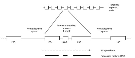
Figure 1.
The ribosomal DNA repeats of fungi and higher organisms. The ribosomal genes are separated by two intergenic regions: the internal transcribed and nontranscribed spacers. Transcription of the 35S pre-RNA is initiated from an external transcribed spacer upstream of the 18S gene (region not shown). The gene for 5S ribosomal DNA, often present in the nontranscribed spacer region, is absent from the Trichophyton rubrum nontranscribed spacer. rRNA indicates ribosomal RNA. (Courtesy of Colin Jackson, PhD, and Richard Barton, PhD.)
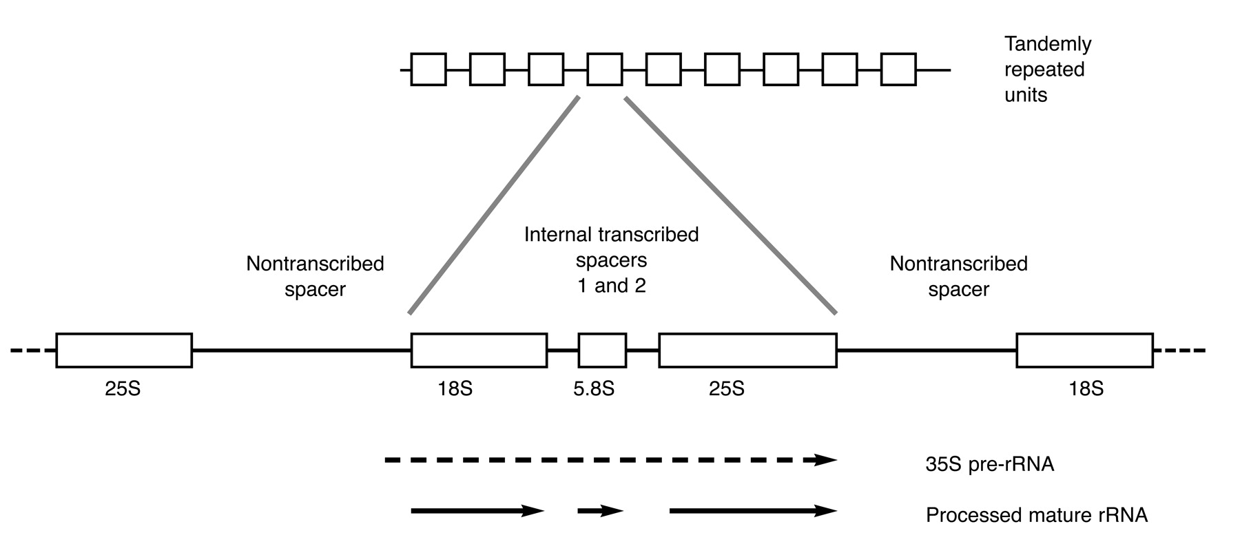
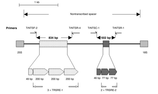
Figure 2.
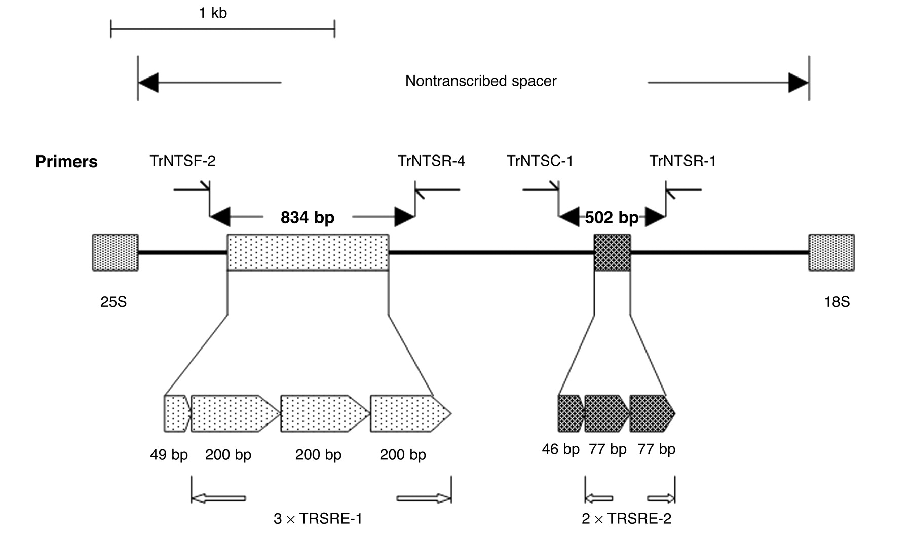
Schematic representation of the nontranscribed spacer region of Trichophyton rubrum showing the location of two sets of repetitive subelements, TRS-1 (TRSRE-1) and TRS-2 (TRSRE-2). The locations of the primer pairs used to amplify these repetitive regions for strain typing are indicated. The expanded section indicates the length in base pairs (bp) and the positions of the five complete and two partial repetitive elements. (Reprinted with permission from Jackson et al.[85])
Results of PCR Studies of Dermatophytes
For the development of specific PCR primers, emphasis has been on the 18S ribosomal RNA gene, the ITS regions, and the NTS sequences. Using the 18S gene, which contains highly variable and highly conserved regions, coupled with restriction fragment length polymorphism analysis, PCR studies have been used for the development of species- and genus-specific markers.[68,69,84] Using a combination of ITS analysis and multiple restriction enzymes to produce different restriction fragment length polymorphism patterns, it has been shown that these DNA techniques can be used as a rapid identification method for most dermatophyte species[93] and were recently used to recognize 23 DNA subtypes of T mentagrophytes.[95]
The NTS region of the T rubrum ribosomal RNA gene also allows for the study of DNA subtypes,[85,86,97] and recent reports have determined that multiple subtypes of T rubrum may be involved in fungal nail infections.[92] One study[86] showed that more than one T rubrum subtype based on restriction fragment length polymorphism was often observed in successive samples from a single patient receiving oral antifungal therapy taken from the same onychomycotic nail during a 1- to 3-year period. Samples taken from different nails of the same patient had either the same or a mixture of the same and different subtypes of T rubrum. In a small study[98] of skin samples from patients with tinea pedis, however, only one subtype of T rubrum was isolated, suggesting a single etiology for this disease.
Although further study is needed, the use of these DNA techniques to study different subtypes of T rubrum should have a major impact on determining whether a relapse after treatment for onychomycosis is due to reinfection with a new dermatophytic strain or reinfection with the same strain and subtype. In addition, these results indicate that when studying antifungal susceptibility, the standard single colony isolated from a clinical sample may not truly represent all of the infecting dermatophytic strains.[92]
Aside from the ribosomal DNA genes, other specific genes are now being studied as potential targets in dermatophyte identification. Topoisomerase II is an enzyme needed to relieve torsional stress in DNA during processes that involve the constant winding and unwinding of DNA, as occurs in replication and transcription. DNA primers targeting the topoisomerase II genes have recently been used in PCR to rapidly identify 18 dermatophytes to the species level based on the PCR or PCR-restriction fragment length polymorphism product size, which varies from species to species.[82,83,97] These authors report that PCR could detect T rubrum and T mentagrophytes by a specific PCR-restriction fragment length polymorphism product size.
Another gene that has been targeted for studied dermatophytic identification is chitin synthase (CHS1),[99] an enzyme implicated in chitin repair. Chitin is a fibrous polysaccharide that constitutes the cell wall of many species of fungi, including dermatophytes. In this study, DNA from skin scrapings and hair samples from animals with clinically identified dermatophytosis were isolated and, using PCR, CHS1 DNA specific for dermatophytes was amplified, demonstrating the potential use of PCR in the clinical diagnosis of dermatophytosis.
Recently, a polymorphic microsatellite marker, T1, which is based on repeats of the nucleotide GT, detected four polymorphic alleles of the T rubrum–Trichophyton violaceum clade using PCR analysis.[65] Using this T1 marker, Kardjeva et al[8] applied this technique to identify and subtype T rubrum directly from clinical samples, finding two of the alleles present in this patient population. Combining repetitive-sequence-based PCR techniques to obtain DNA fingerprinting of dermatophytes, Pounder et al[100] recently combined repetitive-sequence-based PCR with automated separation and identification of the DNA PCR fragments using fluorescence and microfluidic chips, allowing for identification of the dermatophytes in less than 24 hours. This technique, however, depends on first obtaining a pure culture for the analysis.
Use of Reverse Transcriptase–PCR to Assay Viability
Although PCR can detect the presence of dermatophytes, this technique merely detects the presence of DNA in the sample and does not necessarily indicate the viability of the infectious agent. A recent report[78] used reverse transcriptase–PCR to assess the viability of dermatophytes grown from cultures of skin scrapings taken from hyperkeratotic soles. Amplification of the messenger RNA, which represents the viability of the organism from these cultured clinical isolates, allowed for positive detection of dermatophytes in cases that were culture- and PCR-negative. This study was continued using the LightCycler with quantitative reverse transcriptase–PCR, allowing for the quantification of the dermatophyte actin messenger RNA isolated from clinical samples of skin scales and nail plates and thus quantitation of the active dermatophyte.[56] Further reports on following the expression of actin using reverse transcriptase–PCR in T mentagrophytes cultures treated with antifungal agents has shown that this method could be useful for evaluating fungicidal activity against dermatophytes.[79] Thus it may be possible in the future to use this method to assess fungal viability when testing the efficacy of antimycotic agents.[78]
DNA-Based Techniques Compared with Traditional Diagnostic Methods
In comparing methods for the clinical evaluation of onychomycosis, specificity, sensitivity, and time saved are of great importance. The sensitivity of cultures in identifying the causative agents has been historically poor. Multiple reasons for no-growth results on cultures have been enumerated in the literature, some of which include the use of old dermatophyte media, nails stored too long before sending for analysis, insufficient quantity of clinical specimens, and the use of inappropriate specimens that can also result in growth of contaminants.[101]
Reports of positive dermatophyte culture results range from 23%[24] to 77%,[102] with false-negative culture rates of at least 30%.[103] Because the conventional methods of culturing depend on measurement of the phenotypic characteristics of dermatophytes, they can be easily influenced by factors such as temperature and other medications, eg, chemotherapy, which may interfere with the metabolic process of the dermatophyte and affect interpretation of the in vitro culture results.[104] The PCR results of a recent study[8] involving direct analysis of clinically suspected onychomycotic nails detected etiologic fungus in close to 84% of cases in which 72% had failed to be identified using culture. Results of PCR were positive in all of the cases that were culture-positive. Polymerase chain reaction can be used to identify strains that do not present typical morphological characteristics or that will not grow in culture because a significant percentage of clinical samples from patients with dermatophytosis contain nonviable material.[4] In addition, the nonsterile conditions of the nail specimens can lead to the growth of bacterial and fungal contaminants in culture, which may obscure the true etiologic agent. Because PCR directly allows for analysis of the nucleic acid without the necessity of cultures, the problem of interfering organisms may be averted.
The presence of antibiotic-resistant bacteria presents a clinical problem when treating fungal infections. Antibiotic substances have been reported to be produced by fungi in vivo and may result in the selection of resistant bacteria in lesions, with T rubrum seeming particularly likely to be associated with such antibiotic-resistant bacteria. These bacteria tend to prevent pigmentation formation in cultures and, thus, when depending on colony morphology and pigmentation, misidentification in the clinical laboratory is a possible consequence.[4] The problem of identifying nondermatophytes as the causative agent in onychomycosis has been controversial. Even when a nondermatophyte grows out from a specimen that is hyphae-positive on the basis of microscopy, the culture must be secondarily grown to confirm that the nondermatophyte is the etiologic agent. Because a significant portion of clinical specimens (up to 20% in tinea unguium) contain nonviable dermatophyte material, the use of culture as a tool for identification can lead to a confused diagnosis.[4] This problem can be overcome by the use of molecular biology techniques, as identification of the dermatophyte from nonviable tissue using PCR is possible.
The sensitivity of using periodic acid–Schiff is reported to be 4 ng of carbohydrates,[20] whereas that for detecting target DNA using PCR has varied from 10 pg, or approximately 25 fungal genomes,[69] to as little as 1 to 10 fg or 1 to 10 organisms,[50] with similar sensitivity reported using real-time quantitative PCR for dermatophytes.[56] Using the lower limits of PCR detection, there is an increase of 1,000-fold in sensitivity for the PCR method over the periodic acid–Schiff histologic stain.
The PCR technique is not, however, without potential drawbacks. First, owing to the high sensitivity of the reaction, any contamination of the DNA sample and the PCR solutions could lead to additional amplification of other DNA besides the target sample. This procedure must be performed by trained technicians using sterile reagents to prevent false results. Second, although use of the PCR technique is now a basic protocol in all molecular biology laboratories, the cost of the solutions, enzymes, and thermocycler may still pose an obstacle compared with the microscopy, histopathology, and culture techniques commonly used in mycology laboratories. Third, because PCR is based on the enzymatic amplification of target DNA, it is susceptible to any inhibitors that may be present, which would interfere with the PCR reaction.[8,105] Despite these technical difficulties, PCR and related molecular biology techniques will become important tools in diagnosing fungal infections.
Summary
Proper treatment of onychomycosis is possible only with proper diagnosis. An epidemiologic survey[102] of 2,001 patients showed that only 40% of the patients with clinically abnormal nails had onychomycosis based on mycologic evidence. Assuming an 80% cure rate with an oral antifungal agent, it has been estimated that if a physician were to start therapy for every patient with abnormal-appearing nails, then approximately one-third of the patients with abnormal nails would be cured.[106] Because increasingly larger percentages of patients may be presenting with nail dystrophy of nononychomycotic origins,[24] it is important to identify the agent causing the infection before oral antifungal drugs are prescribed. Despite the newer oral antifungal agents that have improved prospects for successful treatment of onychomycosis, studies still show mycologic cure rates ranging from approximately 40% to 80%.[107,108] Not having the identity of the infectious agent may contribute to these therapeutic failures. Restriction enzyme analysis coupled with PCR primers and DNA probes targeted to the ribosomal RNA DNA region or to other genes, such as actin, chitin, and topoisomerase II, have identified and differentiated the two main etiologic agents of onychomycosis—T rubrum and T mentagrophytes—and have identified multiple subtypes of these dermatophytes.
An efficient and quick method for the diagnosis of onychomycosis is necessary before initiating antifungal drug therapy. Although KOH analysis using microscopy can conceivably be performed in the office given the proper instrumentation and histologic training, traditionally cultures and KOH are sent to a specialty laboratory for analysis. This results in a minimum 2- to 4-week time lag, which delays appropriate treatment. The use of molecular biology techniques has made identification of the etiologic agent possible in a much shorter time frame. Whereas the time for detection of the etiologic agent using a molecular biology technique can vary depending on the method and resources, it is usual that identification can be performed within 1 to 2 days, whereas the LightCycler PCR typically allows for DNA amplification to be completed in less than 20 min.[54] Complete identification and quantitation of the viable dermatophytes in skin tissue was recently reported to be less than 4 hours,[56] compared with the 2 to 4 weeks needed for identification using cultures.
Although the advent of new antifungal therapy has improved efficacy and tolerance to many of these drugs, adverse effects of these medications can still occur, and liver function tests are often warranted.[3] Although rare, some of the adverse effects of terbinafine, one of the most common of the antifungal drugs, are diarrhea, pruritus, dyspepsia, abdominal pain, headache, visual disturbance, and neutropenia, as well as a recent observation of facial pigmentation.[109] Liver failure has been described as a possible complication with some antifungal drugs,[110] and their interactions with antidepressant medications, such as paxetine, have also been reported.[108] Given the importance of proper diagnosis and the potency of many of the antifungal drugs, the US Food and Drug Administration[110] now recommends that health-care professionals obtain nail specimens for laboratory testing before prescribing the medications for fungal nail infections, to confirm the diagnosis.
The aging population in the United States is increasing, with a total of approximately 35 million people older than 65 years[111] and projections in 2020 of approximately 55 million people in this age group. Some investigators[112] estimate that up to 30% of the population by age 60 years will have onychomycosis. The need for more sensitive and quicker methods for diagnosing onychomycosis will become even more pressing in the future.
Advances in DNA technologies now allow for the amplification, qualification, and quantitation of DNA isolated from clinical samples. Despite the possible problems with contamination, these protocols will become more widely accepted and used in the diagnostic arena as calibration and training increase. With the explosion of scientific knowledge, the transition from research to clinical application is no longer theoretical. Polymerase chain reaction and DNA technologies are already being used to study dermatophytes and onychomycosis. Podiatric physicians need to be prepared to understand and apply these new techniques as they become available for the diagnosis of diseases, especially as they relate to podiatric medicine.
Funding
This research was supported by the New York College of Podiatric Medicine.
Acknowledgments
Colin Jackson, PhD, and Richard Barton, PhD, for their cooperation and permission to use the figures in this article.
Conflicts of Interest
None reported.
References
- Elewski BE: Onychomycosis: pathogenesis, diagnosis, and management. Clin Microbiol Rev 11: 415, 1998.
- Ghannoum MA, Hajjeh RA, Scher R, et al: A large-scale North American study of fungal isolates from nails: the frequency of onychomycosis, fungal distribution, and antifungal susceptibility patterns. J Am Acad Dermatol 43: 641, 2000.
- Vander Straten MR, Hossain MA, Ghannoum MA: Cutaneous infections dermatophytosis, onychomycosis, and tinea versicolor. Infect Dis Clin North Am 17: 87, 2003.
- Weitzman I, Summerbell RC: The dermatophytes. Clin Microbiol Rev 8: 240, 1995.
- Jousson O, Lechenne B, Bontems O, et al: Multiplication of an ancestral gene encoding secreted fungalysin preceded species differentiation in the dermatophytes Trichophyton and Microsporum. Microbiology 150: 301, 2004.
- Jousson O, Lechenne B, Bontems O, et al: Secreted sub-tilisin gene family in Trichophyton rubrum. Gene 339: 79, 2004.
- Scher RK: Onychomycosis: therapeutic update. J Am Acad Dermatol 40: S21, 1999.
- Kardjeva V, Summerbell R, Kantardjiev T, et al: Forty-eight-hour diagnosis of onychomycosis with subtyping of Trichophyton rubrum strains. J Clin Microbiol 44: 1419, 2006.
- Cribier B, Mena ML, Rey D, et al: Changes in patients infected with human immunodeficiency virus: a prospective controlled study. Arch Dermatol 134: 1216, 1998.
- Akiba H, Motoki Y, Satoh M, et al: Recalcitrant trichophytic granuloma associated with NK-cell deficiency in a SLE patient treated with corticosteroid. J Dermatol 11: 58, 2001.
- Rodriguez-Soto ME, Fernandez-Andreu CM, Moya Duque S, et al: Clinico-mycology study of onychomycosis in elderly patients. Rev Inst Med Trop Sao Paulo 35: 213, 1993.
- National Diabetes Information Clearinghouse Web site. Available at: http://diabetes.niddk.nih.gov/dm/pubs/statistics/index.htm#7. Accessed February 12, 2007.
- Gupta AK, Konnikov N, MacDonald P, et al: Prevalence and epidemiology of toenail onychomycosis in diabetic subjects: a multicentre survey. Br J Dermatol 139: 665, 1998.
- Mullis KB, Faloona FA: Specific synthesis of DNA in vitro via a polymerase-catalyzed chain reaction. Methods Enzymol 155: 335, 1987.
- Foy CA, Parkes HC: Emerging homogeneous DNA-based technologies in the clinical laboratory. Clin Chem 47: 990, 2001.
- Holdane DJ, Robart E: A comparison of calcofluor white, potassium hydroxide, and culture for the laboratory diagnosis of superficial fungal infection. Diagn Microbiol Infect Dis 13: 337, 1990.
- Elewski BE: Diagnostic techniques for confirming onychomycosis. J Am Acad Dermatol 35: S6, 1996.
- Zacharius RM, Zell TE, Morrison JH, et al: Glycoprotein staining following electrophoresis on acrylamide gels. Anal Biochem 30: 148, 1969.
- Maynard JA, Ippolito EG, Ponseti IV, et al: Histochemistry and ultrastructure of the growth plate in metaphyseal dysostosis: further observations on the structure of the cartilage matrix. J Pediatr Orthop 1: 161, 1981.
- Stromqvist M, Gruffman H: Periodic acid/Schiff staining of glycoproteins immobilized on a blotting matrix. Biotechniques 13: 744, 1992.
- Kappe R, Rimek D: Diagnosis of fungal diseases. Prog Drug Res Spec No: 39, 2003.
- Zabawski EJ Jr, Styles AR, Cockerell CJ: Routine periodic acid-Schiff staining of nail plate fragments in fungal cultures for onychomycosis: a method to increase the sensitivity of diagnosis. Cutis 66: 456, 2000.
- Weinberg JM, Koestenblatt EK, Jennings MB: Utility of histopathologic analysis in the evaluation of onychomycosis. JAPMA 95: 258, 2005.
- Lawry MA, Haneke E, Strobeck K, et al: Methods for diagnosing onychomycosis: a comparative study and review of the literature. Arch Dermatol 136: 1112, 2000.
- Borek PP, Albresdki DA, Probolus JA, et al: New methods with potassium hydroxide for diagnosing onychomycosis. Am J Dermatopathol 21: 90, 1999.
- Weinberg JM, Koestenblatt EK, Tutrone WD, et al: Comparison of diagnostic methods in the evaluation of onychomycosis. J Am Acad Dermatol 49: 193, 2003.
- Singh S, Beena PM: Comparative study of different microscopic techniques and culture media for the isolation of dermatophytes. Indian J Med Microbiol 21: 21, 2003.
- Elewski BE, El Charif M, Cooper KD, et al: Reactivity to trichophytin antigen in patients with onychomycosis: effect of terbinafine. J Am Acad Dermatol 46: 371, 2002.
- Yeo SF, Wong B: Current status of nonculture methods for diagnosis of invasive fungal infections. Clin Microbiol Rev 15: 465, 2002.
- Loffler J, Hebart H, Sepe S, et al: Detection of PCR-amplified fungal DNA by using a PCR-ELISA system. Med Mycol 36: 275, 1998.
- Summerbell RC, Cooper E, Bunn U, et al: Onychomycosis: a critical study of techniques and criteria for confirming the etiologic significance of nondermatophytes. Med Mycol 43: 39, 2005.
- Samdani AJ, Dykes PJ, Marks R: The proteolytic activity of strains of T. mentagrophytes and T. rubrum isolated from tinea pedis and tinea unguium infections. J Med Vet Mycol 33: 167, 1995.
- Versalovic J, Koeuth T, Lupski JR: Distribution of repetitive DNA sequences in eubacteria and application to fingerprinting of bacterial genomes. Nucleic Acids Res 19: 6823, 1991.
- Human Genome Project information: DNA forensics. Available at: http://www.ornl.gov/sci/techresources/Human_Genome/elsi/forensics.shtml. Accessed February 9, 2007.
- Ginzinger DG: Gene quantification using real-time quantitative PCR: an emerging technology hits the mainstream. Exp Hematol 30: 503, 2002.
- Ulrichs T, Lefmann M, Reich M, et al: Modified immunohistological staining allows detection of Ziehl-Neelsen-negative Mycobacterium tuberculosis organisms and their precise localization in human tissue. J Pathol 205: 633, 2005.
- Mackay IM, Arden KE, Nitsche A: Real-time PCR in virology. Nucleic Acids Res 30: 1292, 2002.
- Pacheco N, Mago V, Gomez I, et al: Comparison of PCR and common clinical tests for the diagnosis of H. pylori in dyspeptic patients. Diagn Microbiol Infect Dis 39: 207, 2001.
- Nowakowski J, Schwartz I, Liveris D, et al: Laboratory diagnostic techniques for patients with early Lyme disease associated with erythema migrans: a comparison of different techniques. Clin Infect Dis 33: 2023, 2001.
- Glennon M, Cormican M: Detection and diagnosis of mycobacterial pathogens using PCR. Expert Rev Mol Diagn 1: 163, 2001.
- Mohammadi T, Pietersz RN, Vandenbroucke-Grauls CM, et al: Detection of bacteria in platelet concentrates: comparison of broad-range real-time 16S rDNA polymerase chain reaction and automated culturing. Transfusion 45: 731, 2005.
- Balestrino D, Haagensen JA, Rich C, et al: Characterization of type 2 quorum sensing in Klebsiella pneumoniae and relationship with biofilm formation. J Bacteriol 187: 2870, 2005.
- Pilcher CD, McPherson JT, Leone PA, et al: Real-time, universal screening for acute HIV infection in a routine HIV counseling and testing population. JAMA 288: 216, 2002.
- Hess C, Klimkait T, Schlapbach L, et al: Association of a pool of HIV-1 with erythrocytes in vivo: a cohort study. Lancet 359: 2230, 2002.
- Calvario A, Bozzi A, Scarasciulli M, et al: Herpes consensus PCR test: a useful diagnostic approach to the screening of viral diseases of the central nervous system. J Clin Virol 25: (suppl):71, 2002.
- Prado I, Rosario D, Bernardo L, et al: PCR detection of dengue virus using dried whole blood spotted on filter paper. J Virol Methods 125: 75, 2005.
- Radford SA, Johnson EM, Leeming JP, et al: Molecular epidemiological study of Aspergillus fumigatus in a bone marrow transplantation unit by PCR amplification of ribosomal intergenic spacer sequences. J Clin Microbiol 36: 1294, 1998.
- Graser Y, El Fari M, Presber W, et al: Identification of common dermatophytes (Trichophyton, Microsporum, Epidermophyton) using polymerase chain reactions. Br J Dermatol 138: 5762, 1998.
- Velegraki A, Kambouris M, Kostourou A, et al: Rapid extraction of fungal DNA from clinical samples for PCR amplification. Med Mycol 37: 69, 1999.
- Ferrer C, Colom F, Frases S, et al: Detection and identification of fungal pathogens by PCR and by ITS2 and 5.8S ribosomal DNA typing in ocular infections. J Clin Microbiol 39: 2873, 2001.
- Li RY, Li DM, Yu J, et al: Application of molecular biology techniques in the identification of pathogenic fungi and the diagnosis of fungal infection. Beijing Da Xue Xue Bao 36: 536, 2004.
- Heid CA, Stevens J, Livak KJ, et al: Real time quantitative PCR. Genome Res 6: 986, 1996.
- Orlando C, Pinzani P, Pazzagli M: Developments in quantitative PCR. Clin Chem Lab Med 36: 255, 1998.
- Wittwer CT, Ririe KM, Andrew RV, et al: The LightCycler: a microvolume multisample fluorimeter with rapid temperature control. Biotechniques 22: 176, 1997.
- Gutzmer R, Mommert S, Kuttler U, et al: Rapid identification and differentiation of fungal DNA in dermatological specimens by LightCycler PCR. J Med Microbiol 53: 1207, 2004.
- Tsuboi R, Okeke CN, Inoue A, et al: Identification and viability assessment of dermatophytes infecting nail based on quantitative PCR of dermatophyte actin (ACT) mRNA. Nippon Ishinkin Gakkai Zasshi 43: 91, 2002.
- Savolainen V, Cowan RS, Vogler AP, et al: Towards writing the encyclopedia of life: an introduction to DNA barcoding. Philos Trans R Soc Lond B Biol Sci 360: 1805, 2005.
- Summerbell RC, Levesque CA, Seifert KA, et al: Microcoding: the second step in DNA barcoding. Philos Trans R Soc Lond B Biol Sci 360: 1897, 2005.
- Kano R, Nakamura Y, Watari T, et al: Phylogenetic analysis of 8 dermatophyte species using chitin synthase 1 gene sequences. Mycoses 40: 411, 1997.
- Graser Y, El Fari M, Vilgalys R, et al: Phylogeny and taxonomy of the family Arthrodermataceae (dermatophytes) using sequence analysis of the ribosomal ITS region. Med Mycol 37: 105, 1999.
- Graser Y, Kuijpers AF, Presber W, et al: Molecular taxonomy of Trichophyton mentagrophytes and T. tonsurans. Med Mycol 37: 315, 1999.
- Kano R, Nakamura Y, Watari T, et al: Phylogenetic relation of Epidermophyton floccosum to the species of Microsporum and Trichophyton in chitin synthase 1 (CHS1) gene sequences. Mycopathologia 146: 111, 1999.
- Makimura K, Tamura Y, Mochizuki T, et al: Phylogenetic classification and species identification of dermatophyte strains based on DNA sequences of nuclear ribosomal internal transcribed spacer 1 regions. J Clin Microbiol 37: 920, 1999.
- Mochizuki T, Kawasaki M, Ishizaki H, et al: Molecular epidemiology of Arthroderma benhamiae, an emerging pathogen of dermatophytoses in Japan, by polymorphisms of the non-transcribed spacer region of the ribosomal DNA. J Dermatol Sci 27: 14, 2001.
- Ohst T, de Hoog S, Presber W, et al: Origins of microsatellite diversity in the Trichophyton rubrum–T. violaceum clade (dermatophytes). J Clin Microbiol 42: 4444, 2004.
- Bock M, Nickel P, Maiwald M, et al: Diagnosis of dermatomycoses with polymerase chain reaction. Hautarzt 48: 175, 1997.
- Baek SC, Chae HJ, Houh D, et al: Detection and differentiation of causative fungi of onychomycosis using PCR amplification and restriction enzyme analysis. Int J Dermatol 37: 682, 1998.
- El Fari M, Tietz HJ, Presber W, et al: Development of an oligonucleotide probe specific for Trichophyton rubrum. Br J Dermatol 141: 240, 1999.
- Turin L, Riva F, Galbiati G, et al: Fast, simple and highly sensitive double-rounded polymerase chain reaction assay to detect medically relevant fungi in dermatological specimens. Eur J Clin Invest 30: 511, 2000.
- Machouart-Dubach M, Lacroix C, de Chauvin MF, et al: Rapid discrimination among dermatophytes, Scytalidium spp., and other fungi with a PCR-restriction fragment length polymorphism ribotyping method. J Clin Microbiol 39: 685, 2001.
- Arca E, Saracli MA, Akar A, et al: Polymerase chain reaction in the diagnosis of onychomycosis. Eur J Dermatol 14: 52, 2004.
- Howell SA, Barnard RJ, Humphreys F: Application of molecular typing methods to dermatophyte species that cause skin and nail infections. J Med Microbiol 48: 33, 1999.
- Mochizuki T, Sugie N, Uehara M: Random amplification of polymorphic DNA is useful for the differentiation of several anthropophilic dermatophytes. Mycoses 40: 405, 1997.
- Kim JA, Takahashi Y, Tanaka R, et al: Identification and subtyping of Trichophyton mentagrophytes by random amplified polymorphic DNA. Mycoses 44: 157, 2001.
- Liu D, Pearce L, Lilley G, et al: PCR identification of dermatophyte fungi Trichophyton rubrum, T. soudanense and T. gourvilii. J Med Microbiol 51: 117, 2002.
- Nishio K, Kawasaki M, Ishizaki H: Phylogeny of the genera Trichophyton using mitochondrial DNA analysis. Mycopathologia 117: 127, 1992.
- Su CS, Meyer SA: Characterization of mitochondrial DNA in various Candida species: isolation, restriction endonuclease analysis, size, and base composition. Int J Syst Bacteriol 41: 6, 1991.
- Okeke CN, Tsuboi R, Kawai M, et al: Isolation of an intron-containing partial sequence of the gene encoding dermatophyte actin (ACT) and detection of a fragment of the transcript by reverse transcription-nested PCR as a means of assessing the viability of dermatophytes in skin scales. J Clin Microbiol 39: 101, 2001.
- Nimura K, Niwano Y, Ishiduka S, et al: Actin gene-targeted RT-PCR could be a useful method for evaluating in vitro fungicidal activity against dermatophytes. J Int Med Res 31: 407, 2003.
- Kano R, Nakamura Y, Watari T, et al: Molecular analysis of chitin synthase 1 (CHS1) gene sequences of Trichophyton mentagrophytes complex and T. rubrum. Curr Microbiol 37: 236, 1998.
- Kano R, Nakamura Y, Watari T, et al: Species-specific primers of chitin synthase 1 gene for the differentiation of the Trichophyton mentagrophytes complex. Mycoses 42: 71, 1999.
- Kanbe T, Suzuki Y, Kamiya A, et al: PCR-based identification of common dermatophyte species using primer sets specific for the DNA topoisomerase II genes. J Dermatol Sci 32: 151, 2003.
- Kanbe T, Suzuki Y, Kamiya A, et al: Species-identification of dermatophytes Trichophyton, Microsporum and Epidermophyton by PCR and PCR-RFLP targeting of the DNA topoisomerase II genes. J Dermatol Sci 33: 41, 2003.
- Jackson CJ, Barton RC, Evans EG: Species identification and strain differentiation of dermatophyte fungi by analysis of ribosomal-DNA intergenic spacer regions. J Clin Microbiol 37: 931, 1999.
- Jackson CJ, Barton RC, Kelly SL, et al: Strain identification of Trichophyton rubrum by specific amplification of subrepeat elements in the ribosomal DNA nontranscribed spacer. J Clin Microbiol 38: 4527, 2000.
- Gupta AK, Kohli Y, Summerbell RC: Variation in restriction fragment length polymorphisms among serial isolates from patients with Trichophyton rubrum infection. J Clin Microbiol 39: 3260, 2001.
- Bock M, Maiwald M, Kappe R, et al: Polymerase chain reaction-based detection of dermatophyte DNA with a fungus-specific primer system. Mycoses 37: 79, 1994.
- Kappe R, Fauser C, Okeke CN, et al: Universal fungus-specific primer systems and group-specific hybridization oligonucleotides for 18S rDNA. Mycoses 39: 25, 1996.
- Kappe R, Okeke CN, Fauser C, et al: Molecular probes for the detection of pathogenic fungi in the presence of human tissue. J Med Microbiol 47: 811, 1998.
- Summerbell RC, Haugland RA, Li A, et al: rRNA gene internal transcribed spacer 1 and 2 sequences of asexual, anthropophilic dermatophytes related to Trichophyton rubrum. J Clin Microbiol 37: 4005, 1999.
- Gaedigk A, Gaedigk R, Abdel-Rahman SM: Genetic heterogeneity in the rRNA gene locus of Trichophyton tonsurans. J Clin Microbiol 41: 5478, 2003.
- Yazdanparast A, Jackson CJ, Barton RC, et al: Molecular strain typing of Trichophyton rubrum indicates multiple strain involvement in onychomycosis. Br J Dermatol 148: 51, 2003.
- Shin JH, Sung JH, Park SJ, et al: Species identification and strain differentiation of dermatophyte fungi using polymerase chain reaction amplification and restriction enzyme analysis. J Am Acad Dermatol 48: 857, 2003.
- Mochizuki T, Tanabe H, Kawasaki M, et al: Rapid identification of Trichophyton tonsurans by PCR-RFLP analysis of ribosomal DNA regions. J Dermatol Sci 32: 25, 2003.
- Mochizuki T, Ishizaki H, Barton RC, et al: Restriction fragment length polymorphism analysis of ribosomal DNA intergenic regions is useful for differentiating strains of Trichophyton mentagrophytes. J Clin Microbiol 41: 4583, 2003.
- White TJ, Bruns T, Lee S, et al: “Amplification and Direct Sequencing of Fungal Ribosomal RNA Genes for Phylogenetics,” in PCR Protocols, ed by MA Innes, GH Gelfand, JS Sninsky, et al, p 315, Academic Press, London, 1990.
- Kamiya A, Kikuchi A, Tomita Y, et al: PCR and PCR-RFLP techniques targeting the DNA topoisomerase II gene for rapid clinical diagnosis of the etiologic agent of dermatophytosis. J Dermatol Sci 34: 35, 2004.
- Rad MM, Jackson C, Barton RC, et al: Single strains of Trichophyton rubrum in cases of tinea pedis. J Med Microbiol 54: 725, 2005.
- Kano R, Hirai A, Muramatsu M: Direct detection of dermatophytes in skin samples based on sequences of the chitin synthase 1 (CHS1) gene. J Vet Med Sci 65: 267, 2003.
- Pounder JI, Williams S, Hansen D, et al: Repetitive-sequence-PCR-based DNA fingerprinting using the Diversilab system for identification of commonly encountered dermatophytes. J Clin Microbiol 43: 2141, 2005.
- Lemont H: Pathologic and diagnostic considerations in onychomycosis. JAPMA 87: 498, 1997.
- Gupta AK, Jain HC, Lynde CW, et al: Prevalence and epidemiology of unsuspected onychomycosis in patients visiting dermatologists’ offices in Ontario, Canada: a multicenter survey of 2001 patients. Int J Dermatol 36: 783, 1997.
- Fletcher CL, Hay RJ, Smeeton NC: Onychomycosis: the development of a clinical diagnostic aid for toenail disease: part I. Establishing discriminating historical and clinical features. Br J Dermatol 150: 701, 2004.
- Liu D, Coloe S, Baird R, et al: Application of PCR to the identification of dermatophyte fungi. J Med Microbiol 49: 493, 2000.
- Belec L, Authis J, Eliezer-Vanerot MC, et al: Myoglobin as a polymerase chain reaction (PCR) inhibitor: a limitation for PCR from skeletal muscle tissue avoided by the use of Thermus thermophilus polymerase. Muscle Nerve 21: 1064, 1998.
- Gupta AK, Daniel CR III: Factors that may affect the response of onychomycosis to oral antifungal therapy. Australas J Dermatol 39: 222, 1998.
- Drake LA, Scher RK, Smith EB, et al: Effect of onychomycosis on quality of life. J Am Acad Dermatol 38: 702, 1998.
- Molden E, Garcia BH, Braathen P, et al: Co-prescription of cytochrome P450 2D6/3A4 inhibitor-substrate pairs in clinical practice: a retrospective analysis of data from Norwegian primary pharmacies. Eur J Clin Pharmacol 61: 119, 2005.
- Breuer K, Volker B, Gutzmer R, et al: Facial pigmentation following therapy with terbinafine. Hautarzt 56: 1056, 2005.
- US Food and Drug Administration: FDA Talk Paper: FDA Issues Health Advisory Regarding the Safety of Sporanox Products and Lamisil Tablets to Treat Fungal Infections, US Food and Drug Administration, Rockville, MD, May 9, 2001.
- Projected population of the United States, by age and sex: 2000 to 2050. Available at: http://www.census.gov/ipc/www/usinterimproj/natprojtab02a.xls. Accessed February 6, 2007.
- Levy LA: Epidemiology of onychomycosis in special-risk populations. JAPMA 87: 546, 1997.
© 2007 American Podiatric Medical Association