


Abstract
Background: Forefoot adduction is the most common residual deformity in the treatment of pediatric clubfoot. Little documentation exists regarding its late occurrence and early detection. A retrospective analysis was conducted to determine the effect of primary posterior medial release for idiopathic clubfoot that had failed to improve with conservative treatment or had presented after a treatment delay and a subsequent forefoot adduction correction with a cuboid-cuneiform osteotomy. Methods: Radiographic evaluations were conducted of all of the surgical procedures performed at our institution for idiopathic clubfoot during a specified period. Preoperative and postoperative talo–first metatarsal and talocalcaneal angles were measured radiographically. Of 138 patients with clubfoot deformity who met the inclusion criteria, 51 underwent a primary posterior medial release; of these patients, 18 (26 feet) underwent a subsequent cuboid-cuneiform osteotomy. Results: The average preoperative and postoperative talo–first metatarsal anteroposterior angles for patients who underwent primary posterior medial release were 44.6° and 26.8°, respectively. The mean reduction in forefoot adduction was 17.8° (P < .05). After the osteotomy, the average talo–first metatarsal anteroposterior angle was 16°, with an average reduction of 10.8° (P < .05). Mean follow-up was 61.2 months. The average patient age was 3.2 years. Conclusions: Eighteen (35%) of 51 patients who underwent a posterior medial release required a subsequent cuboid-cuneiform osteotomy. The average reduction of 10.8° was statistically significant and has also proved to be clinically significant in the overall correction of the deformity.
Forefoot adduction is the most common residual deformity in the treatment of pediatric clubfoot (Fig. 1).[1–10] As the child grows, this condition can cause functional disability, and in some cases the parents state that the foot looks the same as before the initial club-foot treatment despite the fact that the rearfoot is fully corrected and only the forefoot adduction remains (Fig. 2).[5,11–14]
Although early reports on the natural history of residual forefoot adduction in clubfoot suggested that spontaneous and progressive correction would occur,[15] it is now agreed that such an evolution may be expected in congenital metatarsus adductus but is unlikely in most cases of clubfoot.[16–19] Many authors agree that the goal of any clubfoot deformity correction is a plantigrade foot; hence, when forefoot adduction occurs it is frequently regarded as a less important alteration or deformity. Its incidence varies widely because of many factors, such as the degree of the initial deformity, the quality of the primary correction, the patient’s age, and the surgeon’s level of expertise.
Some key principles involved in understanding the physiopathology of pediatric forefoot adduction state that there is an imbalance between an elongated lateral column and a shortened medial column or that the fibular growth in clubfoot predisposes to the development of an imbalance between the lateral and medial columns.[20] Several surgical procedures have been described to treat forefoot adduction, including soft-tissue releases and osseous procedures.[21–23] Schaefer and Hefti[24] reported using a combined cuboid and medial cuneiform osteotomy to treat residual adductus deformity in cases of idiopathic and secondary clubfoot. Their radiographic criteria were based on the calcaneal–second metatarsal angle. Their mean 5-year follow-up for 27 feet for forefoot adductus showed mean ± SD preoperative and postoperative values of 20.7° ± 2.0° and 8.9° ± 1.8°, respectively (P < .05).
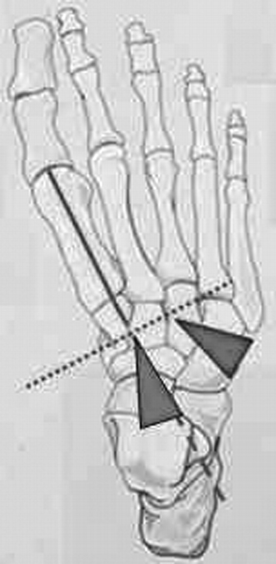
Here we present the results of a retrospective analysis conducted to determine the effect of primary posterior medial release for idiopathic clubfoot that had failed to improve with conservative treatment or had presented after a treatment delay and a subsequent forefoot adduction correction with a combined cuboid–lateral cuneiform osteotomy (Fig. 3). We hope that these study results will improve clinicians’ understanding of many residual and recurrent deformities in pediatric clubfoot.
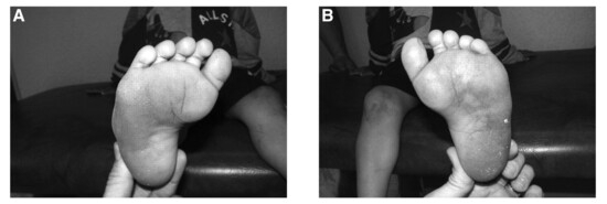
Figure 1.
Forefoot adductus persistence in the right (A) and left (B) feet after posterior medial release.
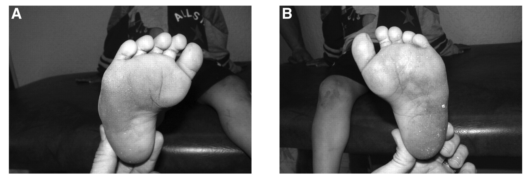
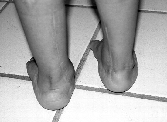
Figure 2.
Perpendicular rearfoot complex alignment in the patient with persistent forefoot adductus from Figure 1.
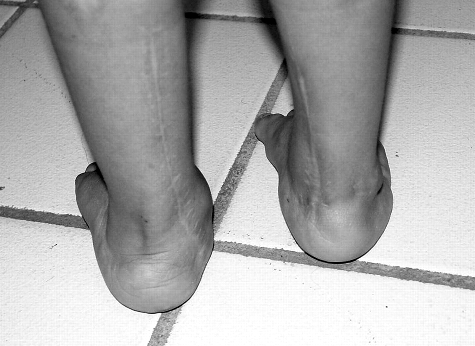
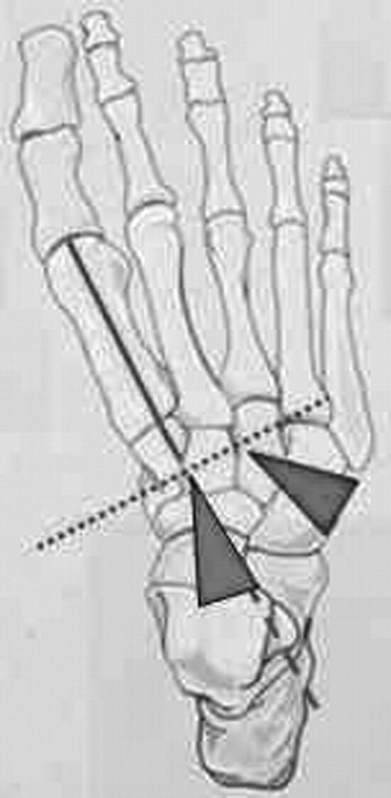
Figure 3.
Diagram of the cuboid-cuneiform osteotomy. Note the matching wedge removal and degree of forefoot adductus.
Materials and Methods
In this study, radiographic and medical chart evaluations with weightbearing anteroposterior and lateral views and clinical evaluation were performed between January 1, 1994, and December 31, 2002, for all idiopathic clubfoot surgical procedures. The radiographic guidelines of Simons[25] were used as the standard to evaluate all of the deformities because all of the preoperative and postoperative radiographic angles were measured using the talo–first metatarsal angle (anteroposterior view) and the talocalcaneal angle (lateral view). Although the lateral talocalcaneal angle was measured, it was not relevant in evaluating the index procedure. A talo–first metatarsal angle greater than 15°, with a talocalcaneal angle less than 15°, indicates talonavicular subluxation as found in clubfoot (Fig. 4).
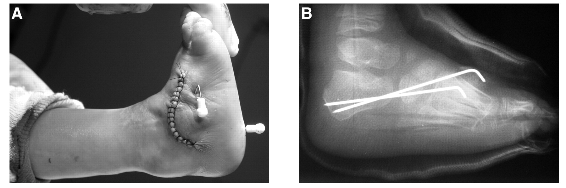
The inclusion criteria were 1) the existence of complete medical charts and radiographs for the entire perioperative period (January 1, 1994, to December 31, 2002); 2) no previous surgical treatment; 3) posterior medial release, including a perisubtalar joint release, as the primary method of correction (Fig. 5)[26,27]; and 4) a subsequent cuboid-cuneiform osteotomy. Only patients who underwent a posterior medial release using a posterior medial jockey-stick incision extension (Fig. 6) and a cuboid-cuneiform osteotomy (Fig. 7) were included.[28] This osteotomy is in the lateral column only, involving the cuboid and the lateral cuneiform, with matching degrees of closing wedge and forefoot adduction.
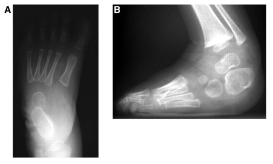
Figure 4.
A talo–first metatarsal anteroposterior angle greater than 15° (A), with a talocalcaneal lateral angle less than 15° (B), indicates talonavicular subluxation as found in clubfoot.
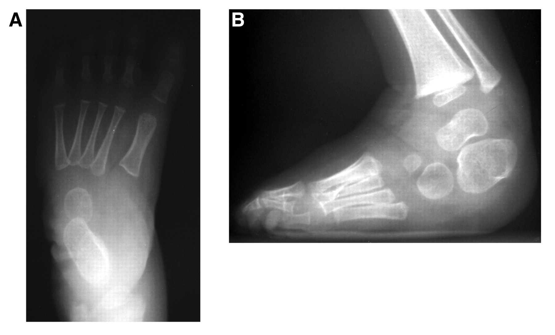
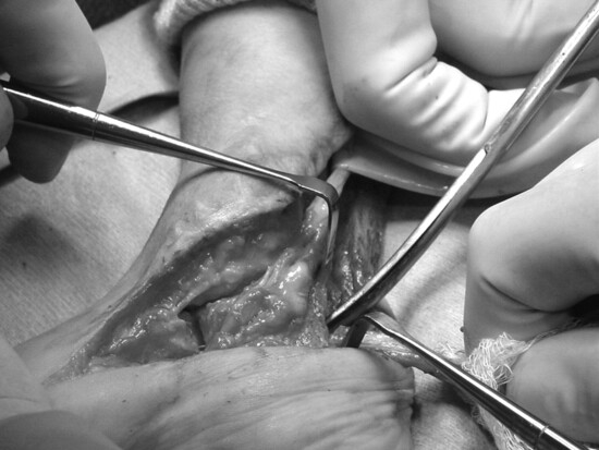
Figure 5.
Initial posterior medial release, including a perisubtalar joint release.
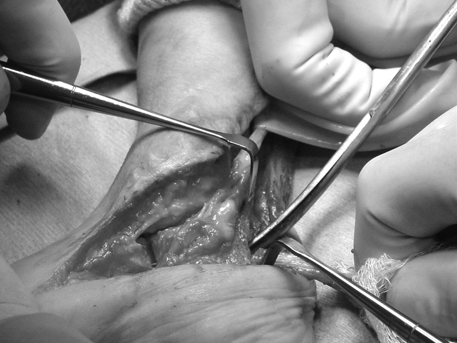
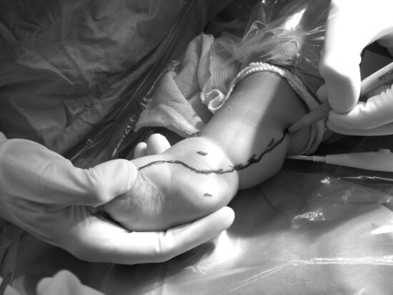
Figure 6.
Posterior medial jockey-stick incision.
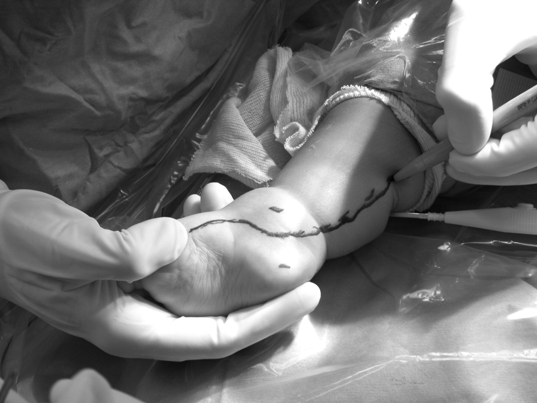
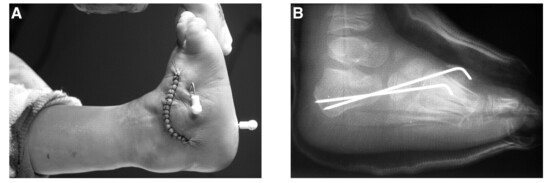
Figure 7.
Clinical (A) and radiographic (B) views of the osteotomy of the cuboid-cuneiform.
Patients were excluded if they 1) underwent other additional procedures for further correction in the frontal and sagittal planes, 2) had medical charts without complete perioperative clinical evaluation data and radiographs, 3) were lost to follow-up within 24 months, and 4) underwent a different soft-tissue release procedure, ie, Cincinnati circumferential release, which would be beyond the scope of this study. Statistical analysis was performed using paired t tests for the difference between the paired data and for the probability that the actual mean difference is consistent with zero. Differences generating P < .05 were considered significant, whereas those generating P = .05 to .10 were considered to suggest a trend. This comparison is aided by the reduction in variance achieved by taking the differences.
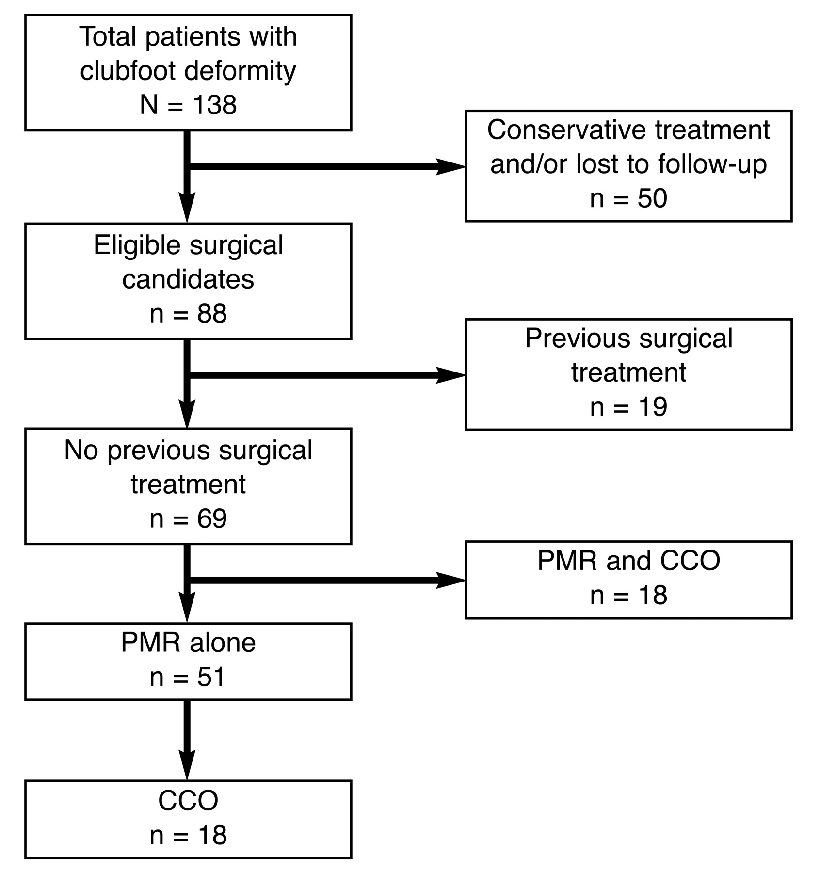
In the present analysis there were 138 patients with clubfoot deformity (Fig. 8). Fifty patients were successfully treated conservatively with manipulation and casting and did not need further surgical intervention or were lost to follow-up. The remaining 88 patients had complete perioperative clinical and radiographic evaluations. Sixty-nine patients had no previous surgical treatment; 19 patients had previous treatment at other institutions and, thus, were excluded from this study. Of the 69 patients who did not respond to conservative treatment of manipulation and casting, 51 underwent a posterior medial release alone and 18 underwent a primary posterior medial release with a cuboid-cuneiform osteotomy. Thus the final 51 patients were used for this evaluation.
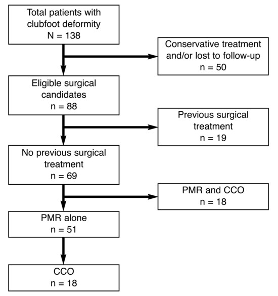
Figure 8.
Patient inclusion and exclusion chart. PMR indicates posterior medial release; CCO, cuboid-cuneiform osteotomy.
Results
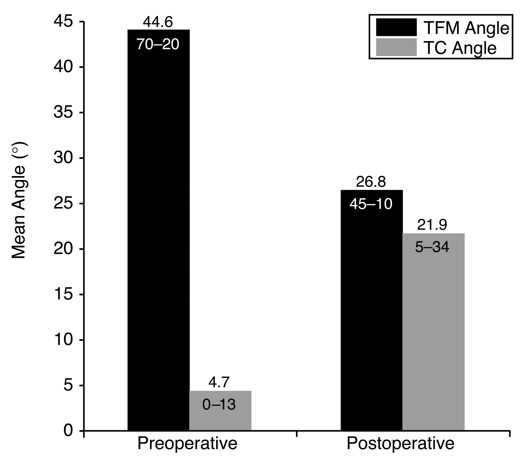
Of the 51 patients who underwent a posterior medial release alone, 18 patients and 26 feet underwent a subsequent cuboid-cuneiform osteotomy. There were 13 right feet and 13 left feet, with 8 cases being bilateral. For these 18 patients, Group 1 values were obtained after the posterior medial release alone and Group 2 values were obtained after the osteotomy. In Group 1, the average talo–first metatarsal angle reduction was from 44.6° (range, 70° to 20°) to 26.8° (range, 45° to 10°), whereas the talocalcaneal angle increased from 4.7° (range, 0° to 13°) to 21.9° (range, 5° to 34°). The mean reduction in forefoot adduction was 17.8°, with an SD of 15.2°, a median of 18.5°, and an average absolute deviation from median of 11.3°. There was a statistically significant (P < .05) difference between the preoperative and postoperative talo–first metatarsal angle values in Group 1 (Fig. 9).
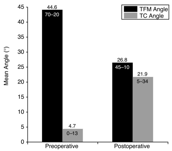
Figure 9.
Comparison of Group 1 preoperative and postoperative talo–first metatarsal (TFM) and talocalcaneal (TC) angles. The mean reduction in the TFM angle was 17.8° (P < .05).
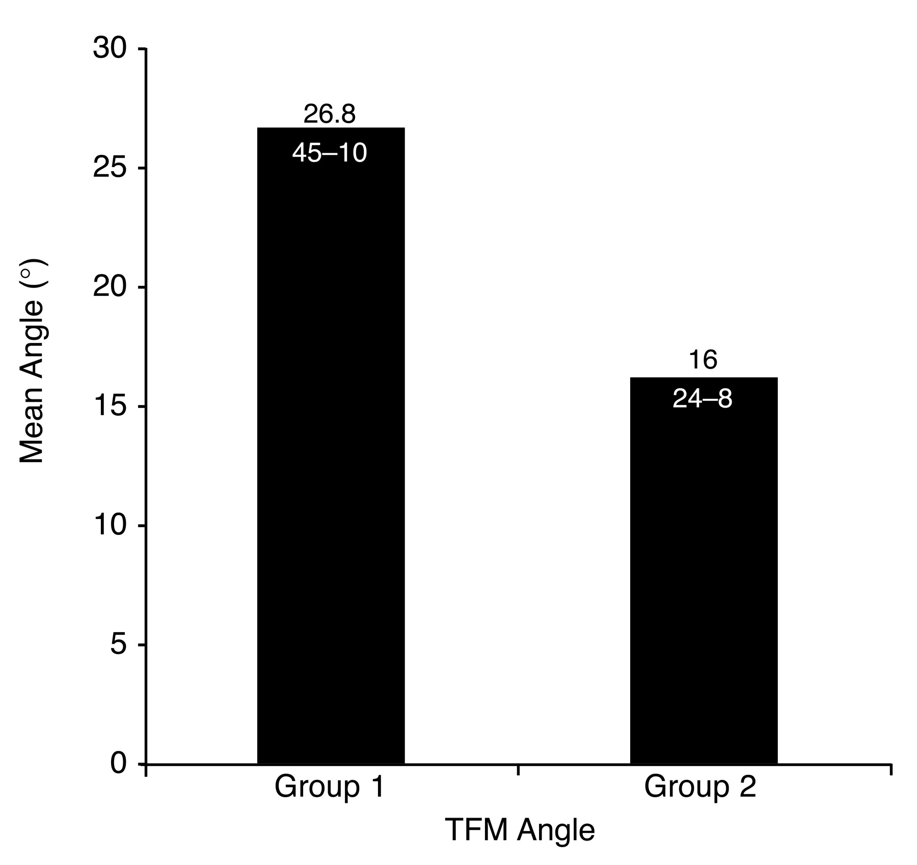
After the cuboid-cuneiform osteotomy (Group 2), the average talo–first metatarsal angle was 16° (range, 24° to 8°), which reflected an average reduction of 10.8° in forefoot adduction, with an SD of 9.12°, a median of 8°, and an average absolute deviation from median of 7.77°. There was a statistically significant (P < .05) difference between the preoperative and postoperative talo–first metatarsal angle values in these groups (Fig. 10). The average time between the posterior medial release and the osteotomy was 10.6 months (range, 3–47 months). Mean follow-up was 61.2 months (range, 32–103 months). The average patient age was 3.2 years (range, 1–6 years). There were eight boys and ten girls.
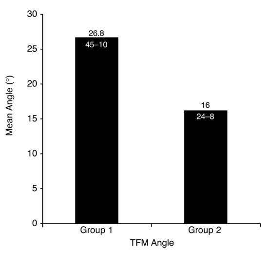
Figure 10.
Comparison of talo–first metatarsal (TFM) angles in Groups 1 and 2. The mean reduction in the TFM angle was 10.8° (P < .05).
Discussion
In severe deformities, neglected clubfoot, or relapsed clubfoot, osseous procedures have been widely used and have been preceded by soft-tissue procedures. Soft-tissue procedures have been performed mainly to allow stretching, elongation, and relaxation of vital structures before the dramatic osseous procedures.
In many situations, soft-tissue releases are insufficient in the correction of clubfoot deformities. To date, it has been very difficult to catalog and evaluate any long-term studies of the success of a posterior medial release performed alone as a primary correction procedure. The lack of information in the literature on the predictability of the amount of correction achieved by a posterior medial release alone has led to a second-stage procedure for correction of a persistent forefoot adduction.
The literature from the 1970s to 2002 contains enthusiastic reports on the correction of congenital club-foot through extensive surgical release procedures.[27,28] Over time, we have recognized the complications of such surgery, including recurrence, overcorrection, stiffness, and pain.[29]
For patients who are older than 4 years, many procedures have been described, including excision of the distal part of the calcaneus, fusion of the calcaneo-cuboid joint, opening-wedge osteotomy of the first cuneiform, metatarsal osteotomies, and tarsometatarsal capsulotomies. Lichtblau,[23] in 1973, described a medial soft-tissue release and an osteotomy of the distal end of the calcaneus in which 1 cm of the distal lateral border and 2 mm of the distal medial border are removed. He claimed that the resected calcaneal articular surface was replaced by fibrocartilage, and he demonstrated mobility at the calcaneocuboid joint up to 6 years after surgery.
Evans,[22] in 1961, described a procedure consisting of posteromedial releases in conjunction with lateral calcaneocuboid wedge resection and fusion. The procedure is not recommended for children younger than 4 years because of possible overcorrection. The correction of adduction occurs at the level of the midfoot, not distal to the navicular. Accurate reduction of the navicular on the talus is essential because the position of the navicular is permanently stabilized by the procedure. Only a narrow wedge from the calcaneocuboid joint should be removed; otherwise, overcorrection into valgus may occur. The operation reduces growth of the lateral column of the foot. Satisfactory long-term functional results have been documented in 60% to 80% of the patients undergoing the procedure.[30]
Fowler,[31] in 1959, described an opening-wedge osteotomy of the medial cuneiform, and Hofmann et al,[32] in 1984, reported on this procedure for the treatment of residual adduction in clubfoot. McHale and Lenhart[12] described a procedure for an adducted forefoot and a supinated midfoot with rearfoot varus. Köse et al,[33] in 1999, described trans-midtarsal osteotomy. Metatarsal osteotomies were described first by Steytler and Van der Walt[34] in 1966 and are indicated when the adduction deformity originates distal to the navicular. Heyman et al[11] described release of the tarsometatarsal joints for correction of resistant metatarsus adductus or for treating residual clubfoot adduction deformity. Through a dorsal incision, complete capsulotomies and ligament releases were performed. Because of reports of frequent postoperative stiffness and pain, this procedure is not recommended.
In our experience and on the basis of the results of this study, when persistent forefoot adduction is found in patients who are younger than 2 years, a repeated complete soft-tissue release is usually recommended over osseous correction owing to the possibility of arrest of osseous growth. In patients who are 2 to 4 years old, osteotomies may be recommended with caution. Excision of the calcaneocuboid joint cartilage and cuboid enucleation are other options. These procedures must be combined with a medial soft-tissue release. Cuboid decancellation preserves the articular surface of the cuboid surface proximally and distally, whereas “crushing” of the bone shortens the lateral column and corrects adduction.
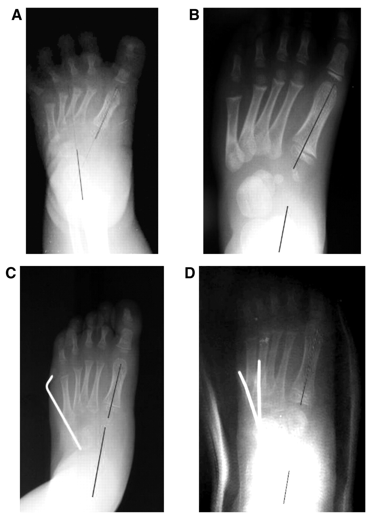
To date, the lack of an agreed-on and reproducible post-treatment evaluation system still hinders outcome studies of the treatment of clubfoot. The analysis of other persistent deformities with various other clubfoot etiologies is beyond the scope of this article. On the basis of the results of this study, it is very likely that a small number of idiopathic clubfeet will require an additional surgery even after expertly performed posterior medial release. The present study shows that 18 (35%) of 51 patients who underwent a primary posterior medial release alone required a subsequent cuboidcuneiform osteotomy owing to forefoot adduction. The average reduction of the preoperative talo–first metatarsal angle of 10.8° was statistically significant and correlated with a clinical improvement in the overall correction of the deformity (Fig. 11). Using the previously mentioned study criteria and an average follow-up of 61.2 months (range, 32–103 months), it was possible to evaluate the persistence of this deformity after a posterior medial release procedure. Further research is required to ascertain specific radiographic values and surgical guidelines for considering an osteotomy for the idiopathic clubfoot that does not respond to conservative treatment or presents with a delay in the start of treatment.
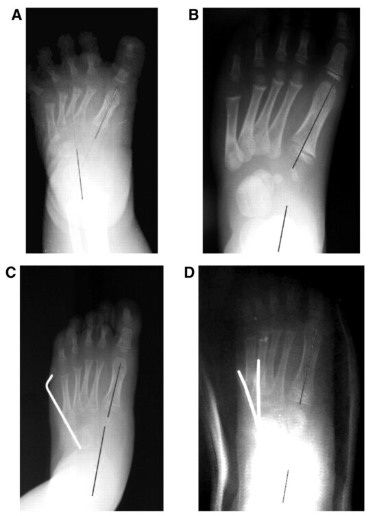
Figure 11.
Preoperative (A and B) and postoperative (C and D) radiographs showing the reduction in forefoot adductus in two different left feet.
Conclusion
Forefoot adduction is the most commonly recurring deformity in pediatric clubfoot surgery. This study evaluated the rate of forefoot adduction persistence and the effects of cuboid-cuneiform osteotomy after a specific single primary procedure, a posterior medial release, in patients with idiopathic clubfoot who did not respond to conservative treatment. This study may help the clubfoot surgeon determine the need for an osseous procedure in cases of residual or persistent forefoot adduction.
Funding
None reported.
Acknowledgments
All of the volunteer attending surgeons, the Red Cross Hospital in Mexicali, Mexico, and the Department of Orthopaedics at the University of California, San Diego, for their assistance.
Conflicts of Interest
None reported.
References
- Asperheim MS, Moore C, Carroll NC, et al: Evaluation of residual clubfoot deformities using gait analysis. J Pediatr Orthop B 4: 49, 1995.
- Atar D, Lehman WB, Grant AD, et al: Revision surgery in clubfoot. Clin Orthop 283: 223, 1992.
- Berman A, Gartland JJ: Metatarsal osteotomy for the correction of adduction of the fore part of the foot in children. J Bone Joint Surg Am 53: 498, 1971.
- Kling TF, Conklin MJ, Schmidt TL: “Opening Wedge First Cuneiform Osteotomy (Usually With 2d Through 4th Metatarsal Osteotomies) for Resistant Metatarsus Adductus Following Clubfoot Release,” in Clubfoot, ed by GW Simons, p 404, Springer-Verlag, New York, 1994.
- Lehman WB, Atar D, Grant AD, et al: Re-do clubfoot surgical approach and long-term results. Bull N Y Acad Med 66: 601, 1990.
- Lloyd-Roberts GC, Swann M, Catterall A: Medial rotational osteotomy for severe residual deformity in club-foot. J Bone Joint Surg Br 56: 37, 1974.
- Otremski I, Salama R, Khermosh O, et al: Residual adduction of the forefoot. J Bone Joint Surg Br 69: 832, 1987.
- Prichasuk S, Mulpruek P: Problems in surgical treatment of congenital clubfoot. J Med Assoc Thai 75: 106, 1992.
- Tarraf YN, Carroll NC: Analysis of the components of clubfeet presenting for reoperation. J Pediatr Orthop 12: 207, 1992.
- Vizkelety T, Szepesi K: Reoperation in treatment of club-foot. J Pediatr Orthop 9: 144, 1989.
- Heyman CH, Herndon CH, Strong JM: Mobilization of the tarsometatarsal and intermetatarsal joints for the correction of resistant adduction of the fore part of the foot in congenital clubfoot or congenital metatarsus varus. J Bone Joint Surg Am 40: 299, 1958.
- McHale KA, Lenhart M: Treatment of residual clubfoot deformity—the “bean-shaped” foot—by opening wedge medial cuneiform osteotomy and closing wedge cuboid osteotomy: clinical review and cadaver correlations. J Pediatr Orthop 11: 374, 1991.
- Schoenecker PL, Anderson DJ, Blair VP III, et al: “Combined Lateral Column Shortening and Medial Column Lengthening of Severe Forefoot Adductus,” in Clubfoot, ed by GW Simons, p 412, Springer-Verlag, New York, 1994.
- Yamamoto H, Muneta T, Furuya K: Cause of toe-in gait after posteromedial release for congenital clubfoot. J Pediatr Orthop 14: 369, 1994.
- Wynne-Davies R: Talipes equinovarus: a review of eighty-four cases after completion of treatment. J Bone Joint Surg Br 46: 464, 1964.
- Lowe LW, Hannon MA: Residual adduction of the forefoot in treated congenital clubfoot. J Bone Joint Surg Br 55: 809, 1973.
- Main J, Crider RJ: An analysis of residual deformity in clubfeet submitted to early operation. J Bone Joint Surg Br 60: 536, 1978.
- Porat S, Kaplan L: Critical analysis of results in clubfeet treated surgically along the Norris Carroll approach: seven years of experience. J Pediatr Orthop 9: 137, 1989.
- Rushforth GF: The natural history of hooked forefoot. J Bone Joint Surg Br 60: 530, 1978.
- Victoria-Diaz A, Victoria-Diaz J: Pathogenesis of idiopathic clubfoot. Clin Orthop Relat Res 185: 5, 1984.
- Ghali NN, Abberton MJ, Silk FF: The management of metatarsus adductus et supinatus. J Bone Joint Surg Br 66: 376, 1984.
- Evans D: Relapsed clubfoot. J Bone Joint Surg Br 43: 722, 1961.
- Lichtblau S: A medial and lateral release operation for clubfoot. J Bone Joint Surg Am 55: 1377, 1973.
- Schaefer D, Hefti F: Combined cuboid/cuneiform osteotomy for correction of residual adductus deformity in idiopathic and secondary club feet. J Bone Joint Surg Br 82: 881, 2000.
- Simons GW: Analytical radiography and the progressive approach in talipes equinovarus. Orthop Clin North Am 9: 187, 1978.
- Turco VJ: Surgical correction of the resistant club foot: one-stage posteromedial release with internal fixation: a preliminary report. J Bone Joint Surg Am 53: 477, 1971.
- Simons GW: Complete subtalar release in clubfeet: part I. A preliminary report. J Bone Joint Surg Am 67: 1044, 1985.
- Simons GW: The complete subtalar release in clubfeet. Orthop Clin North Am 18: 667, 1987.
- Shaughnessy WJ, Dechet P, Kitaoka HB: Posteromedial release for idiopathic clubfoot: sixteen-year follow-up study. Paper presented at the Annual Meeting of the Pediatric Orthopaedic Society of North America, May 1–4, 2000, Vancouver, British Columbia.
- Abrams RC: Relapsed clubfoot: the early results of an evaluation of Dillwyn Evans’ operation. J Bone Joint Surg Am 51: 270, 1969.
- Fowler AW: A method of forefoot reconstruction. J Bone Joint Surg Br 41: 507, 1959.
- Hofmann AA, Constine RM, McBride GG, et al: Osteotomy of the first cuneiform as treatment of residual adduction of the fore part of the foot in club foot. J Bone Joint Surg Am 66: 985, 1984.
- Kose N, Gunal I, Gokturk E, et al: Treatment of severe residual clubfoot deformity by trans-midtarsal osteotomy. J Pediatr Orthop B 8: 251, 1999.
- Steytler JC, Van der Walt ID: Correction of resistant adduction of the forefoot in congenital club-foot and congenital metatarsus varus by metatarsal osteotomy. Br J Surg 53: 558, 1966.
© 2007 American Podiatric Medical Association