


Abstract
Although many reports have been published on the usefulness of costal cartilage grafting in the reconstruction of interphalangeal joints of fingers, there are only a few published reports on the reconstruction of interphalangeal joints of toes. We describe a 21-year-old woman with a tissue defect of the dorsum pedis and a partial defect of the interphalangeal joint of the great toe caused by a motor-vehicle accident. We attempted arthroplasty using a free latissimus dorsi myocutaneous flap and a costal osteochondral graft. The grafted rib and cartilage survived, allowing the patient to resume functional ambulation for day-to-day activities. Arthroplasty using costal osteochondral grafts seems to be an effective means of reconstructing the interphalangeal joints of toes. (J Am Podiatr Med Assoc 96(6): 508–512, 2006)
Arthroplasty by costal cartilage grafting has been used often to treat injured interphalangeal joints of fingers. However, when dealing with injured interphalangeal joints of toes, procedures such as arthrodesis are usually used. To treat an extensive defect of the soft tissue of the dorsum pedis with a partial defect of the interphalangeal joint of the great toe caused by a motor-vehicle accident, we recently used a free latissimus dorsi myocutaneous flap and a costal osteochondral graft.
Case Report
A 21-year-old woman operating a motorcycle was hit by a truck that was turning to the left. She was admitted to the emergency department with an extensive tissue defect visible in the dorsum pedis and bone exposure (Fig. 1 [). Radiographs and computed tomographic scans revealed defects in half of the interphalangeal joint and proximal phalanx of the first toe and a defect on the dorsal side of the metatarsal bone of the same toe. In the second through fifth toes, defects on the dorsal side of the metatarsal bone were noted, although the joints were intact (Fig. 2 [).
We planned reconstructive surgery for the extensive tissue defect and arthroplasty using a free latissimus dorsi myocutaneous flap and a costal osteochondral graft. An incision measuring approximately 3 cm was made just below the inframammary fold on the left side. Costal components were obtained from the left seventh costal cartilage, including the costo-osteochondral junction. The obtained rib and costal cartilage were processed to a form tailored to the shape of the joint cartilage and bone in the defective areas. They were grafted into the proximal phalanx of the great toe using a 1-mm-diameter Kirschner wire (Fig. 3 [). The rib and costal cartilage were grafted and compressed against the phalanx. Because instability of the blood supply was expected in the injury-related defective areas, we covered the grafts with a free latissimus dorsi myocutaneous flap. End-to-end anastomosis was performed between the anterior tibial artery and the dorsal thoracic artery and between the anterior tibial vein and the dorsal thoracic vein.
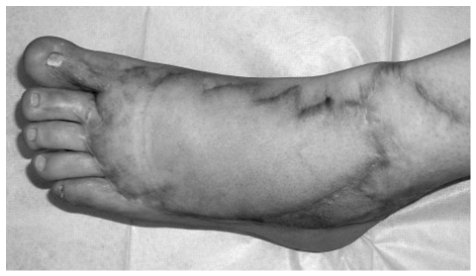
The flap and costal osteochondral graft survived satisfactorily (Fig. 4 [). At the last follow-up examination, 3 years and 4 months after surgery, passive flexion and extension of the surgical area were possible, although the range of motion of the interphalangeal joint showed some restriction (Fig. 5 [). The patient did not complain of pain during walking, and she had no problem with the transition from the stance phase to the swing phase. Moreover, she could wear high-heeled and narrow-toed shoes (Fig. 6 [).
Discussion
Since Blair [1]. suggested the usefulness of costal cartilage in treating ankylosis of the jaw joint, several reports have been published on costal cartilage grafting in the field of oral surgery [2]. Subsequently, costal cartilage grafting was applied by orthopedic surgeons in surgery for interphalangeal joints of fingers [3,4]. Although interphalangeal joints of toes were often used as material for the reconstruction of interphalangeal joints of fingers [5]. the use of costal cartilage for the reconstruction of interphalangeal joints of toes has rarely been reported. Our literature search revealed no reports on toe arthroplasty using rib or costal cartilage grafting.
Toes, especially the great toe, are involved in the important functions of body weight support and walking. When the stance phase of walking changes to the swing phase, the ground is hit by the anterior part of the foot, especially the first toe [6]. Therefore, if a defect in the interphalangeal joint of the great toe is left untreated or if the joint is fixed by arthrodesis, smooth walking and the fitting of high-heeled shoes in female patients will be difficult.
Reconstruction of the interphalangeal joint of the great toe using costal osteochondral grafts allows reconstruction of the joint by means of a single-stage operation without sacrificing any other joints. If only the costal cartilage is grafted, the graft survival rate is low. It is, therefore, advisable to use the costo-osteochondral junction so that the graft contains more rib, which markedly increases the survival rate. Because the costal cartilage is fibrous, it is unlikely that regeneration of hyaline cartilage will occur in the costal cartilage–grafted articular plane. Therefore, it should be used in cases in which the contralateral articular plane remains intact [3]. When this procedure is used in cases with extensive soft-tissue defects, the area surrounding the graft needs to be covered with a tissue rich in blood supply. In the present case, extensive soft tissue was covered with a free latissimus dorsi myocutaneous flap. We also succeeded in reconstructing the dorsal artery of the foot. In this way, we could prepare a recipient site rich in blood supply before costal osteochondral grafting. At the last follow-up examination, the reconstructed interphalangeal joint of the great toe retained good morphology. The patient could smoothly transition from the stance phase to the swing phase during walking and did not complain of pain or other discomfort. Our experience with the present case suggests that arthroplasty for toes is useful in allowing patients to resume walking smoothly and in alleviating pain during walking.
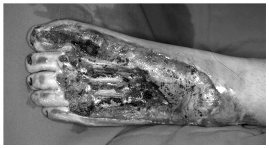
Figure 1.
Extensive tissue defect visible in the dorsum pedis accompanied by bone exposure.
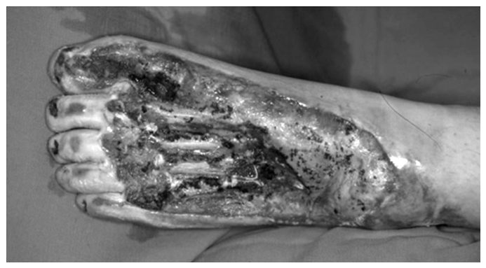
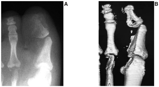
Figure 2.
Preoperative radiograph (A) and computed tomographic scan (B). The interphalangeal joint and phalanx of the great toe are partially defective. A defect is also visible on the dorsal side of the metatarsal bone of the great toe, but the joint is preserved relatively well.
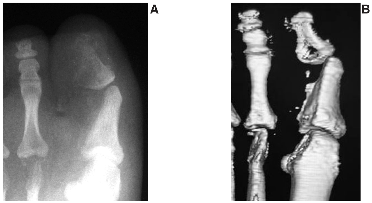
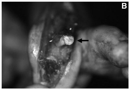
Figure 3.
Costal cartilage components, including the costo-osteochondral junction, were obtained from the seventh costal cartilage. The graft was fixed on the phalanx of the great toe using a 1-mm-diameter Kirschner wire (arrows), as shown in these dorsal (A) and anterior (B) views.
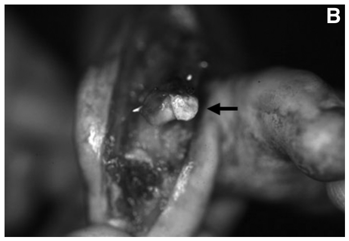
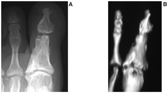
Figure 4.
Postoperative radiograph (A) and computed tomographic scan (B) showing good bone survival and shape of the articular plane.
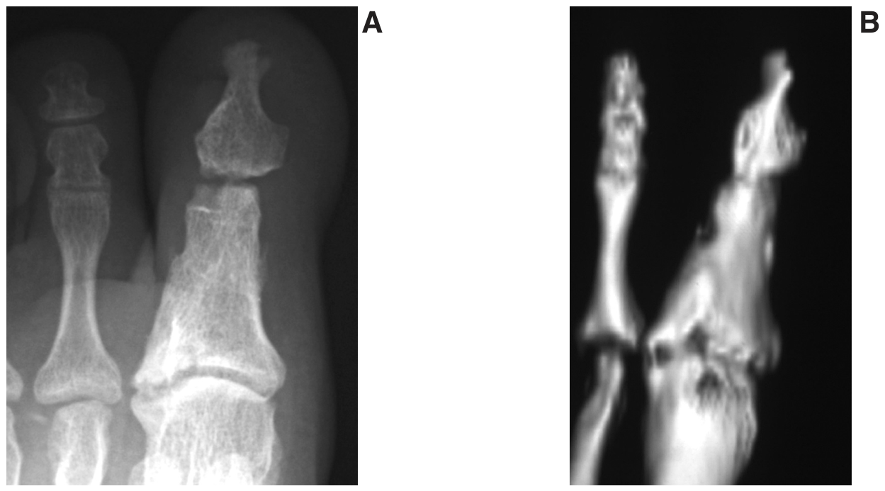
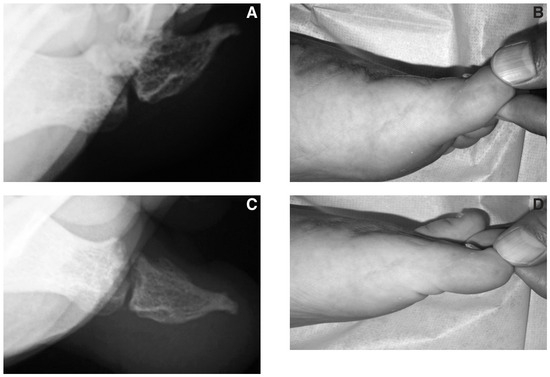
Figure 5.
Postoperative assessment. The angles formed where two lines, the long axis of the proximal phalanx and distal phalanx, crossed each other represented the range of motion of the interphalangeal joint. Postoperative radiograph (A) and photograph (B) reveal that the angle was 25° in passive extension. Postoperative radiograph (C) and photograph (D) show that the angle was 7° in passive flexion.
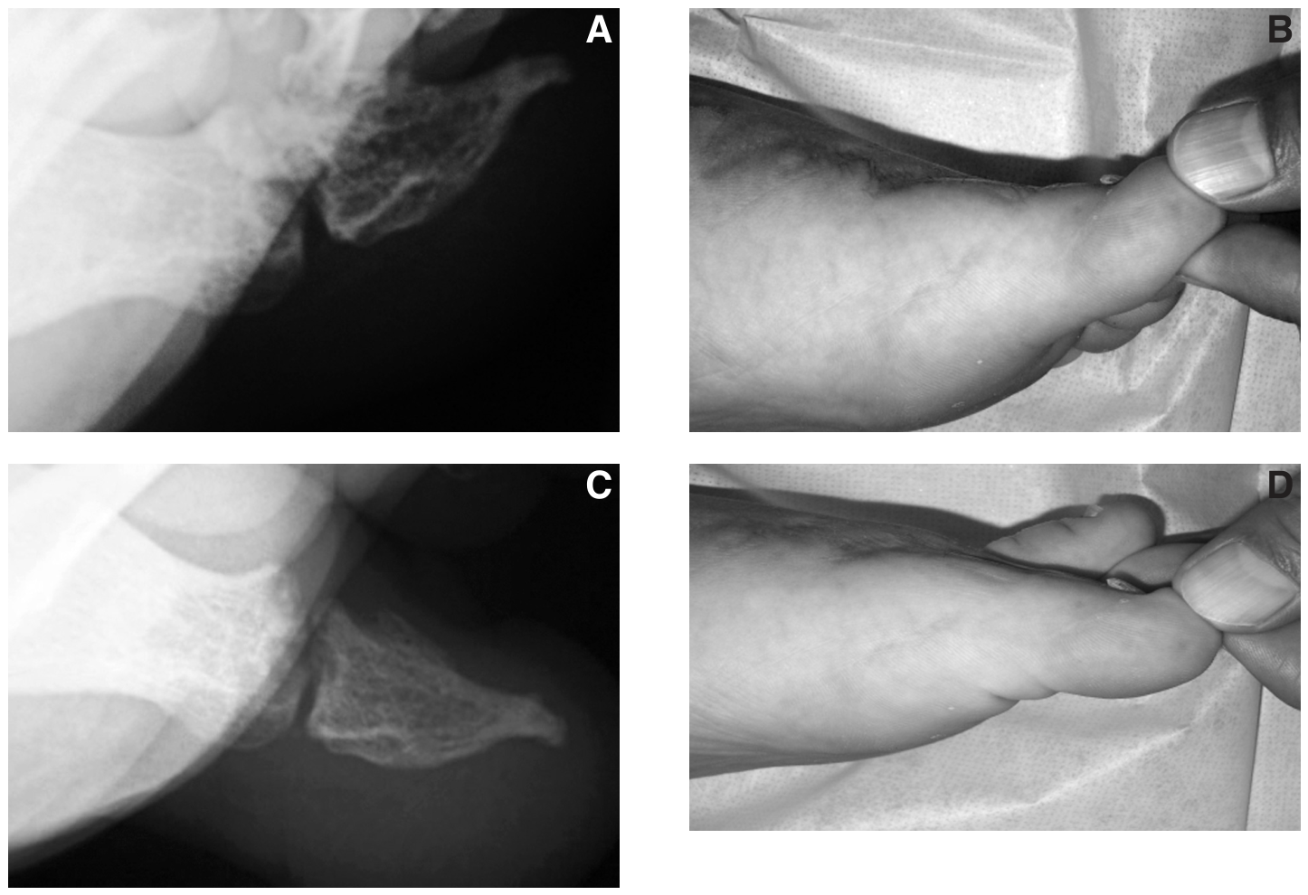
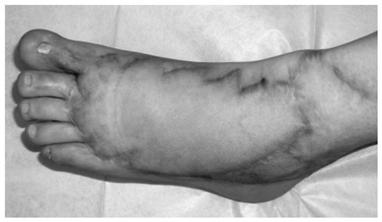
Figure 6.
Appearance of the foot 3 years and 4 months after surgery.
References
- Blair VP: The consideration of contour as well as functions for organic ankylosis of the lower jaw. .Surg Gynecol Obstet46::167. ,1928. .
- Figueroa AAA, Gans BJ, Pruzansky S: Long term follow-up of a mandibular costochondral graft. .J Oral Surg58::257. ,1984. .
- Hasegawa T, Yamano Y, Akatsukasa K: Primary reconstruction with costal cartilage graft in the interphalangeal joint. .J Jpn Soc Surg Hand8::723. ,1991. .
- Hasegawa T, Yamano Y: Arthroplasty of the proximal interphalangeal joint using costal cartilage grafts. .J Hand Surg Br17::583. ,1992. .
- Tsai TM, Singer R, Elliott E, et al: Immediate free vascularized joint transfer from second toe to index finger proximal interphalangeal joint: a case report. .J Hand Surg Br10::85. ,1985. .
- Ito T: “Lower Limb,” in Human Anatomy, ed by T Ito, p 137, Nanzando, Tokyo. ,1991. .
© 2006 American Podiatric Medical Association