Syndactyly is derived from the Greek word
syn, meaning together, and
dactyly, meaning digit [
1–
3]. The art of skin grafting has been documented in Hindu Sanskrit textbooks as far back as 3,000 years ago [
4,
5]. It was not until the 19th century that surgical approaches to finger and toe desyndactylization appeared in the literature, with Zeller (1810) [
6] and Dieffenbach (1834) [
7] paving the way for subsequent techniques [
8–
17]. We sought to develop a streamlined surgical procedure that uses donor grafts from the foot and ankle in toe desyndactylization. Weinstock et al [
18] described the procedure using redundant skin harvested from the dorsum of the foot. Giorgini et al [
19] later modified the procedure for repair of a cleft foot by debulking the skin graft taken from the ankle region of subcutaneous tissue.
Syndactylism may be classified anatomically. If the toes are fused to the tips of the digits, they are termed “completely syndactylized.” If the toes are not fused to the tips of the digits, they are termed “incompletely syndactylized.” When the phalangeal bones are normal, the type is simple; when they are abnormal in size, shape, number, and arrangement, the type is complicated.
When syndactyly is inherited (congenital syndactyly), there is intrauterine failure of cell development whereby programmed cell death occurs between the digits, causing them to be joined together or webbed. Syndactyly occurs in 1 of every 2,000 births, with males having a slightly higher incidence than females [
8,
9]. Bilateral syndactyly occurs more than 36% of the time.
10 Syndactylization of the second and third toes has the greatest prevalence because the webbing between these toes is the last to develop [
2,
11]. If syndactylized toes occur in individuals who have syndromes such as Apert’s syndrome, Poland’s syndrome, Jarcho-Levin syndrome, brachydactyly, polysyndactyly, and talipes equinovarus, these individuals are said to have “syndromic syndactyly” [
12,
20]. Syndactylism that follows severe burns or trauma to the hands or feet is known as “acquired syndactyly.” Often, destruction of the tissues of the fingers or toes results in subsequent fusion of granulating surfaces. In the acquired type, the web generally consists of dense scar tissue, and there is usually an associated contracture or distortion of the digits, but there is seldom, if ever, fusion of the bones.
Surgical Indications and Preoperative Considerations
The indication for surgical separation of the digits is usually related to cosmesis, although pain and functional digital contractures can cause a patient to seek treatment. Nonosseous syndactyly of the toes usually has no functional consequence, although osseous involvement implies shared myotendinous units and, therefore, functional alteration. Concerns may range from the functional inability to wear certain types of shoes to cosmetic appearance.
Surgical treatment requires preoperative planning. The goals of surgical desyndactylization include complete release of the affected digits, adequate soft-tissue coverage, an acceptable cosmetic appearance, and prevention of contracture and recurrence of the deformity. A complete physical examination should include assessment of the neurovascular supply to each digit. Local skin tension and mobility become an issue if the surgical plan includes rotational or advancement flaps. Digital contractures place tension on the skin and require more cutaneous skin coverage when contractures are released and lengthened. Flexion and extension functions of each toe should also be assessed, as this may reveal absent or weakened myotendinous input to the fused digit.
Case Report
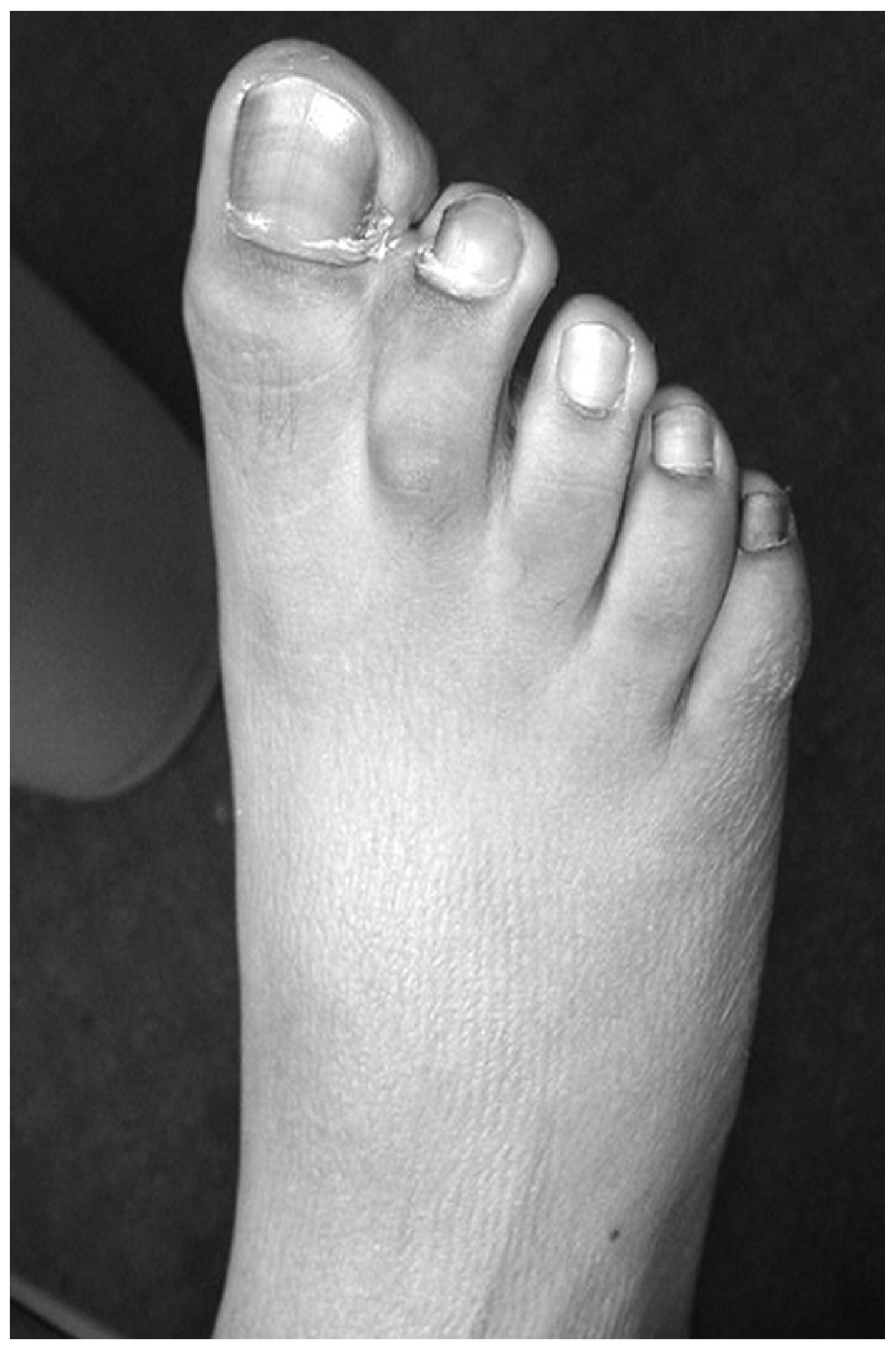
A 13-year-old girl presented to Good Samaritan Hospital, West Islip, New York, with a chief complaint of bilateral, complete, simple syndactyly of the first digital interspace (
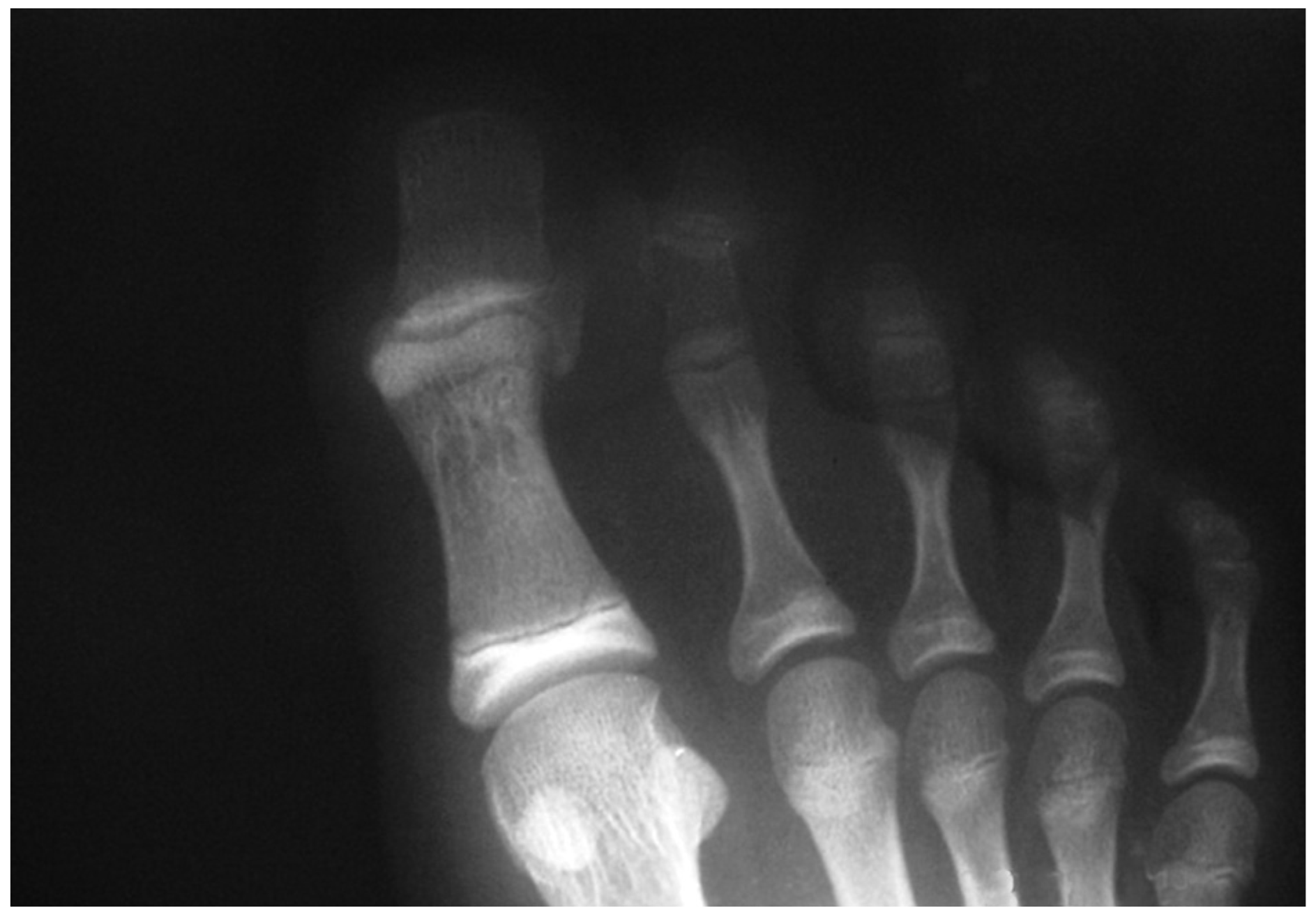
Fig. 1). Preoperative radiographs revealed no bony attachments (
Fig. 2). There was evidence of incomplete bridging at the interphalangeal joint. The child’s medical history was unremarkable. Findings from the physical examination were otherwise normal. There was a family history of syndactyly (maternal grandmother).
Surgical Technique
The following is a stepwise technique that uses a full-thickness autologous skin graft from the lateral aspect of the ankle, which obviates surgery elsewhere on the body, eliminates the need for an additional surgical team, and saves the patient anesthesia time, operating room time, and cost.
The procedure is performed with the patient supine and under intravenous sedation with local anesthesia to the affected digits and graft donor site (if required). A tourniquet is not used. The foot is prepared with 10% povidone-iodine solution and is then draped in the usual sterile manner to just above the ankle.
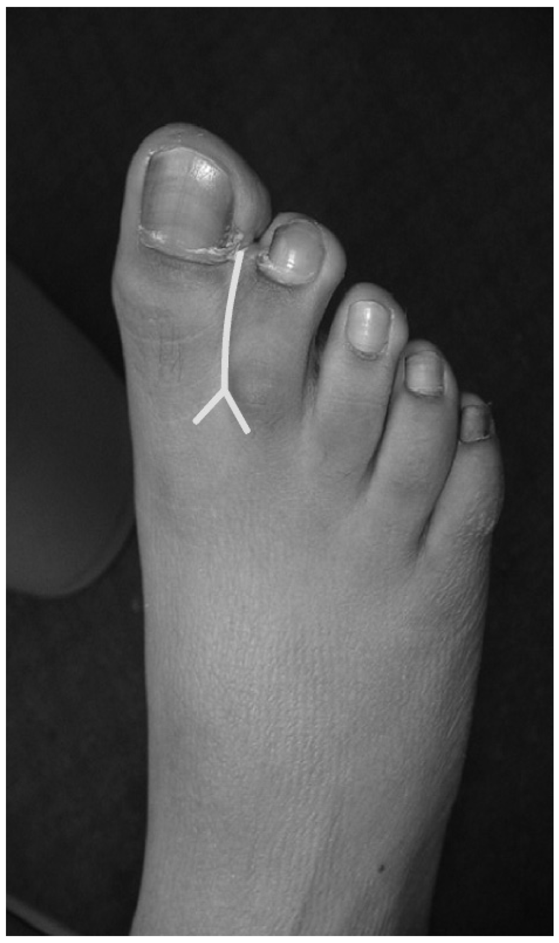
The syndactylized toes are addressed first to prepare the recipient site. The orientation of the adjacent sulci with respect to the syndactylized web space is determined dorsally and plantarly, and an arc is formed from the boundaries to define the proximal extent of the incision. Using sharp dissection, a linear-shaped incision is made separating the first and second digits. The incision extends proximally to the base of the proximal phalanx and from dorsal to plantar. At the proximal end of this incision, a V-shaped incision extension is made for use as the interspace flap (
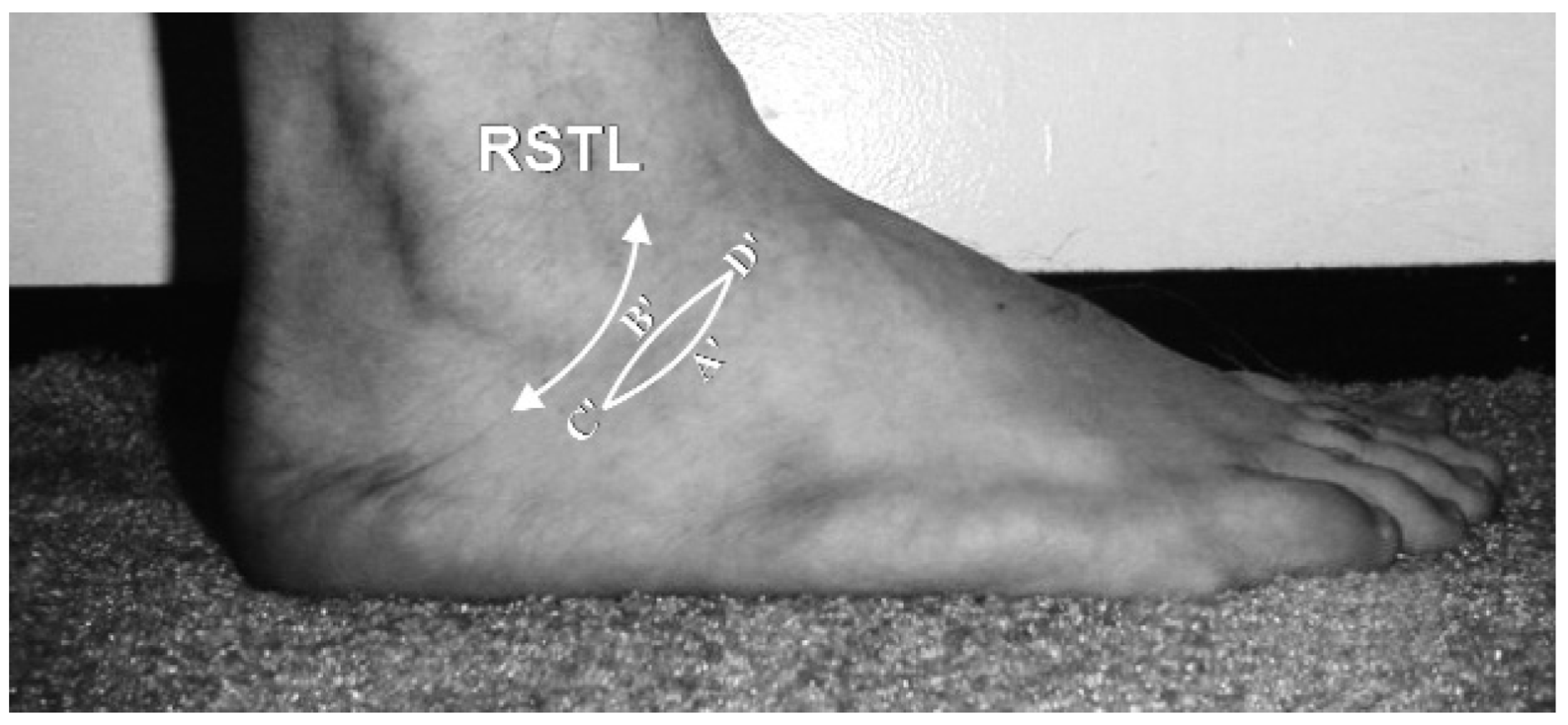
Fig. 3). The wound is measured along the sagittal and transverse axes of each toe, and a moist sponge is then applied in the newly created sulcus. The transverse axis has two “wings,” with each wing being oriented on either side of the commissure. Next, a full-thickness skin graft from the lateral aspect of the foot just inferior to the lateral malleolus is harvested by making two semi-elliptical incisions (
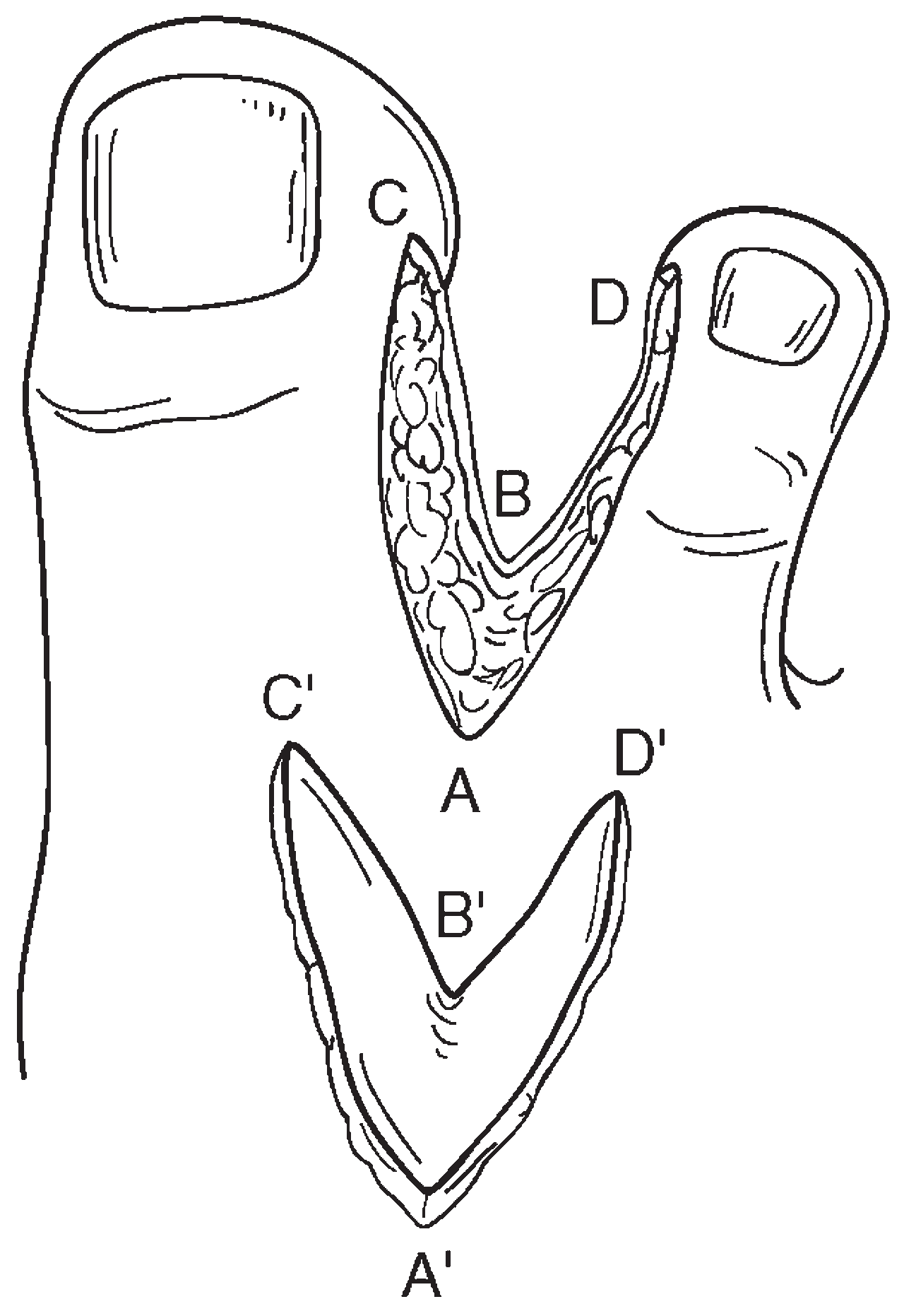
Fig. 4). Graft size is determined by measuring the length of the inner voided borders of the first and second digits (
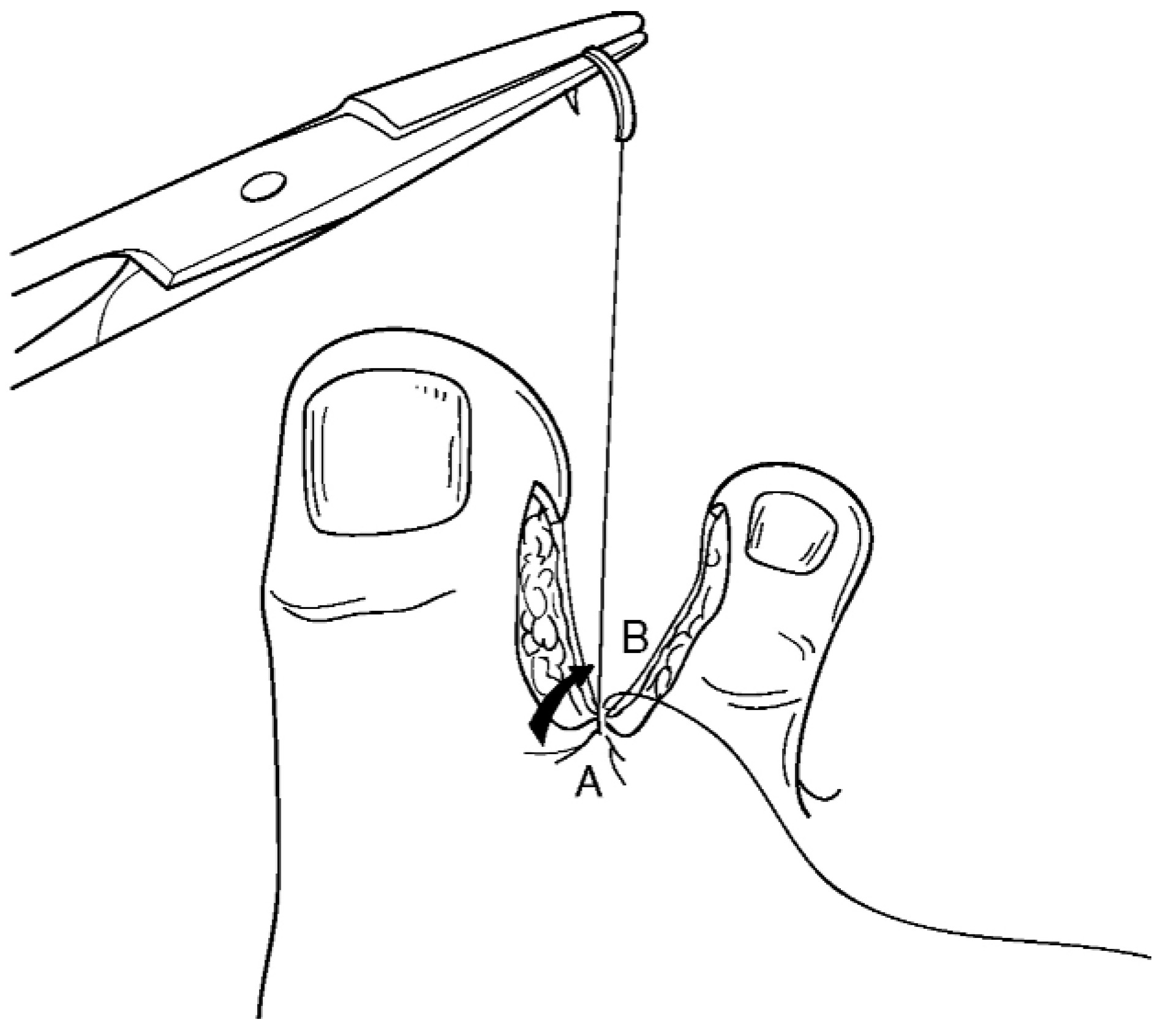
Fig. 5). The graft is then harvested and placed in saline for later use. With the ankle mildly pronated, the ankle defect is closed using 3-0 nylon sutures. Next, the V-shaped incision flap is drawn plantarly across the proximal commissure, and the tip is sutured plantarly to create the interspace (
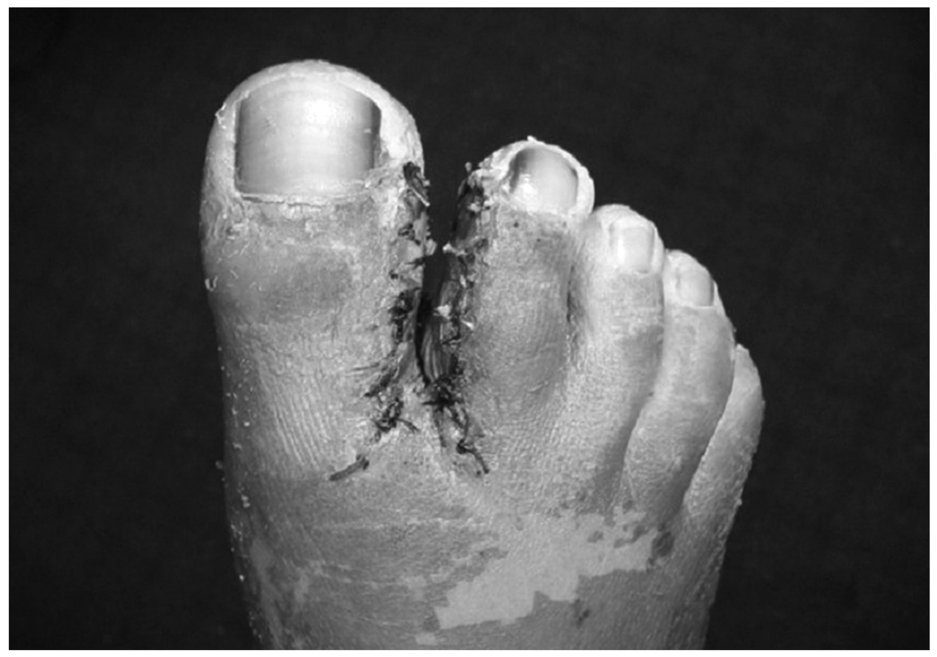
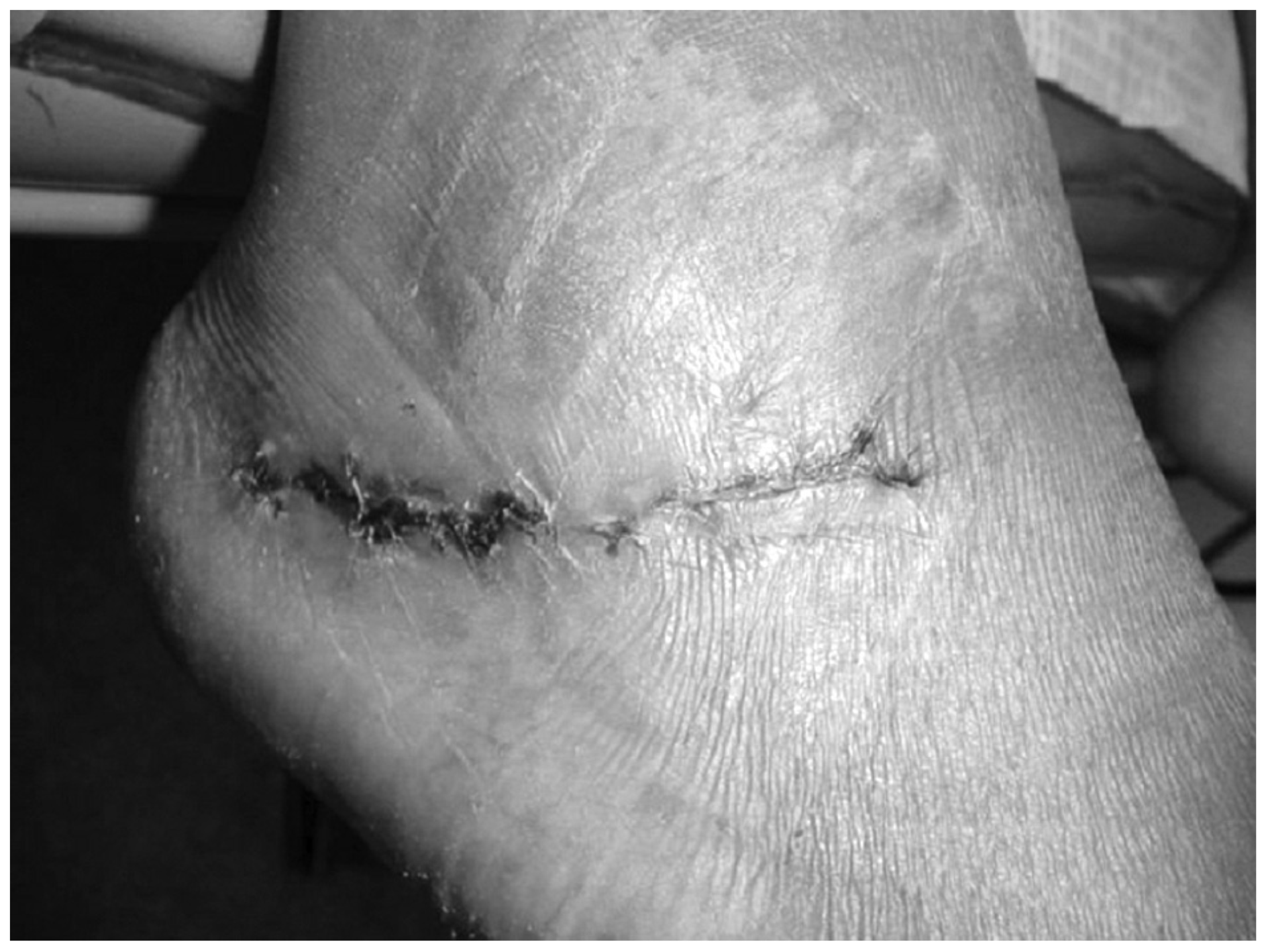
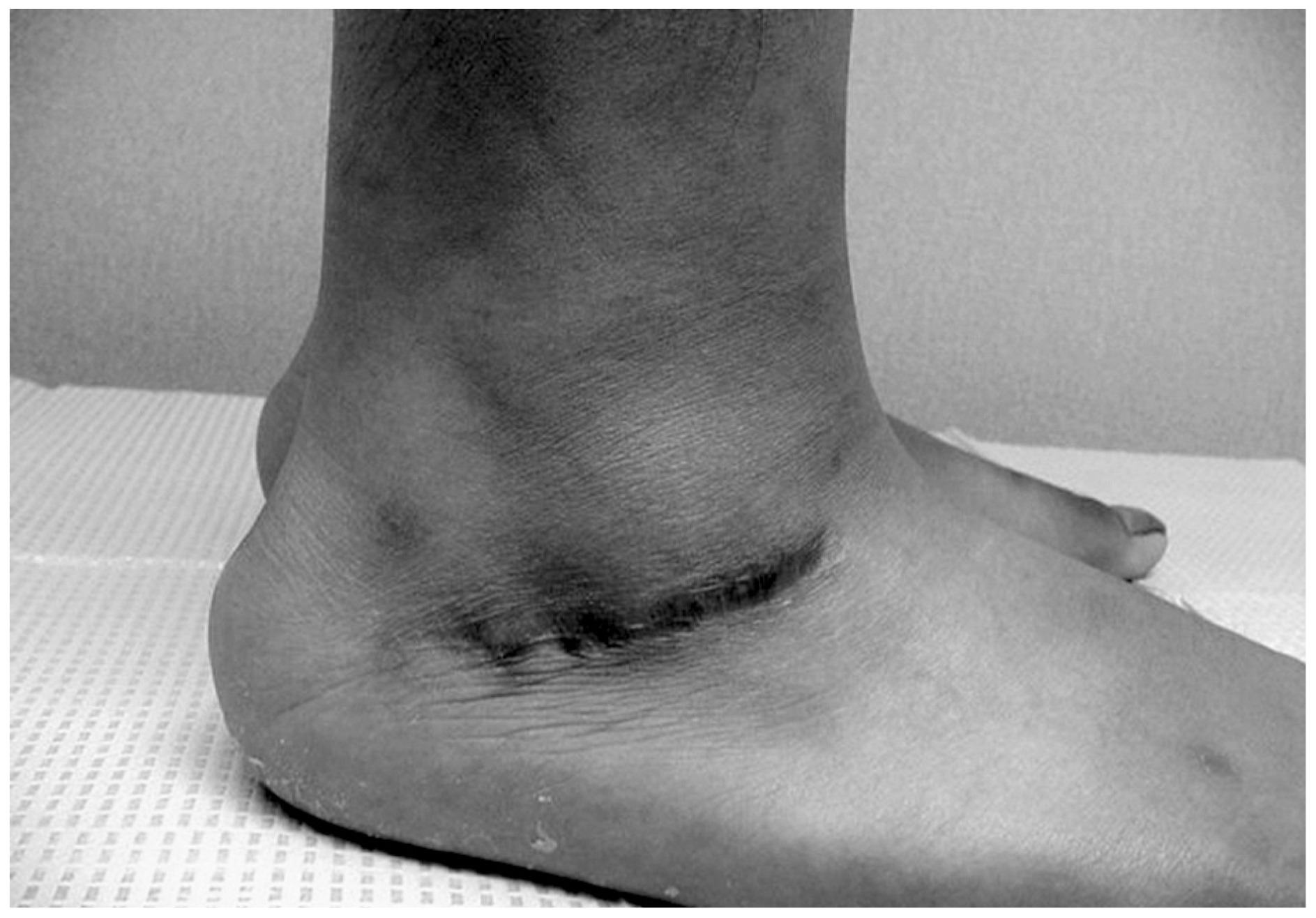
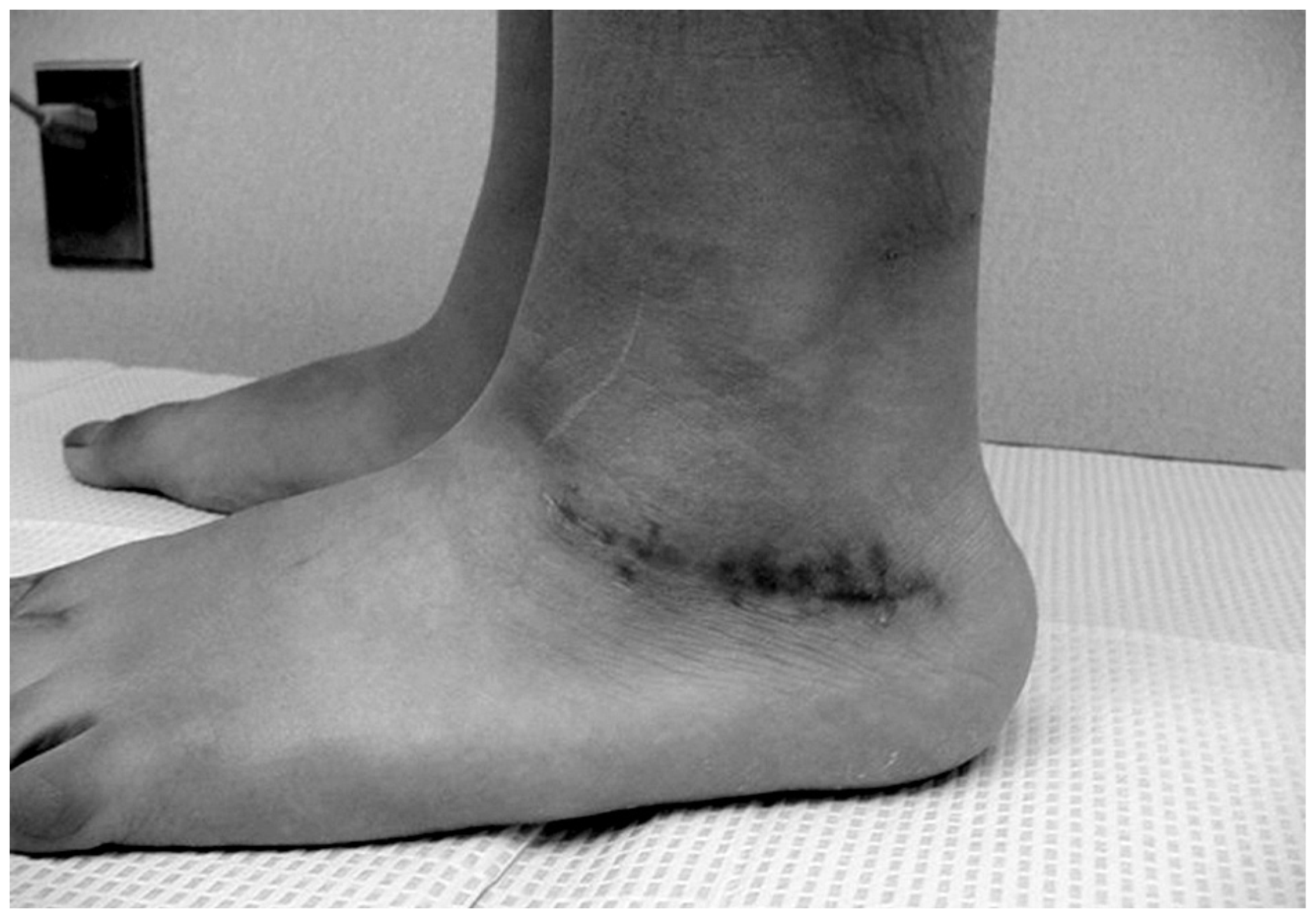
Fig. 6). The harvested full-thickness skin graft is then debulked of subcutaneous tissue placed between the first and second interspace using an interrupted simple suture technique with 4-0 Vicryl sutures (Ethicon, Inc, Somerville, New Jersey). A well-padded postoperative dressing is applied along with a nonweightbearing below-the-knee fiberglass cast to optimize the integrity of the graft and donor site. The initial surgical dressing should be removed and evaluated 5 to 7 days postoperatively. A short-leg fiberglass cast is recommended for 2 weeks, followed by a sterile compression dressing for another 2 to 4 weeks (
Figs. 7–
10). Keeping the surgical site free of edema, underlying hematoma, and infection, as well as clean and dry, is essential for graft survival [
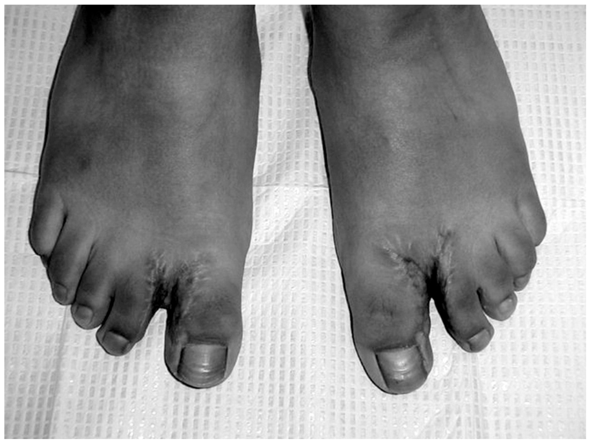
21]. When syndactyly is bilateral, we recommend that only one side be treated at a time (
Fig. 11).
Discussion
There are various reported techniques for correcting syndactylized toes. The present method eliminates the need for local flaps, whether rotational or advancement flaps, thereby eliminating excessive tension on digital circulation. Excess skin tension may lead to complications such as hypertrophic scar formation, tissue contracture, and graft failure.
Another advantage of this method is the need for only one surgical team to harvest and apply the full-thickness skin graft, saving the patient time in the operating room and the cost of another surgical team to harvest the donor graft. In addition, this surgical approach eliminates scar formation above the ankle. This is an important cosmetic factor to consider, especially in a pediatric patient.
Figure 1.
Right foot with syndactyly of the first interspace.
Figure 1.
Right foot with syndactyly of the first interspace.
Figure 2.
Radiograph of the right foot revealing no osseous abnormalities contributing to syndactyly of the first interspace.
Figure 2.
Radiograph of the right foot revealing no osseous abnormalities contributing to syndactyly of the first interspace.
Figure 3.
Right foot with syndactyly of the first interspace. The white line is the planned initial incision site. The V-shaped proximal extension of the initial dorsal incision is used to help with plastic closure of the first interspace.
Figure 3.
Right foot with syndactyly of the first interspace. The white line is the planned initial incision site. The V-shaped proximal extension of the initial dorsal incision is used to help with plastic closure of the first interspace.
Figure 4.
Right lateral ankle skin graft donor site. The white line, A′B′C′D′, is the planned incision site for the full-thickness skin graft harvested along the relaxed skin tension line (RSTL).
Figure 4.
Right lateral ankle skin graft donor site. The white line, A′B′C′D′, is the planned incision site for the full-thickness skin graft harvested along the relaxed skin tension line (RSTL).
Figure 5.
Donor graft A′B′C′D′ is previously measured to fit the newly created interspace recipient site ABCD.
Figure 5.
Donor graft A′B′C′D′ is previously measured to fit the newly created interspace recipient site ABCD.
Figure 6.
The V-shaped dorsal proximal extension (A) is pulled plantarly to meet the plantar proximal extension (B), filling the interspace sulcus. This creates a lateral hallux skin defect and a medial interspace defect on the second toe. Subsequently, these defects are filled with the skin graft material harvested from the ankle.
Figure 6.
The V-shaped dorsal proximal extension (A) is pulled plantarly to meet the plantar proximal extension (B), filling the interspace sulcus. This creates a lateral hallux skin defect and a medial interspace defect on the second toe. Subsequently, these defects are filled with the skin graft material harvested from the ankle.
Figure 7.
Right foot 2 weeks after desyndactylization showing the first interspace.
Figure 7.
Right foot 2 weeks after desyndactylization showing the first interspace.
Figure 8.
Right foot 2 weeks after desyndactylization showing the lateral ankle donor site.
Figure 8.
Right foot 2 weeks after desyndactylization showing the lateral ankle donor site.
Figure 9.
Right foot 4 weeks after desyndactylization showing the lateral ankle donor site.
Figure 9.
Right foot 4 weeks after desyndactylization showing the lateral ankle donor site.
Figure 10.
Left foot 4 weeks after desyndactylization showing the lateral ankle donor site.
Figure 10.
Left foot 4 weeks after desyndactylization showing the lateral ankle donor site.
Figure 11.
Right foot 12 weeks after desyndactylization and left foot 4 weeks after desyndactylization.
Figure 11.
Right foot 12 weeks after desyndactylization and left foot 4 weeks after desyndactylization.