


Abstract
A pronated foot posture is considered to be a factor in limitation of dorsiflexion at the first metatarsophalangeal joint during weightbearing. Customized foot orthoses are widely used to increase dorsiflexion at the first metatarsophalangeal joint in people with pronated feet. However, the effect of foot posture and customized foot orthoses on maximum first metatarsophalangeal joint dorsiflexion has not been widely investigated. This study sought to determine 1) the relationship between foot posture and static maximum first metatarsophalangeal joint dorsiflexion and 2) the effect of customized foot orthoses on static maximum first metatarsophalangeal joint dorsiflexion in people with pronated feet. Foot posture was assessed using the Foot Posture Index. Static maximum first metatarsophalangeal joint dorsiflexion of the right foot was determined using a goniometer while participants stood relaxed with and without Blake-style inverted (30°) foot orthoses positioned under their feet. There was a significant negative correlation between Foot Posture Index and static maximum first metatarsophalangeal joint dorsiflexion (r = −0.587). Inverted (30°) foot orthoses increased the magnitude of static maximum first metatarsophalangeal joint dorsiflexion from 83.4° to 85.3° in participants with an excessively pronated foot posture. However, this difference was not statistically significant. People with pronated feet are more likely to exhibit limitation of dorsiflexion at the first metatarsophalangeal joint during gait, and inverted foot orthoses are unlikely to be effective in increasing dorsiflexion at the first metatarsophalangeal joint in these people. (J Am Podiatr Med Assoc 96(1): 32–37, 2006).
Dorsiflexion at the first metatarsophalangeal joint during the propulsive period of the gait cycle is important for efficient foot function.[1–5] During the propulsive period of the gait cycle, the first metatarsophalangeal joint dorsiflexes 45° to 65° as the body pivots over the hallux, which is fixed on the ground.[6–8] This dorsiflexion is responsible for establishing the windlass mechanism, originally described by Hicks,[5] who demonstrated that dorsiflexion of the first metatarsophalangeal joint generated tension within the plantar aponeurosis (a fibrous tissue band originating at the calcaneus and inserting into the proximal phalanges of the foot) as it was pulled distally through its attachment to the proximal phalanx of the hallux. This tension caused an increase in the medial longitudinal arch height through first-ray plantarflexion and midtarsal and subtalar joint supination, resulting in the foot being a stable structure during propulsion.[9] Inadequate dorsiflexion at the first metatarsophalangeal joint during propulsion, therefore, has been cited as a major cause of abnormal foot function.[1–5,10]
Functional and structural deformities, including functional hallux limitus,[2–4,11] hallux rigidus,[12–18] and hallux abducto valgus,[12,13,19–21] have been reported to develop as a result of a restriction of dorsiflexion at the first metatarsophalangeal joint. Limited dorsiflexion at the first metatarsophalangeal joint may have numerous causes. An excessively pronated foot type has been named as a major cause of limited dorsiflexion at the first metatarsophalangeal joint during gait.[1,22,23] Harradine and Bevan[22] demonstrated that lateral foot wedging to induce pronation caused a significant reduction in dorsiflexion at the first metatarsophalangeal joint. To our knowledge, however, no reported studies have investigated whether there are differences in first metatarsophalangeal joint dorsiflexion among people with different foot postures.
Functional foot orthoses have been used successfully to alleviate symptoms associated with limited hallux dorsiflexion caused by excessive pronation.[24,25] A major rationale for their use is that they increase the magnitude of dorsiflexion at the first metatarsophalangeal joint by restricting excessive pronation[26–33] and facilitating first-ray plantarflexion.[7,24,25] Two studies[7,34] have investigated the effect of foot orthoses on the magnitude of first metatarsophalangeal joint dorsiflexion in people with excessively pronated feet. In these studies, the use of customized foot orthoses did not increase the magnitude of first metatarsophalangeal joint dorsiflexion, either statically[34] or dynamically.[7] However, the orthoses used were standard midfoot-controlling (Root-style) devices that may not be as effective in controlling excessive pronation as more rearfoot-controlling inverted orthoses, such as the Blake-style device.[26]
The aim of the present study, then, was to determine whether a correlation existed between foot posture (pronated, neutral, or supinated) and static maximum first metatarsophalangeal joint dorsiflexion. A secondary aim was to determine whether inverted foot orthoses increased static first metatarsophalangeal joint dorsiflexion in participants with an excessively pronated foot posture.
Materials and Methods
Participants
Participants were recruited from the staff and undergraduate student population of La Trobe University, Victoria, Australia. For the aspect of the study investigating the relationship between foot posture and static maximum first metatarsophalangeal joint dorsiflexion, 36 individuals (16 men and 20 women) aged 20 to 40 years (mean ± SD age, 22.3 ± 4.1 years) with a mean ± SD body weight of 69.5 ± 13.6 kg were recruited. From this sample, 12 individuals (7 men and 5 women) aged 20 to 40 years (mean ± SD age, 22.8 ± 5.8 years) with a mean ± SD body weight of 70.7 ± 14.4 kg and an excessively pronated foot type (Foot Posture Index ≥+4)[35] were used to investigate the effect of Blake-style inverted (30°) foot orthoses on static maximum first metatarsophalangeal joint dorsiflexion. Approval for the study was obtained from the Faculty of Health Sciences Human Ethics Committee of La Trobe University, and informed consent was obtained from all participants. Individuals were excluded from the study if they had any acute or chronic lower-limb pathology or a history of trauma or surgery of the right foot. Individuals were also excluded if they demonstrated less than 65° of passive nonweightbearing dorsiflexion at the first metatarsophalangeal joint of the right foot.
Equipment
A goniometer with 1° increments was used to measure static maximum first metatarsophalangeal joint dorsiflexion. Goniometer readings were covered with opaque tape to conceal them from the examiner who performed the measurements of static maximum first metatarsophalangeal joint dorsiflexion. A rigid shaft (4 × 35 × 150 mm) made of polypropylene and beveled 45° at one end was used to maximally dorsiflex the first metatarsophalangeal joint. Emery paper, affixed to the weightbearing surface under the participant’s right foot, was used to create friction between the polypropylene shaft and the weightbearing surface (Fig. 1).
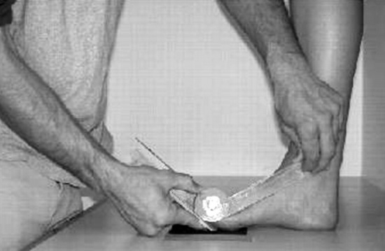
Figure 1.
Technique used to measure static maximum first metatarsophalangeal joint dorsiflexion.
The orthoses used in this study were custom-made Blake-style inverted functional foot orthoses posted to 30°.[36] Participants with an excessively pronated foot type (Foot Posture Index ≥+4)[35] were placed in a cast at the start of the study using a neutral casting technique[37] and were issued a pair of orthoses generated with a computer-aided design/computer-aided manufacturing system (Computer Orthotic Laboratory, Mudjimba, Queensland, Australia). The manufacture of the orthoses with such a system ensured a very high level of consistency in the orthoses prescriptions.[38]
Procedure
Data for this study were collected during two test sessions, separated by at least 1 week. During the first session, foot posture and static maximum first metatarsophalangeal joint dorsiflexion were assessed. The postures of the participants’ right feet were determined using the Foot Posture Index, as described by Redmond et al.[35] The Foot Posture Index consists of eight specific criteria: talar head palpation, supra-lateral and infralateral malleolar curvature, Helbing’s sign, calcaneal frontal plane position, prominence in the region of the talonavicular joint, congruence of the lateral border of the foot, and abduction and adduction of the forefoot on the rearfoot. Each Foot Posture Index criterion was scored on a 5-point scale (range, −2 to +2) by each examiner. The eight scores obtained were then summed to give an overall foot posture score. The summed score had the potential to range from −16 (highly supinated) to +16 (highly pronated).[35] The Foot Posture Index measurements were performed by two independent examiners, with the average score being used for analysis. Previous studies[35,39,40] indicate that the clinical measurement of Foot Posture Index has moderate-to-high reliability when assessing adult populations.
Static maximum first metatarsophalangeal joint dorsiflexion of the participants’ right feet was measured using a modified version of the technique originally described by Hogan and Kidd.[34] Their technique for the measurement of dorsiflexion at the first metatarsophalangeal joint assessed the angulation between the hallux and the plantar plane of the foot. Because participants with differing foot postures and, thus, different arch heights and metatarsal declination angles[41] were being studied, the first metatarsal shaft was used as the reference rather than the plantar plane of the foot. Participants were asked to stand in their normal angle and base of gait with their buttocks or shoulders just touching the wall behind them. Emery paper was positioned under the first metatarsophalangeal joint of the right foot of the participant and was affixed to the weightbearing surface using adhesive tape to prevent sliding of the poly-propylene shaft as the hallux was dorsiflexed. The center of the first metatarsophalangeal joint and the proximal medial aspect of the first metatarsal were located and marked with a fine felt-tipped marker. The medial side of the first metatarsal was then bisected by drawing a straight line connecting the proximal metatarsal to the center of the metatarsophalangeal joint. The bisection of the first metatarsal was performed while the participants were weightbearing to avoid error associated with soft-tissue movement occurring when participants move from a nonweight-bearing position to a weightbearing position.[42]
A piece of polypropylene was then placed under the hallux, with its proximal beveled end positioned under the center of the first metatarsophalangeal joint. A goniometer was positioned with its distal arm resting on the piece of polypropylene and its proximal arm parallel to the first metatarsal bisection line. Participants were then again reminded to stand erect in their normal angle and base of gait with their shoulders or buttocks just touching the wall. This was done to ensure that the participants maintained a constant body posture because there was a tendency for the participants to alter their body posture as the first metatarsophalangeal joint was dorsiflexed. The shaft of polypropylene was then used to maximally dorsiflex the proximal phalanx of the hallux. Care was taken to ensure that the distal arm of the goniometer was kept in contact with the polypropylene shaft and that the proximal arm aligned with the first metatarsal bisection as the first metatarsophalangeal joint was dorsiflexed (Fig. 1). The angle formed between the bisection of the first metatarsal shaft and the polypropylene used to dorsiflex the proximal phalanx was then measured by an examiner using the goniometer; the goniometer was then passed to an independent person not involved in any measurements for the study who read and recorded the value and replaced the arms to a parallel, 0° position. The measurement was completed three times by two independent examiners, and the average score was documented as the test result.
During the second test session, foot posture and static maximum first metatarsophalangeal joint dorsiflexion were assessed again to enable reliability calculations to be performed. Static maximum first metatarsophalangeal joint dorsiflexion in the presence of inverted (30°) foot orthoses was also assessed during the second session. The inverted (30°) foot orthoses were positioned under both feet of the participants, and the static maximum first metatarsophalangeal joint dorsiflexion of the participants’ right feet was measured as previously described. The measurement was performed three times by two independent examiners, and the average score was used for analysis.
Data Analysis
The intrarater and interrater reliability of Foot Posture Index and static maximum first metatarsophalangeal joint dorsiflexion measurements were determined using the type (2,1) intraclass correlation coefficient (ICC) statistic with 95% confidence intervals (CIs). The relationship between Foot Posture Index and static maximum first metatarsophalangeal joint dorsiflexion was assessed using the Pearson product moment correlation coefficient. Differences in static maximum first metatarsophalangeal joint dorsiflexion in the presence and absence of inverted (30°) foot orthoses were investigated using a paired-samples t-test. In all of the analyses, P < .05 was considered statistically significant. SPSS (version 10.1; SPSS Science, Chicago, Illinois) software was used for the analyses.
Results
The intrarater and interrater reliability of the measurements of Foot Posture Index and static maximum first metatarsophalangeal joint dorsiflexion were assessed using ICCs. For the Foot Posture Index measurement, the ICCs for the intrarater reliability of investigators 1 and 2 were found to be 0.95 (95% CI, 0.90–0.98) and 0.95 (95% CI, 0.88–0.98), respectively. The ICC for the interrater reliability of the Foot Posture Index was 0.95 (95% CI, 0.90–0.97). For the measurement of static maximum first metatarsophalangeal joint dorsiflexion, the ICCs for the intrarater reliability of investigators 1 and 2 were found to be 0.90 (95% CI, 0.80–0.95) and 0.87 (95% CI, 0.74–0.94), respectively. The ICC for the interrater reliability of the measurement of static maximum first metatarsophalangeal joint dorsiflexion was 0.89 (95% CI, 0.79–0.96). These results suggest that the measurements of Foot Posture Index and static maximum first metatarsophalangeal joint dorsiflexion were highly reliable.
For the aspect of the study investigating the relationship between Foot Posture Index and static first metatarsophalangeal joint dorsiflexion, the mean ± SD Foot Posture Index was 6 ± 3 (range, 1–12). The mean ± SD static maximum first metatarsophalangeal joint dorsiflexion measurement was 84.7° ± 8° (range, 61°–96°). The correlation (r) between Foot Posture Index and static maximum first metatarsophalangeal joint dorsiflexion was −0.587 (P < .01). This finding indicates that a more pronated foot posture was associated with a reduced magnitude of static maximum first metatarsophalangeal joint dorsiflexion.
For the aspect of the study investigating the effect of inverted (30°) foot orthoses on static maximum first metatarsophalangeal joint dorsiflexion in participants with an excessively pronated foot posture (Foot Posture Index ≥+4), the mean ± SD Foot Posture Index was 7 ± 3 (range, 4–12). The mean ± SD static maximum first metatarsophalangeal joint dorsiflexion measurements were 83.4° ± 8.96° (range, 64°–94°) and 85.3° ± 8.75° (range, 66°–99°) in the absence and presence of the orthoses, respectively (P = .061). These findings indicate that the inverted (30°) foot orthoses did not significantly affect the magnitude of static maximum first metatarsophalangeal joint dorsiflexion in participants with an excessively pronated foot posture.
Discussion
The results of this study show that a more pronated foot posture (increased Foot Posture Index) was associated with a reduced magnitude of static maximum first metatarsophalangeal joint dorsiflexion. Furthermore, inverted (30°) foot orthoses, first described by Blake,[36] did not significantly affect the magnitude of static maximum first metatarsophalangeal joint dorsiflexion in participants with an excessively pronated foot posture (Foot Posture Index ≥+4).
The finding that a more pronated foot posture is associated with a reduced magnitude of static maximum first metatarsophalangeal joint dorsiflexion is novel but supports the original theory of Root et al[1] that a pronated foot type may cause reduced first metatarsophalangeal joint dorsiflexion during gait by interfering with normal first-ray mechanics. To achieve more than 20° dorsiflexion at the first metatarsophalangeal joint, the first ray must plantarflex as the hallux dorsiflexes on the first metatarsophalangeal joint.[1] First-ray plantarflexion allows the first metatarsal head to glide in a plantar direction relative to the base of the proximal phalanx, which shifts the transverse axis of the first metatarsophalangeal joint dorsally, allowing the base of the proximal phalanx to articulate with the dorsal articular surface of the first metatarsal head, providing greater range of motion.[1,22,23,43] Furthermore, plantarflexion of the first ray reduces the distance between the origin and the insertion of the medial slip of the plantar aponeurosis, resulting in reduced tension on the plantar aponeurosis and allowing dorsiflexion at the first metatarsophalangeal joint.[5,9] However, closed-kinetic-chain foot pronation causes the first ray to dorsiflex as a result of increased ground reaction force,[44–46] which impedes first-ray plantarflexion, increases tension within the plantar fascia, and limits the range of first metatarsophalangeal joint dorsiflexion.[9,23]
Functional foot orthoses have been used successfully to manage symptoms associated with limited hallux dorsiflexion in people with excessively pronated feet.[24,25] In our study, inverted (30°) foot orthoses did not significantly affect the magnitude of static maximum first metatarsophalangeal joint dorsiflexion in participants with excessively pronated feet (Foot Posture Index ≥+4). These results are in agreement with those of previous studies[7,34] that demonstrated that midfoot-controlling (Root-style) foot orthoses do not significantly increase first metatarsophalangeal joint dorsiflexion. It is possible that a significant increase in dorsiflexion at the first metatarsophalangeal joint in the presence of inverted orthoses may have been achieved if participants with more pronated foot types were used. Furthermore, the orthoses used here did not have any additional features, such as a plantar fascial groove or a kinetic wedge,[47] that have also been suggested to facilitate first metatarsophalangeal joint dorsiflexion. Such features could be investigated in future studies. Finally, it is possible that foot orthoses have been effective in managing symptoms associated with limited hallux dorsiflexion by influencing other parameters at the first metatarsophalangeal joint, such as the force required to dorsiflex the hallux. In this regard, a recent study[48] demonstrated that the use of foot orthoses significantly reduced the force required to dorsiflex the hallux in people with symptoms associated with excessive pronation.
Our study should be interpreted in view of the limitation of being a static study of a dynamic structure. Nawoczenski et al[49] demonstrated that static maximum first metatarsophalangeal joint dorsiflexion was only moderately correlated to dynamic first metatarsophalangeal joint dorsiflexion (r = 0.61). A moderate correlation was also found between foot posture and static maximum first metatarsophalangeal joint dorsiflexion (r = 0.587). Other factors, such as first metatarsal length, metatarsal head shape, joint capsule mobility, and muscular flexibility, may also be important in influencing the range of dorsiflexion at the first metatarsophalangeal joint during weightbearing.[25] Furthermore, it was not possible to blind the examiners who were assessing static maximum first metatarsophalangeal joint dorsiflexion to the foot postures of the participants or the orthotic intervention. Although the examiners were not aware of the participants’ specific Foot Posture Index scores, they observed the feet of the participants with and without the orthotic intervention. Finally, further research is required to determine when a decrease in hallux dorsiflexion becomes sufficient to cause symptoms. Variations in other factors, such as stride length, activity level, heel height, and surface angulation, are likely to be important.[22]
Our research has potential clinical implications in that it demonstrates that people with pronated foot types are more likely to exhibit limitation of dorsiflexion at the first metatarsophalangeal joint. This may predispose these people to deformities such as functional hallux limitus, hallux rigidus, and hallux abducto valgus. Furthermore, the results of our study are in agreement with those of previous studies that suggest that customized foot orthoses are unlikely to be effective in increasing the range of dorsiflexion at the first metatarsophalangeal joint in people with pronated feet.
Acknowledgments
Adam Bird, BPod(Hons), and Craig Payne, DipPod(NZ), MPH, of La Trobe University, Victoria, Australia, for their assistance in the preparation of this article.
References
- Root ML, Orien WP, Weed JH: Normal and Abnormal Function of the Foot, Clinical Biomechanics Corp, Los Angeles, 1977.
- Dananberg HJ: Functional hallux limitus and its relationship to gait efficiency. JAPMA 76: 648, 1986.
- Dananberg HJ: Gait style as an etiology to chronic postural pain: part II. Postural compensatory process. JAPMA 83: 615, 1993.
- Dananberg HJ: Gait style as an etiology to chronic postural pain: part I. Functional hallux limitus. JAPMA 83: 433, 1993.
- Hicks JH: The mechanics of the foot: part 2. The plantar aponeurosis and the arch. J Anat 88: 25, 1954.
- Hetherington VJ, Johnson RE, Albritton JS: Necessary dorsiflexion of the first metatarsophalangeal joint during gait. J Foot Surg 29: 218, 1990.
- Kilmartin TE, Wallace WA, Hill TW: Orthotic effect on metatarsophalangeal joint extension: a preliminary study. JAPMA 81: 414, 1991.
- Phillips RD, Law EA, Ward ED: Functional motion of the medial column joints of the foot during propulsion. JAPMA 86: 474, 1996.
- Fuller EA: The windlass mechanism of the foot: a mechanical model to explain pathology. JAPMA 90: 35, 2000.
- Hall C, Nester CJ: Sagittal plane compensations for artificially induced limitation of the first metatarsophalangeal joint: a preliminary study. JAPMA 94: 269, 2004.
- Cohn I, Kanat IO: Functional limitation of motion of the first metatarsophalangeal joint. J Foot Surg 23: 477, 1984.
- McGlamry ED, Banks AS, Downey MS: Comprehensive Textbook of Foot Surgery, Vol 2, Williams & Wilkins, Baltimore, 1992.
- Gerbert J: Textbook of Bunion Surgery, 2nd Ed, Futura Publishing, New York, 1991.
- Jack EA: The aetiology of hallux rigidus. Br J Surg 27: 49, 1940.
- Bingold AC, Collins DH: Hallux rigidus. J Bone Joint Surg Br 32: 214, 1950.
- Kashuk KB: Hallux rigidus, hallux limitus, and other functionally limiting disorders of the great toe joint: background, treatment, and case studies. J Foot Surg 14: 45, 1975.
- Drago JJ, Oloff L, Jacobs AM: A comprehensive review of hallux limitus. J Foot Surg 23: 213, 1984.
- McMaster MJ: The pathogenesis of hallux rigidus. J Bone Joint Surg Br 60: 82, 1978.
- Brahm SM: Shape of the first metatarsal head in hallux rigidus and hallux valgus. JAPMA 78: 300, 1988.
- Shereff MJ, Baumhauer JF: Hallux rigidus and osteoarthrosis of the first metatarsophalangeal joint. J Bone Joint Surg Am 80: 898, 1998.
- Donick II, Berlin SJ, Block LD, et al: An approach for hallux valgus surgery: fifteen-year review: part I. J Foot Surg 19: 113, 1980.
- Harradine PD, Bevan LS: The effect of rearfoot eversion on maximal hallux dorsiflexion: a preliminary study. JAPMA 90: 390, 2000.
- Roukis TS, Scherer PR, Anderson CF: Position of the first ray and motion of the first metatarsophalangeal joint. JAPMA 86: 538, 1996.
- Shrader JA, Siegel KL: Nonoperative management of functional hallux limitus in a patient with rheumatoid arthritis. Phys Ther 83: 831, 2003.
- Grady JF, Axe TM, Zager EJ, et al: A retrospective analysis of 772 patients with hallux limitus. JAPMA 92: 102, 2002.
- Baitch SP, Blake RL, Fineagan PL, et al: Biomechanical analysis of running with 25° inverted orthotic devices. JAPMA 81: 647, 1991.
- Bates BT, Osternig LR, Mason B, et al: Foot orthotic devices to modify selected aspects of lower extremity mechanics. Am J Sports Med 7: 338, 1979.
- Nawoczenski DA, Cook TM, Saltzman CL: The effect of foot orthotics on three-dimensional kinematics of the leg and rearfoot during running. J Orthop Sports Phys Ther 21: 317, 1995.
- McCulloch MU, Brunt D, Vander Linden D: The effect of foot orthotics and gait velocity on lower limb kinematics and temporal events of stance. J Orthop Sports Phys Ther 17: 2, 1993.
- Novick A, Kelley DL: Position and movement changes of the foot with orthotic intervention during the loading response of gait. J Orthop Sports Phys Ther 11: 301, 1990.
- Rodgers MM, Leveau BF: Effectiveness of foot orthotic devices used to modify pronation in runners. J Orthop Sports Phys Ther 4: 86, 1982.
- Nester CJ, Hutchins S, Bowker P: Effect of foot orthoses on rearfoot complex kinematics during walking gait. Foot Ankle Int 22: 133, 2001.
- Nester CJ, van der Linden ML, Bowker P: Effect of foot orthoses on the kinematics and kinetics of normal walking gait. Gait Posture 17: 180, 2003.
- Hogan D, Kidd R: Do functional foot orthoses change the range of motion of the first metatarsophalangeal joint of hallux limitus/hallux rigidus?. Australas J Podiatr Med 35: 39, 2001.
- Redmond AC, Crosbie J, Peat J, et al: “A New Criterion Based, Composite Clinical Rating System for the Quantification of Foot Posture: Its Validation and Use in Clinical Trials,” in 19th Australian Podiatry Conference, p 55, National Convention Centre, Canberra, 2001.
- Blake RL: Inverted functional orthosis. JAPMA 76: 275, 1986.
- Valmassy RL: Clinical Biomechanics of the Lower Extremities, CV Mosby, St Louis, 1996.
- Bird AR: Computer generated orthoses. Australas Podiatrist 30: 79, 1996.
- Payne CB, Oates MJ, Mitchell A: The response of the foot to prefabricated foot orthoses of different arch heights. Australas J Podiatr Med 36: 7, 2002.
- Evans AM, Copper AW, Scharfbillig RW, et al: Reliability of the foot posture index and traditional measures of foot position. JAPMA 93: 203, 2003.
- Lutter LD: Atlas of Adult Foot and Ankle Surgery, CV Mosby, St Louis, 1997.
- Transberg R, Karlsson D: The relative skin movement of the foot: a 2-D roentgen photogrammetry study. Clin Biomech (Bristol, Avon) 13: 71, 1998.
- Ahn TK, Kitaoka HB, Luo ZP, et al: Kinematics and contact characteristics of the first metatarsophalangeal joint. Foot Ankle Int 18: 170, 1997.
- Kelso SF, Richie DH Jr, Cohen IR, et al: Direction and range of motion of the first ray. JAPA 72: 600, 1982.
- D’Amico JC, Schuster RO: Motion of the first ray: clarification through investigation. JAPA 69: 17, 1979.
- Ebisui JM: The first ray axis and the first metatarsophalangeal joint: an anatomical and pathomechanical study. JAPA 58: 160, 1968.
- Dananberg HJ: The Kinetic Wedge. JAPMA 78: 98a, 1988.
- Payne CB: “Foot Orthoses Reduce the Force Needed to Establish the Windlass Mechanism, but Do Not Change Calcaneal Angle,” in The Foot in Sport, p 48, New Zealand Society of Podiatrists, Queenstown, New Zealand, 2003.
- Nawoczenski DA, Baumhauer JF, Umberger BR: Relationship between clinical measurements and motion of the first metatarsophalangeal joint during gait. J Bone Joint Surg Am 81: 370, 1999.
© 2006 American Podiatric Medical Association