


Abstract
Osteochondroma of the talus is rare but must be considered as part of the differential diagnosis of any swelling in this part of the skeleton. This article describes the differential diagnosis and treatment of osteochondroma of the talus in a male adolescent. Careful surgical planning in all cases of benign-appearing lesions of small bones will prevent the recurrence of this lesion and its complications. (J Am Podiatr Med Assoc 95(5): 494–496, 2005)
Many benign tumors and tumor-like bony conditions arise from various parts of the skeleton. Most of these swellings have a predilection for a particular part of the skeleton. Occasionally, however, they present at unfamiliar sites, creating a dilemma for the treating clinician.
Osteochondromas are cartilage-capped benign projections affecting the bones that arise from cartilage. They are hamartomas derived from an aberrant subperiosteal germ of physeal cartilage, which grows and matures according to normal enchondral ossification. Osteochondromas are the most common benign tumors, accounting for 50% of benign bone tumors and 10% to 15% of all bone tumors.[1] They can be solitary or multiple and have an autosomal dominant nature. Osteochondromas most commonly develop at the growing ends of long bones, especially where such growth is most active. They result from an aberration of the growth plate, in which a few cells from the plate grow centrifugally as a separate lump of bone.
Osteochondroma is usually diagnosed in patients before the third decade of life and mostly affects the metaphyses of long bones, particularly around the knee and proximal humerus. This type of lesion is uncommon in tubular bones of the hands and feet, and it does not occur in bones that arise from membranous ossification (skull bones). Nor does it occur in the epiphysis or in carpal and tarsal bones (except in the calcaneus). Rarely, exostoses compress a peripheral nerve or dural sac, causing neurologic symptoms, or they rub against a large artery, producing a false aneurysm. In a deep location, an exostosis may be totally asymptomatic and detected by chance on radiographs. The lesion, which has its own growth plate, usually stops growing at skeletal maturity.[2] It rarely affects the small bones of the hands and feet.[3] Exostoses of the tarsus account for 12% to 25% of all exostoses of the foot, and most are in the calcaneus.[4] Osteochondroma should always be considered in the differential diagnosis of benign swellings arising from the short bones of the hands and feet, because extraperiosteal complete excision is key to its successful eradication and to preventing recurrence. In 1978, Dahlin reported 15 single exostoses of the hands and feet, 4 of which were in tarsal bones. Rook and Milch each described one case in the talus in 1953 and 1954, respectively.[5] Geschichter and Copeland[6] tabulated 310 cases, which included solitary and multiple osteochondromas, with 27 occurring in the calcaneus, 3 in the talus, and 1 each in the carpal and scaphoid bones.
Osteochondromas may be complicated by pressure on adjacent nerves or blood vessels, fracture of the adjacent bone or of the lesion itself, or inflammatory changes in the overlying adventitious bursa. Malignant transformation occurs in less than 1% of solitary lesions.[2,7,8] Whenever the patient is symptomatic or the diagnosis is questionable, complete extraperiosteal excision of the lesion should be performed to prevent recurrence.
Various other differential diagnoses must be kept in mind. One of the most common such conditions that is histologically similar to osteochondroma is dysplasia epiphysealis hemimelica (Trevor’s disease).[1] This condition is sometimes undetectable at skeletal maturity. However, the characteristic radiologic finding in dysplasia epiphysealis hemimelica is the presence of small, multiple, irregular ossifications adjacent to one side of an ossifying epiphysis.[1,9,10] Myositis ossificans often presents as a tumor-like projection that can occur at any age; trauma is often a precipitating factor. It is often painful, is firm to hard, has characteristic cortical bone, and has a cancellous core on histologic examination.[1] Tumoral calcinosis is another rare condition that is a differential diagnosis of benign swellings in this region. It is characterized by the presence of calcium salt deposition in extra-articular soft tissues around the joints. Radiographs show homogeneous calcific nodules around joints that are histologically multilocular.1] The present case report discusses an osteochondroma arising from the talus in a male adolescent and its differential diagnosis.
Case Report
An adolescent boy was referred to the Central Institute of Orthopaedics, Safdarjang Hospital, New Delhi, India, with a gradually progressive painless mass on the medial side of the right ankle (Fig. 1). The patient noticed a pea-sized swelling below the ankle at age 10 years, which had grown rapidly during the past 3 years (ages 14–17 years), when he reached puberty. Besides the swelling, he also had functional limitation in the form of episodes of pain and mechanical block in terms of limitation of movement, especially during playing and running, making him reluctant to participate in such activities.
A detailed clinical examination revealed a bony swelling (3 × 2 cm) below the medial malleolus. The surface was smooth and tender, although no signs of acute inflammation were found. The mass arose from the medial side of the body of the talus, with no fixity to overlying skin. Plantarflexion of the right ankle was normal, whereas dorsiflexion was limited to 10° and was less than that on the contralateral side. The power of the tibialis posterior muscle was decreased, with no neurovascular deficit. Detailed examination of the right hip, right knee, and spine revealed normal findings. No other similar mass or skeletal abnormality was found elsewhere on the body. Radiographs revealed a trumpet-like, smooth, sessile swelling arising from the talus. On clinical and radiographic examination, the following differential diagnoses were considered: 1) osteochondroma, 2) myositis ossificans, 3) tumoral calcinosis, and 4) dysplasia epiphysealis hemimelica.
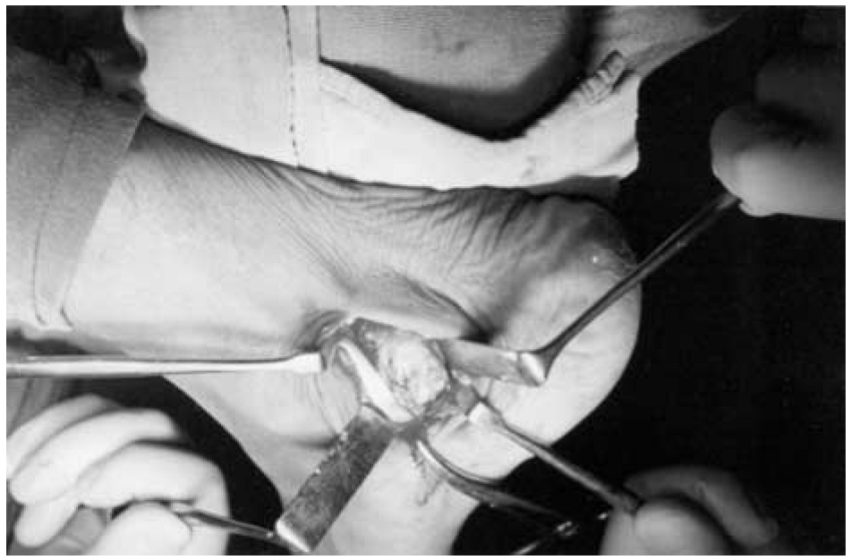
Needle biopsy was performed because histopathologic diagnosis was required before treatment could be planned. Histopathologic analysis revealed suspected osteochondroma, and surgical treatment was planned. The suspected osteochondroma was approached by medial incision and was found to be pressing on the tibialis posterior tendon (Fig. 2). Excisional biopsy along the overlying periosteum (to avoid recurrence) was performed. On histologic examination, a cartilaginous cap was seen covering the cancellous bone core, with a peripheral fibrous perichondrium. There was no sign of anaplasia. Thus a final diagnosis of osteochondroma, a rare entity in this part of the skeleton, was made.
The patient was followed up for 2 years. All of his symptoms subsided. He could now actively take part in sports and other outdoor activities. Repeated radiographs and clinical examinations have shown no evidence of recurrence. The postoperative power of the tibialis posterior muscle was normal. Dorsiflexion had improved to 20°.
Discussion
When an osteochondroma is present at an uncommon location such as the talus, it may be asymptomatic, or the only symptom may be a skeletal lump. Most pediatric patients with this condition are seen by a physician because their parents suspect a tumor. Rarely, the lesion may cause discomfort during exercise and limitation of joint motion. Very rarely, a large bursa with rapid accumulation of fluid may arouse suspicion of malignancy. Sometimes, as a result of traumatic fracture, an osteochondroma may become painful and mobile, clinically simulating myositis ossificans or a loose articular body.[11]
Solitary osteochondromas do occur in small bones, such as the talus, although they are rare in this part of the skeleton. Careful planning of surgery in all cases of benign-appearing lesions of small bones will prevent recurrence of this lesion and its complications.
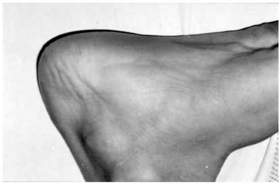
Figure 1.
Preoperative photograph showing a mass on the medial aspect of the ankle.
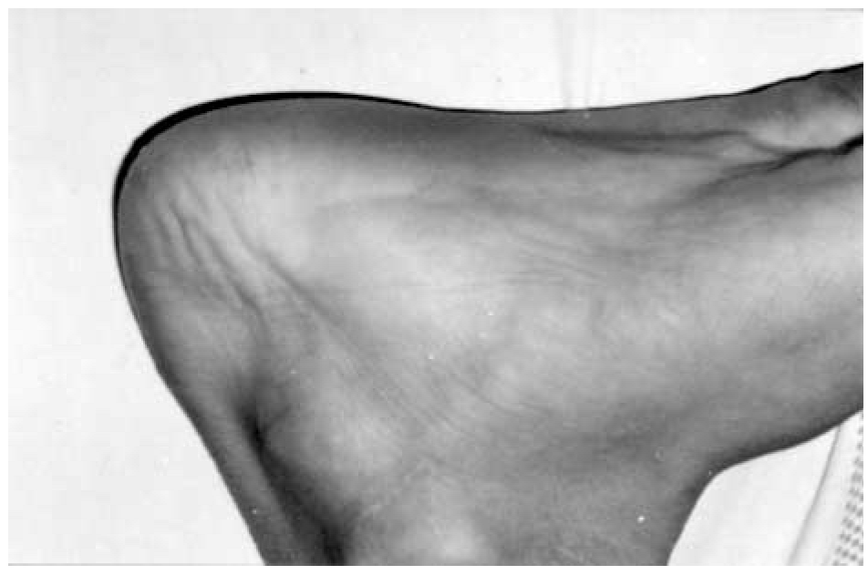
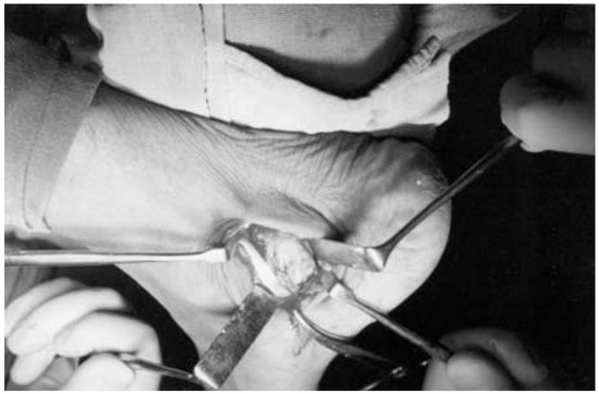
Figure 2.
Intraoperative photograph showing an osteochondroma of the talus.
References
- Kawai A, Mitani S, Okuda K, et al: Ankle tumor in a 5-year old boy. .Clin Orthop406::308. ,2003. .
- Schramm G: Pathogenesis of cartilaginous exostoses and enchondromas. .Arch Orthop27::421. ,1929. .
- Karakurun G, Onzur A, Atilla B: Bilateral peroneal tubercle osteochondroma of calcaneus: case report. .Foot Ankle Int19::259. ,1998. .
- Farrett WD, Stone PA, McGarry JJ: Rare presentation of hereditary multiple exostoses: a case report. .JAPMA88::135. ,1998. .
- Wilner D: “Osteochondroma,” in Radiology of Bone Tumors and Allied Disorders, p 305, WB Saunders, Philadelphia. ,1982. .
- Geschichter CF, Copeland MM: “Osteochondroma,” in Tumors of Bone, 3rd Ed, p 37, JB Lippincott, Philadelphia. ,1949. .
- Jaffe HI: Hereditary multiple exostosis. .Arch Pathol36::335. ,1943. .
- Bennet GE, Berkheimer GA: Malignant degeneration in a case of multiple benign exostoses. .Surgery10::781. ,1991. .
- Cruz-Conde R, Amaya S, Valdivia P, et al: Dysplasia epiphysealis hemimelica. .J Pediatr Orthop4::625. ,1989. .
- Kettelkamp DB, Campbell CJ, Bonfiglio M: Dysplasia epiphysealis hemimelica: a report of fifteen cases and a review of the literature. .J Bone Joint Surg Am48::746. ,1966. .
- Campanacci M: “Exostoses,” in Bone and Soft Tissue Tumors: Clinical Features, Imaging, Pathology, and Treatment, 2nd Ed, p 179, Piccin Nuova Libraría, Padova, Italy, and Springer-Verlag, Vienna and New York. ,1999. .
© 2005 American Podiatric Medical Association