


Abstract
We describe the management of a patient who presented to a family-practice clinic with gangrenous digits. After a thorough evaluation, she was found to have protein C deficiency, which produced a hypercoagulable state. Differential diagnosis in the evaluation of the coagulopathic patient with appropriate hematologic tests is briefly discussed. (J Am Podiatr Med Assoc 95(5): 491–493, 2005)
Coagulation is a complex process that requires a precise balance of many mechanisms. The thrombomodulin–protein C–protein S system is an important element of the anticoagulation arm of this process. Thrombomodulin enhances the activation of thrombin. Protein C is activated by thrombin to form a serine protease. Protein S is a required cofactor of protein C for the inactivation of factor Va. Specifically, protein C is a vitamin K–dependent proenzyme that exerts its anticoagulative properties by inactivating factors Va and VIIIa. Protein C also blocks receptors for factor Xa located on platelets. Hereditary or acquired deficiency states of protein C may predispose individuals to thrombosis, especially venous thrombosis of the lower extremity.[1] Arterial thrombosis seems to be rare, unless some other predisposing factor is present.[2] There is evidence of warfarin sodium–induced skin necrosis due to decreased protein C levels resulting from this drug.[3] The underlying pathology involves small dermal vessels filled with thrombi and overlying necrosis.[1
In 1998, Huseyin et al[4] reported progressive gangrene of the hand associated with hereditary deficiency of protein C. Onwuanyi et al[5] described a case of thromboembolism of the mesenteric artery. In their report, multiple thrombi associated with a protein C deficiency were subsequently found in the aorta and the right-sided heart chambers. Cho et al6] described two patients with peripheral arterial insufficiency associated with protein C deficiency. They noted that the exact incidence of protein C deficiency in patients with peripheral arterial insufficiency has not been established. Our report further underscores the importance of obtaining protein C and other coagulation factor levels in patients with dry gangrene without an obvious etiology.
Case Report
A 42-year-old woman presented to the John Peter Smith Primary Care Clinic, Fort Worth, Texas, with complaints of swelling of the face and legs. She was seen a month earlier at a different clinic for a left foot infection, at which time she was given antibiotics and was sent home. At the time of presentation to our institution, she was not taking medications for this infection. Her medical history was positive for hypertension of 4 years’ duration and iron-deficiency anemia of 9 years’ duration, for which she was taking iron supplements. The patient’s surgical history was positive for a cesarean section. She denied having allergies, previous hospitalizations, and trauma. Her family history was unremarkable, and her social history was positive for smoking (10 pack-years) and occasional alcohol consumption.
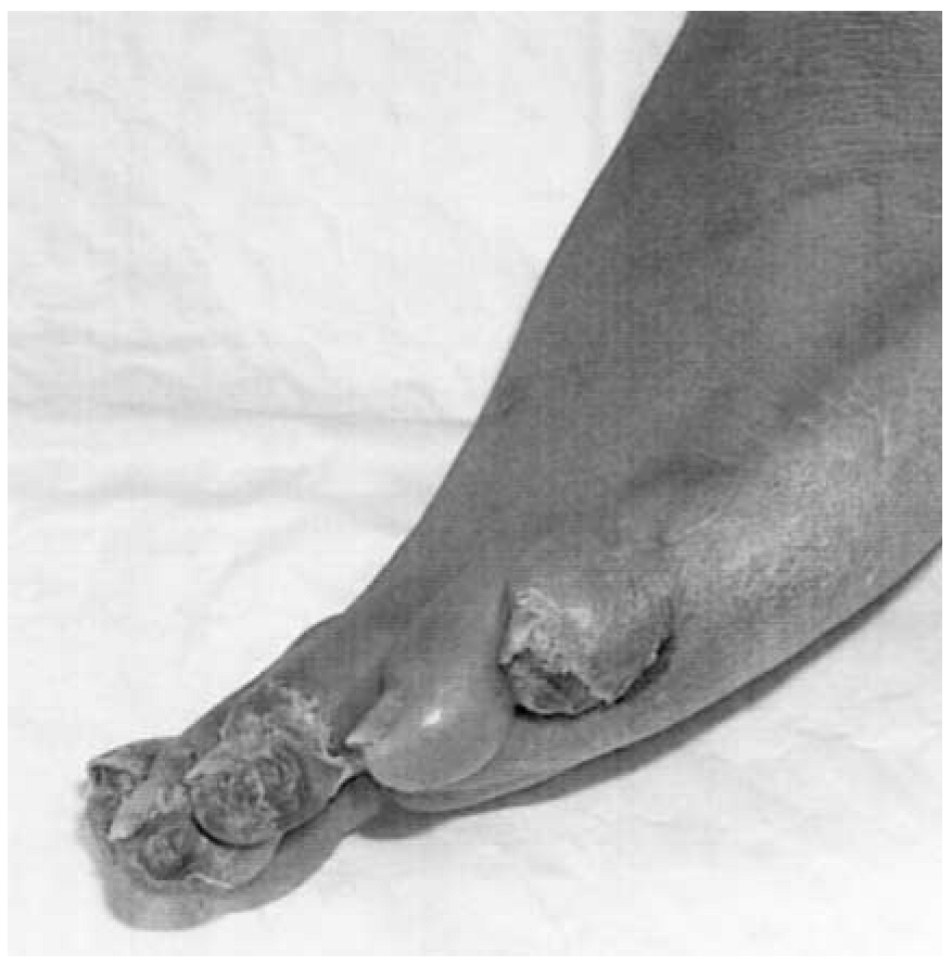
The initial physical examination revealed the following values: blood pressure, 138/68 mm Hg; pulse rate, 100/min; respirations, 16/min; temperature, 97°F; and weight, 60 kg. The conjunctivae and oral mucosa were found to be pale. The patient’s neck, heart, and lungs were unremarkable. Her abdomen was found to be distended but nontender, with no guarding, rebound, or fluid wave. The lower-extremity examination revealed 1 to 2+ pitting edema of the tibial tuberosity bilaterally. The left foot was not erythematous, but there was a dark, dry, and gangrenous appearance to digits 1, 2, 3, and 5 (Fig. 1). Homan’s sign was negative, no splinter hemorrhages were noted, and the extremities were nontender on palpation. The initial laboratory work-up included a complete blood cell count, chemistry panel, coagulation profile, and liver enzyme testing. The findings included the following values: hemoglobin, 6.3 g/dL; hematocrit, 21.5%; and sodium, 129 mmol/L. Prothrombin time was 12.4 sec, international normalized ratio was 1.070, and partial thromboplastin time was short at 24.0 sec. All other laboratory values were within normal ranges. The initial assessment by the internal medicine service was anemia secondary to iron deficiency, lower-extremity edema secondary to cardiac or liver disease, and gangrenous digits secondary to microvascular disease. The initial plan included an anemia work-up, evaluation of glycosylated hemoglobin (5.7%), urinalysis (unremarkable), and measurement of erythrocyte sedimentation rate (66 mm/h). A podiatric consultation for gangrenous digits was requested, and lower-extremity Doppler studies were ordered. The patient underwent a transfusion with 2 U of packed red blood cells, followed by 20 mg of intravenous furosemide therapy, and was supplemented with iron (325 mg by mouth 3 times daily).
On hospital day 2, a podiatric medical resident examined the patient and evaluated the lower-extremity Doppler studies. The patient denied any history of coagulopathy, immobility, arrhythmias, malignancy, oral contraceptive use, current pregnancy, sickle cell disease, venous thrombosis, or trauma. The distal aspects of digits 1, 2, 3, and 5 were black, dry, necrotic, and painful on palpation. Pedal pulses were found to be nonpalpable. Lower-extremity Doppler studies revealed a significant decline in segmental pressures of the left leg between the popliteal artery (144 mm Hg) and the dorsalis pedis artery (79 mm Hg). All of the veins compressed evenly and uniformly in both lower extremities. Protein C and S profiles were ordered, lower-extremity angiography was scheduled, and a surgical consultation was requested for possible revascularization.
On day 3, the white blood cell count increased to 14,496/μL, but the hemoglobin and hematocrit values stabilized after transfusion. Her iron level was found to be low (14 μg/dL), and her transferrin level was found to be within the normal range. The patient was given 3.375 g of piperacillin/tazobactam intravenously every 8 hours, and daily complete blood cell counts were ordered.
Protein C and S results were posted on hospital day 4. Protein C activity was low (61%), and protein S activity was within normal limits (79.4%). Antithrombin III activity was normal (90.7%). The aortogram with bilateral runoff revealed mild disease in the renal arteries and in the mid–left popliteal artery. There was two-vessel runoff from the trifurcation involving the anterior tibial and posterior tibial arteries, with good flow to the proximal left foot. Minimal flow was present to all digits of the left foot. On the right side, there was good flow to the anterior and posterior tibial arteries as well as to the entire foot and the toes. It was agreed among all specialties involved that an emergency vascular reconstruction was not warranted. The patient was administered an intravenous heparin drip for anticoagulation owing to a protein C deficiency that resulted in a hypercoagulable state.
The patient was treated in the hospital for 1 week with heparin and was discharged with long-term warfarin sodium therapy. After 3 days of intravenous antibiotic therapy, the patient’s white blood cell count returned to normal limits. Eventually, the patient underwent an echocardiogram that ruled out a cardiac etiology. Computed tomography of the chest, abdomen, and pelvis ruled out aortic or femoral thrombi. Antithrombin III, CIII, and CIV levels were found to be within normal limits. Antinuclear antibody testing was unremarkable. The lupus anticoagulant antibody and the factor V gene R506Q mutation test results were both negative.
The patient was lost to follow-up and was not evaluated again for 2 months, when she presented to the emergency department after autoamputation of the distal phalanx of the third digit. Digits 1, 2, and 5 had improved, with only a superficial crust remaining on the distal aspect of the hallux. There were no signs of acute infection, and laboratory values were within normal limits. The patient consented to amputation of the proximal phalanx of the third digit. Her amputation site healed without incident. Currently, the patient remains compliant with her iron supplements and warfarin sodium therapy.
Discussion
There are multiple etiologies of hypercoagulable states that must be considered when caring for a patient with dry gangrene or signs of vascular compromise (Table 1).[7] Although venous thromboembolism is the most frequent manifestation of protein C deficiency, arterial thrombosis is not uncommon. Protein C–deficient states have recently been associated with peripheral arterial disease and have been reported in numerous cases of myocardial infarction and stroke. In the event of idiopathic arterial or venous thromboembolism, protein C deficiency must be considered so that appropriate therapy can be instituted to prevent further sequelae or even death.

Table 1.
Hypercoagulable States
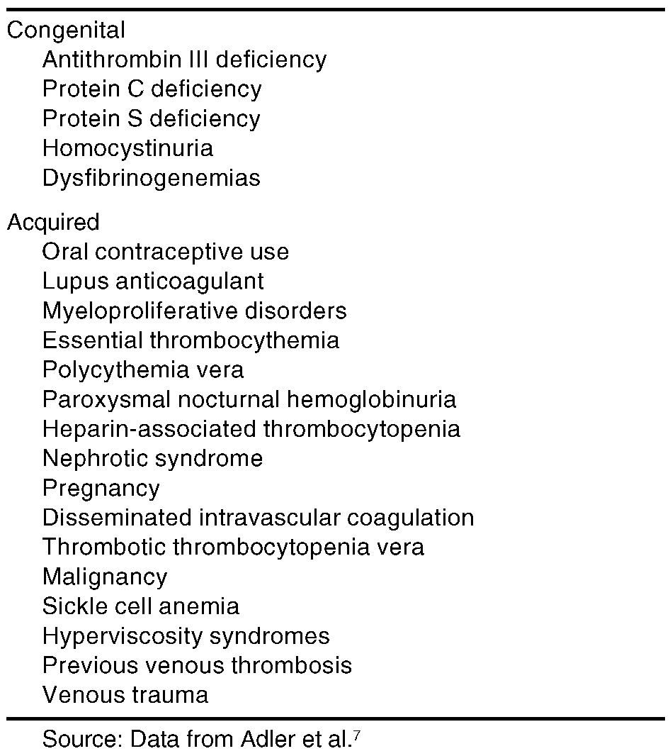
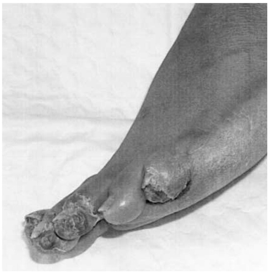
Figure 1.
Clinical appearance of gangrenous digits (lateral view).
References
- Nizzi FA Jr, Kaplan HS: Protein C and S deficiency. .Semin Thromb Hemost25::265. ,1999. .
- Sadiq A, Ahmed S, Karim A, et al: Acute myocardial infarction: a rare complication of protein C deficiency. .Am J Med110::414. ,2001. .
- Warkentin TE, Sikov WM, Lillicrap DP: Multicentric warfarin-induced skin necrosis complicating heparin-induced thrombocytopenia. .Am J Hematol62::44. ,1999. .
- Huseyin B, Dogan T, Anil A, et al: Progressive gangrene of the hand following extravasation of antibiotics associated with hereditary resistance to activated protein C. .Ann Plast Surg41::194. ,1998. .
- Onwuanyi A, Sachdeva R, Hamirani K, et al: Multiple aortic thrombi associated with protein C and S deficiency. .Mayo Clin Proc76::319. ,2001. .
- Cho YP, Lee DH, Jang HJ, et al: Peripheral arterial insufficiency associated with protein C deficiency. .Br J Radiol75::843. ,2002. .
- Adler SN, Adler-Klein D, Gasbarra DB: “Hematologic System,” in Pocket Manual of Differential Diagnosis, 4th Ed, p 223, Lippincott Williams & Wilkins, Philadelphia. ,1999. .
© 2005 American Podiatric Medical Association