


Abstract
Understanding the dynamic function of the rearfoot is necessary for recognizing and treating several types of mechanical foot dysfunction. Although the motion of the rearfoot is often measured during treadmill locomotion, the effect of different types of treadmills on the motion of the foot is unclear. In this study, the kinematics of the right subtalar joint in 24 volunteers walking at three speeds on two motorized treadmills were examined. The two treadmills (a wide width and a soft surface versus a narrow width and a hard surface) were selected to maximize motion differences. Maximal change in angular position (positive: supination; negative: pronation) about each volunteer’s subtalar joint axis was estimated during three gait phases: weight acceptance, midstance, and push-off. A factorial, repeated-measures analysis of variance determined that the treadmill design had a significant effect on subtalar joint position (F = 5.423; P = .029), albeit with moderate power (0.61). Descriptively, collapsed over all speeds, the subject’s feet on the narrow/hard compared with the wide/soft treadmill showed more pronation (0.44°), less pronation (0.46°), and more supination (1.44°) during weight acceptance, midstance, and push-off, respectively. We conclude that treadmill design can affect an individual’s rearfoot kinematics.
Understanding the anatomical structure and function of the foot during use is a necessary foundation for recognizing and treating mechanical foot dysfunction. Often, the dynamic function of the rearfoot, considered a key element of overall foot function, is measured during treadmill locomotion. [1-6] Although treadmills vary in running surface size, hardness, smoothness, power output, railing placement, and noise, the influence of different treadmills on the foot’s motion is unclear. Nigg [7] examined dynamic foot function in 22 subjects running on three different treadmills that varied in running surface size and power output. Substantial kinematic differences were reported for each subject on the different treadmills, but the differences depended on each individual subject’s running style and could not be consistently attributed to treadmill design. It was concluded that it is not yet understood how the human locomotor system adapts to a particular treadmill with specific design features.
Savelberg et al [8] closely examined the influence of intrastride treadmill belt-speed variations on locomotor kinematics. Examining nine volunteers walking and running on a high- and low-powered treadmill, they reported that the treadmill power rating significantly affected locomotor kinematics and coordination. They concluded that if a treadmill is used as a coordination or kinematics training device, the treadmill design will interfere with the aim of its use.
Many investigators have measured the frontal plane motion of the rearfoot (inversion/eversion) during treadmill locomotion in an attempt to understand the influence of foot orthoses [9] and pathology. [10] Recently, the motion of the rearfoot, partitioned into components about the anatomical talocrural and subtalar joint axes (dorsiflexion/plantarflexion and supination/pronation, respectively), has been explored because these measures are better representations of the true anatomical or functional motions within the rearfoot. [11,12] In none of these studies, however, were the design parameters of the treadmill considered with respect to its influence on rearfoot motion. Mechanically, it could be argued that an individual on a narrow treadmill, walking with a more in-line foot-placement pattern, would have increased supination/pronation owing to a longer lever arm system. Similarly, an individual walking on a hard treadmill would realize increased supination/pronation during weight acceptance as a result of a stiffer lever arm system. [7] Combining these two design features suggests that a narrow and hard treadmill surface would be associated with increased supination/pronation compared with a wide and soft treadmill surface.
The purpose of this study was to determine whether the design of a treadmill influences normal rearfoot kinematics during gait. Specifically, it was hypothesized that an individual walking on a narrow and hard compared with a wide and soft treadmill would demonstrate increased supination/pronation during weight acceptance. In addition, changes in supination/pronation within the gait cycle (midstance and push-off) and at various walking speeds were explored. Such data would help with the assessment of patients with mechanical rearfoot dysfunction.
Materials and Methods
A three-dimensional (3-D) kinematic data–acquisition system (OptoTrak; Northern Digital Inc, Waterloo, Ontario, Canada) was used to measure the motion of 24 subjects’ instrumented right lower leg and rearfoot while they stood quietly, circumducted the foot, and walked on a treadmill. These data were analyzed to provide the true supination/pronation about each subject’s estimated subtalar joint axis.
Subjects
We tested a convenience sample of 24 university undergraduate and graduate students, faculty members, and staff (12 men and 12 women; height, 1.50–1.85 m; mass, 48–88.9 kg). Their current foot orthoses use (n = 6), current ankle injuries (none), and previous rearfoot injuries (two ankle sprains and three fractures) were recorded, and each subject signed a consent form approved by the Hamilton Health Sciences/McMaster University ethics review board. Each subject wore shorts and was barefoot.
Instrumentation and Reference System Definition
The 3-D position of each subject’s right rearfoot was measured using a kinematic data–acquisition system. This device records the 3-D spatial location of special markers (infrared light–emitting diodes, <1 g) affixed to an object of interest. All data-capture rates were set at 50 Hz.
Knowing the location of three or more markers on an object at a given instant in time provides enough information to calculate the orientation and position of that object. [13] If the orientation and position of two objects are known, one can calculate the orientation and location of one of the objects in relation to the other [14]; therefore, with appropriate numeric-processing techniques, the 3-D positions of the markers can provide the orientation and position of the lower leg relative to the rearfoot, irrespective of rearfoot position.
For this study, four markers were placed on the lower leg (approximately 5 cm proximal to the medial and lateral malleoli and over the Achilles tendon, approximately 10 and 20 cm proximal to its insertion), and four markers were placed on the rear aspect of the calcaneus in a 10-cm diamond configuration to define a rigidly modeled lower leg and rearfoot. [15] For each subject, the 3-D positions of each of these eight markers, collected for 5 sec, were averaged while the subject stood with his or her feet positioned in a frame that held the medial foot borders parallel and 8.5 cm apart. In this manner, two local coordinate systems were defined, one for the lower leg and the other for the rearfoot, with axes directed laterally, anteriorly, and vertically.
Each subject was then instructed to circumduct the foot, making sure that an adequate range of dorsiflexion/plantarflexion and supination/pronation was achieved. This trial was processed to obtain an estimate of the orientation and location of the talocrural and subtalar joint axes relative to the surface markers. A modification of the two-axis model method presented by van den Bogert et al [12] was used to specify these joint axes.
Knowing the locations and orientations of the talocrural and subtalar joint axes relative to the surface markers, whose positions during the walking trial were known relative to the laboratory-fixed reference system, allowed calculation of the time-varying talocrural dorsiflexion/plantarflexion and subtalar supination/pronation motion patterns. For this study, only the supination/pronation patterns were of interest.
Treadmills
Two different treadmills were used in this study: the True S.O.F.T System 500 (True Fitness Technology, O’Fallon, Missouri) and the Quinton model 4-44B (Quinton Instruments, Seattle, Washington). Both treadmills had a 2.2-kW motor. The True treadmill had a soft and even running surface and was wider (50.5 cm), whereas the Quinton treadmill was harder and felt “bumpy” and was narrower (34.5 cm). In this article, the True and Quinton treadmills will be referred to as the wide/soft and narrow/hard treadmills, respectively.
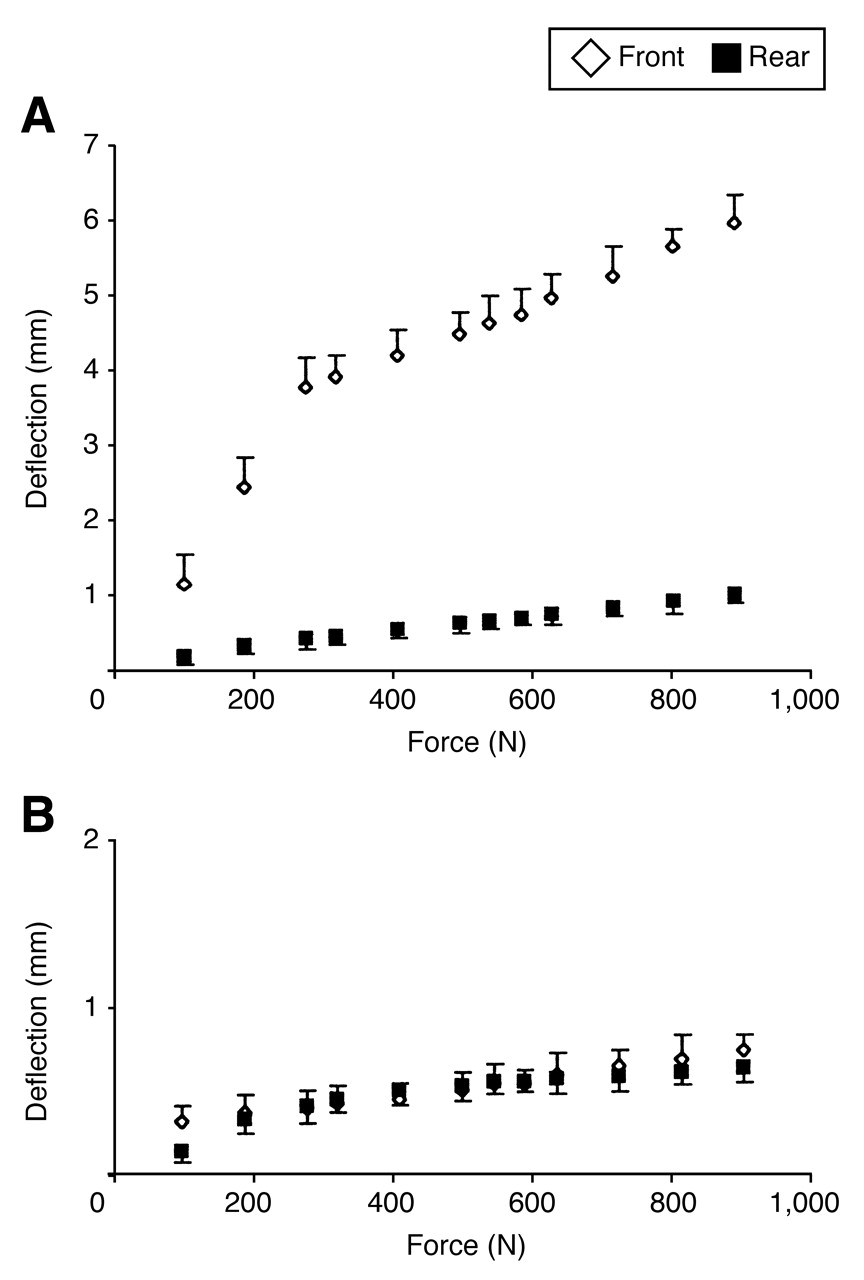
To quantify the “softness” of the wide/soft and narrow/hard treadmills, static load versus vertical deflection data were collected. Twelve static loads ranging from 90.5 to 905 N were applied to the front and back of the treadmill belts. These locations were selected to correspond to the approximate locations where the foot made contact with the treadmill belt during weight acceptance and push-off. The downward deflection of the treadmill was recorded using the kinematic data–acquisition system (Fig. 1). These data demonstrate that the softness of the wide/soft and narrow/hard treadmills is similar, except that the front of the wide/soft compared with the narrow/ hard treadmill is approximately ten times softer.
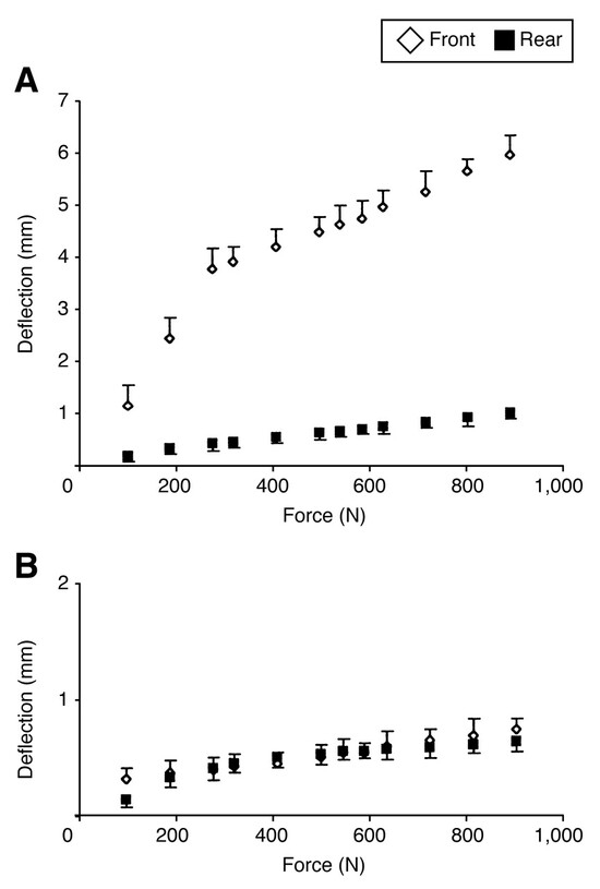
Figure 1.
Mean downward deflection of the wide/soft (A) and narrow/hard (B) treadmills to a set of static loads (force) applied to the front (average foot weight-acceptance location) and rear (average foot push-off location) of their belts. The narrow/hard treadmill is approximately ten times stiffer than the wide/soft treadmill during weight acceptance (treadmill front; 1,000-N load). Error bars represent SEM.
Walking Trials
Before any formal testing, each subject walked for 5 to 8 min on each treadmill until he or she was walking comfortably (per self-report and as assessed by visual observation). Each subject then walked three times on each treadmill, set at three different velocities (0.66, 0.89, and 1.11 m/sec). For each subject, the treadmill and velocity presentation order were randomized to minimize the influence of a potential learning effect on the results. During these six walking trials, the 3-D motions of the eight surface markers were captured for 30 sec. In this manner, approximately 25 complete walking cycles for each subject on each treadmill and at each velocity were collected. The use of a treadmill easily allowed the collection of many gait cycles to provide better estimates of a subject’s true motion pattern. [16] Use of the vertical motion of the inferior rearfoot marker indicated the time of rearfoot contact with the floor for each subject during the walking trials. It was, therefore, possible to average the multiple gait cycles for each subject’s walking trial. In this manner, mean ± 1 SD curves, defined from right rearfoot–ground contact to the next right rearfoot–ground contact, normalized to 100% of the gait cycle were generated for each subject.
To determine whether the treadmill influenced the subject’s rearfoot motion pattern during gait, critical phases of the gait cycle were selected for analysis. A priori it was decided that differences were meaningful only during the weight-acceptance (0%–12% of the gait cycle), midstance (>12%–42% of the gait cycle), and push-off (>42%–60% of the gait cycle) phases. During these phases, the treadmill and foot are in direct contact, and the treadmill may influence the foot’s motion. To quantify the effect of the treadmill on foot motion, the changes in each subject’s supination/ pronation angle during the aforementioned gait cycle phases were calculated. Positive values indicate increased rearfoot position toward supination.
Statistical Analyses
Initially, descriptive statistics provided summary data and were used to check whether the data were normally distributed. Next, a four-factor (sex, phase, treadmill type, and speed) analysis of variance (ANOVA) with repeated measures on the last three factors was used to examine whether the supination/pronation values exhibited statistically significant differences. The ANOVAs were performed at the 5% level of significance. Post hoc analyses (least significant difference, P < .05) were then performed to further investigate whether the rearfoot kinematics exhibited statistically significant differences between the two treadmills at similar speeds and stance phases. These calculations were performed using SPSS version 10.1 for Windows (SPSS Science, Chicago, Illinois).
Results
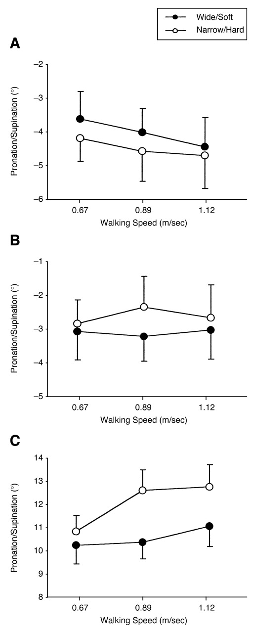
The four-way ANOVA revealed that the main effect “sex” was not statistically significant (F1,22 = 1.705; P = .21) and did not participate in any interaction effects; therefore, a three-way repeated-measures ANOVA, omitting the main effect “sex,” was performed (Table 1). This analysis found that the main effects “phase” and “treadmill type” and the interaction effects “phase × treadmill type” and “phase × speed” were significant. The main effect “speed” was not significant (F = 1.065; P = .36). A post hoc analysis of the main effect “phase” determined that each of the three phases was significantly different from the others (means: −4.2°, −2.9°, and 11.3°; least significant difference test, P < .001). As hypothesized, treadmill design had a significant effect on supination/pronation (F = 5.423; P = .03), albeit with moderate power (0.61). Specifically, collapsed over all speeds, the subjects’ feet on the narrow/hard compared with the wide/soft treadmill showed more pronation (0.44°), less pronation (0.46°), and more supination (1.44°) during the weight-acceptance, midstance, and push-off gait phases, respectively (Fig. 2).

Table 1.
Repeated-Measures Analysis of Variance Examining the Effect of Phase, Treadmill Type, and Speed on Rearfoot Supination/Pronation
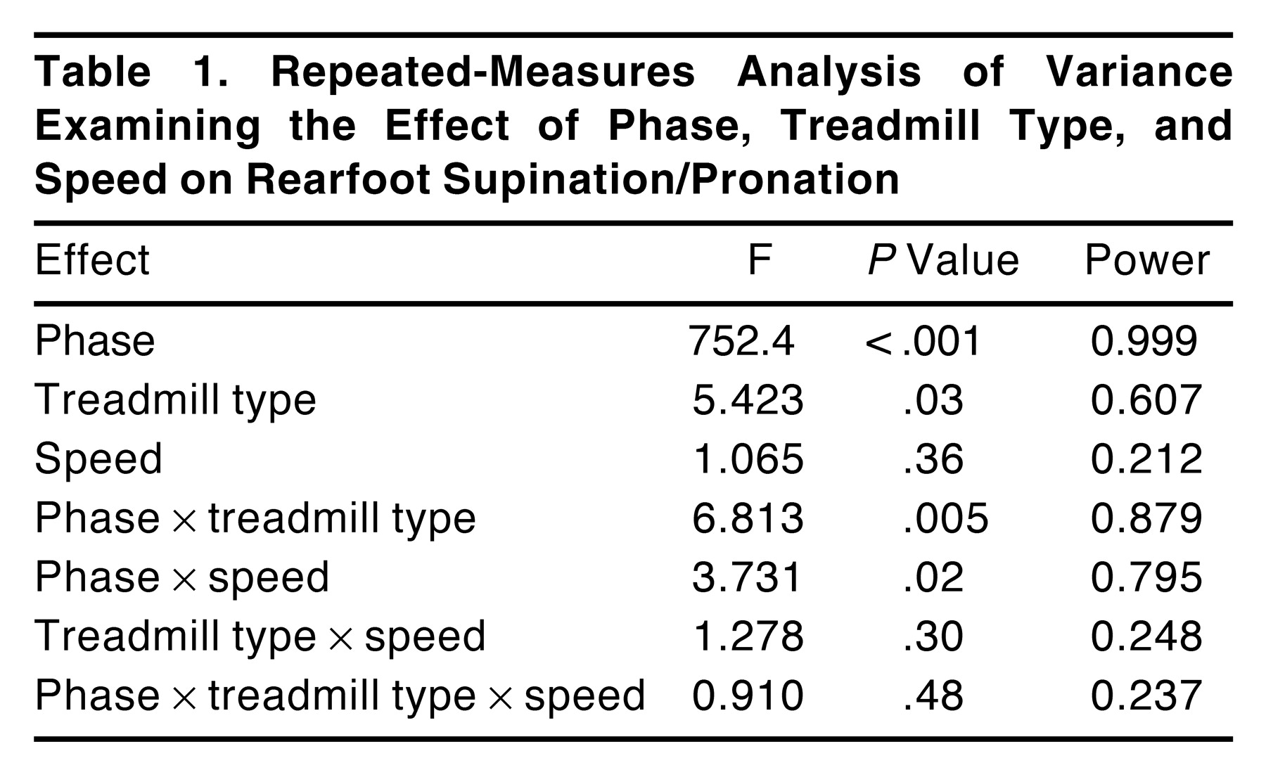
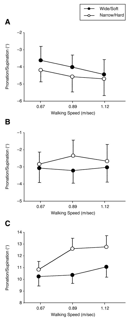
Figure 2.
Effect of walking speed and treadmill design on mean rearfoot pronation (negative) or supination (positive) during the weight-acceptance (A), midstance (B), and push-off (C) phases of the gait cycle. Error bars represent SEM.
Discussion
Clinicians often use a motorized treadmill to help them observe the motion of the foot during gait. Atypical rearfoot motion patterns are sometimes interpreted as indicative of an underlying mechanical foot dysfunction requiring intervention. Rarely, however, has the treadmill been considered to contribute to these altered rearfoot patterns. In this study, we observed small-magnitude but statistically significantly increased rearfoot pronation during weight acceptance in volunteers walking on a narrow/hard versus a wide/soft treadmill.
This study was designed to examine the influence of treadmill design on rearfoot kinematics. It was hypothesized that volunteers walking on a narrow and hard surface would exhibit increased rearfoot pronation. First, subjects may have altered their gait patterns owing to the visually perceived differences in the treadmill belt width. The primary sensory modality used for adapting the gait pattern is vision, and it is the visual system that allows us to make anticipatory adjustments to the gait patterns. [17] Although subjects were asked to walk with their heads up and to maintain their gaze facing forward throughout the trials, they may have thought that the narrow belt width required a gait pattern with a narrow gait width. This narrow gait width directly leads to increased supination/pronation. Second, volunteers walking on a hard surface have increased supination/pronation. The design of the narrow/hard treadmill was a consecutive set of hard and unyielding rollers. These stiff rollers did not attenuate the force applied to the foot during gait. Given the same treadmill-to-foot force lever arm (point of force application relative to the subtalar joint axis), the applied moment of force would be greater, which would lead to increased supination/ pronation.
This study determined that treadmill design influences rearfoot kinematics during gait. Each volunteer dynamically adapted his or her gait pattern to a new preferred pattern on the two treadmills used in this study. Although the rationale behind the selection of the new neuromusculoskeletal control pattern objective is presently unknown, one possibility may be offered. Donelan et al, [18] who examined the influence of gait width on locomotion economy, reported that any deviation from a preferred gait pattern has an elevated metabolic cost. However, Krebs et al [19] observed that patients with an unsteady gait pattern increase their gait width at the expense of metabolic cost. Although globally this inefficient gait pattern is unwelcome, it may be that the system is attempting to maximize stability at the expense of metabolic economy. Further investigations using different treadmill designs may have the potential to characterize the relationships among preferred motor control patterns, stability, and economy.
Although our data support the hypothesis that walking on a narrow and hard treadmill surface increases rearfoot supination/pronation during stance, the design of our experiment could not independently examine the influence of treadmill width or hardness. This limitation was partially dictated by feasibility issues such as treadmill availability. Further investigations that explore the influence of treadmill width and hardness, individually and in combination, have the potential to suggest design parameters to beneficially modify rearfoot kinematics during gait. Furthermore, because treadmills are frequently used for prolonged training sessions at running speeds, the interaction of width, hardness, speed, and test duration may lead to designs that permit a more typical pattern of rearfoot motion to be observed or recorded.
Conclusion
In this study, healthy volunteers walking at three different speeds on two different treadmills were tested to determine whether they had similar rearfoot kinematics. Statistical examination showed that the volunteers walked differently on the narrow and hard treadmill compared with the wide and soft treadmill. We conclude that a volunteer’s rearfoot kinematics can be altered by the design of the treadmill and that if clinical decisions depend on small rearfoot angular changes, the treadmill effect must be noted.
References
- Lemke K, Cornwall MW, McPoil TG, et al: Comparison of rearfoot motion in overground versus treadmill walking. .JAPMA85::243. ,1995. .
- Nilsson J, Thorstensson A: Adaptability in frequency and amplitude of leg movements during human locomotion at different speeds. .Acta Physiol Scand129::107. ,1987. .
- Pierrynowski MR, Smith SB: Rear foot inversion/eversion during gait relative to the subtalar joint neutral position. .Foot Ankle Int17::406. ,1996. .
- Stolze H, Kuhtz-Buschbeck JP, Mondwurf C, et al: Gait analysis during treadmill and overground locomotion in children and adults. .Electroencephalogr Clin Neurophysiol105::6. ,1997. .
- White SC, Yack HJ, Tucker CA, et al: Comparison of vertical ground reaction forces during overground and treadmill walking. .Med Sci Sports Exerc30::1537. ,1998. .
- Yamasaki M, Sasaki T, Torii M: Sex difference in the pattern of lower limb movement during treadmill walking. .Eur J Appl Physiol62::2. ,1991. .
- Nigg BM: Biomechanics of Running Shoes, Human Kinetics Publishers, Champaign, IL. ,1986. .
- Savelberg HH, Vorstenbosch MA, Kamman EH, et al: Intra-stride belt-speed variation affects treadmill locomotion. .Gait Posture7::26. ,1998. .
- Brown GP, Donatelli R, Catlin PA, et al: The effect of two types of foot orthoses on rearfoot mechanics. .J Orthop Sports Phys Ther21::258. ,1995. .
- McClay I, Manal K: A comparison of three-dimensional lower extremity kinematics during running between excessive pronators and normals. .Clin Biomech13::195. ,1998. .
- Siegler S, Chen J, Schneck CD: The three-dimensional kinematics and flexibility characteristics of the human ankle and subtalar joints: part I. Kinematics. .J Biomech Engineer110::364. ,1988. .
- van den Bogert AJ, Graham DS, Nigg BM: In vivo determination of the anatomical axes of the ankle joint complex: an optimization approach. .J Biomech27::1477. ,1994. .
- Veldpaus FE, Woltring HJ, Dortmans LJ: A least-squares algorithm for the equiform transformation from spatial marker co-ordinates. .J Biomech21::45. ,1988. .
- Soderkvist I, Wedin PA: Determining the movements of the skeleton using well-configured markers. .J Biomech26::1473. ,1993. .
- Pierrynowski MR, Smith SB: Effect of patient position on the consistency of placing the rearfoot at subtalar neutral. .JAPMA87::399. ,1997. .
- Taylor JR: An Introduction to Error Analysis, University Science Books, Mill Valley, CA. ,1982. .
- Patla AE: “Visual Control of Human Locomotion,” in Adaptability of Human Gait: Implications for the Control of Locomotion, Elsevier Science Publishers, New York. ,1991. .
- Donelan JM, Kram R, Kuo AD: Mechanical and metabolic determinants of the preferred step width in human walking. .Proc R Soc Lond B Biol Sci268::1985. ,2001. .
- Krebs DE, Goldvasser D, Lockert JD, et al: Is base of support greater in unsteady gait?. Phys Ther82::138. ,2002. .
© 2005 American Podiatric Medical Association