


Abstract
A prospective analysis was conducted of the amount of correction obtained and number of complications resulting from 31 endoscopic gastrocnemius recessions in 28 patients. The average amount of increase in ankle dorsiflexion was 18°. Few complications were encountered, with mild “soreness” and distal “bruising” being reported by four patients. The only other complaint was “weakness.” The endoscopic gastrocnemius recession is a minimally invasive technique with a low rate of complications that offers a comparable amount of correction to that of traditional open procedures in the treatment of gastrocnemius equinus.
Gastrocnemius equinus develops when there is contracture or shortening of the gastrocnemius muscle. A Silfverskiöld test may be performed to differentiate between gastrocnemius equinus and other types of posterior equinus.[1] With the patient in a supine position, the examiner extends and flexes the patient’s knee and passively dorsiflexes the ankle with the subtalar joint in a neutral position and the midtarsal joint adducted. The sagittal plane relationship of the bisection of the leg to the rearfoot is measured. Because the gastrocnemius muscle crosses the knee joint and the soleus does not, the gastrocnemius is shortened when the knee is flexed. Gastrocnemius equinus is present if there is less than 10° of dorsiflexion with the knee extended and an increase in ankle joint range of motion with the knee flexed. According to Root,[2] “ten degrees of dorsiflexion range of the foot at the ankle is required when the knee extends, just before heel lift, in the stance phase of gait.” DiGiovanni et al[3] suggested that “the gastrocnemius muscle and paratenon is the predominant deforming force in patients with structural breakdown and chronic pathological changes related to the foot and ankle.” Subotnick[4] stated that “gastrocnemius or soleus equinus is the greatest symptom producer in the human foot.”
Conservative treatment for equinus deformity has consisted of stretching exercises, orthoses, padding, molded footwear, and, in younger patients, serial casting or bracing. Surgery is indicated if conservative treatment fails. Various surgical procedures have been described for correction of gastrocnemius equinus.[5]-[10] Spastic gastrocnemius equinus has been treated with selective neurectomy of branches of the tibial nerve supplying the gastrocnemius muscle. Other procedures have consisted of proximal release of the muscular heads of the gastrocnemius and distal recession of the gastrocnemius aponeurosis. The distal gastrocnemius recession should be performed for correction of nonspastic gastrocnemius equinus. We describe an endoscopic technique for performing a gastrocnemius recession for nonspastic gastrocnemius equinus.
Materials and Methods
Endoscopic gastrocnemius recession was performed on 31 lower extremities in 28 people (surgery was performed bilaterally in 3 people). The average age of the 11 males and 17 females was 41 years (range, 10–83 years). The degree of ankle joint dorsiflexion was measured preoperatively using a standard tractograph (average: −9°). Two lines were placed along the lateral lower extremity and foot to serve as reference points. One line was drawn from the midshaft of the fibula along the distal one-third and the other along the plantarmost aspect of the calcaneus to prevent measuring errors from inadvertent dorsiflexion at the Lisfranc and midtarsal joints (Fig. 1). Measurements were taken using the reference lines with the subtalar joint in neutral and the midtarsal joints adducted. Passive range-of-motion measurements were taken after anesthesia induction and performed twice, with an average value calculated. After completion of the procedure, the measurement process was repeated and an average value calculated.
Surgical Procedure
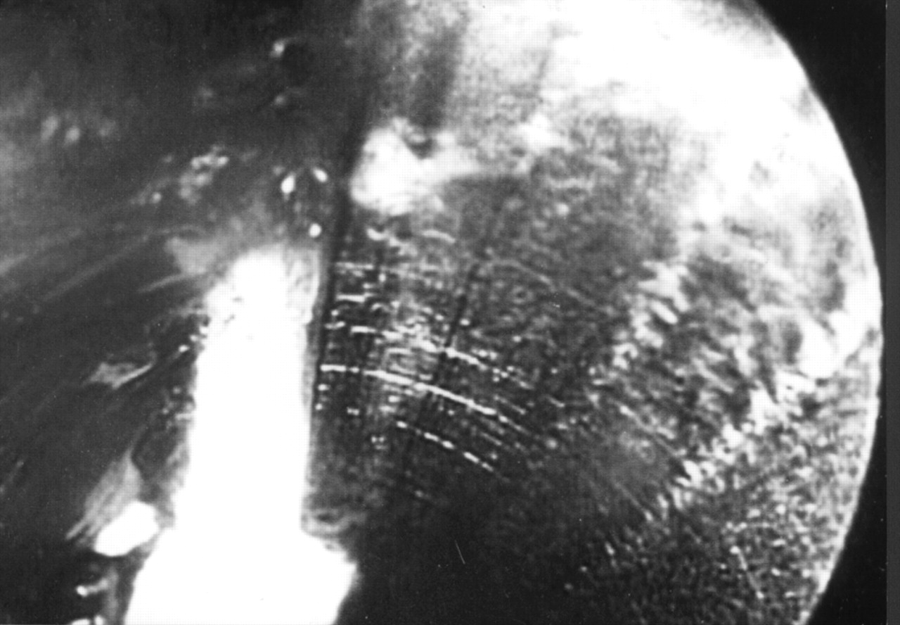
The patient is placed in a supine position and administered general anesthesia. The gastrocnemius soleus complex just inferior to the gastrocnemius muscle bellies is palpated for orientation. A small stab incision is made posteromedially with a No. 15 blade, and a blunt hemostat is used to dissect down to the gastrocnemius paratenon. A blunt trochar is introduced into the medial incision, with care taken not to disrupt the saphenous nerve or vein (Figs. 2 and 3). A distinction between the deep fascia and the posterior aspect of the gastrocnemius is made, and the blunt trochar is advanced laterally through the deep tissue, “tenting” the skin at the lateral aspect of the leg. A small stab incision is then made posterolaterally with a No. 15 blade to allow for further advancement of the trochar through the skin. An obturator, with the barrel oriented laterally, is placed over the trochar and swiveled to allow advancement medially through the skin (Fig. 4). The slotted aspect of the cannula faces anteriorly, allowing the gastrocnemius aponeurosis to be seen. The endoscopic camera is placed into the lateral portal to allow visualization of the entire gastrocnemius complex (Fig. 5). The fibers of the gastrocnemius aponeurosis can be viewed while plantarflexing and dorsiflexing the ankle. Care must be taken so that the gastrocnemius aponeurosis is the only structure visualized, ensuring that the sural nerve and other neurovascular structures are free from transection. A triangle blade is introduced into the medial portal and used to puncture the gastrocnemius paratenon on the lateralmost edge. The hook blade is inserted into the puncture hole through the medial portal. With the ankle dorsiflexed, the gastrocnemius is released (Fig. 6). At this time, it is important to ensure that the longitudinal bands of the intramuscular septum are also released, as this will generally offer a great deal of additional correction. Caution must be exercised to prevent the blade from violating the underlying muscle. Once adequate release is attained, all equipment is removed from the leg and the skin is closed with a few simple interrupted sutures. Postoperatively, patients are allowed immediate weightbearing in a CAM Walker (Zinco Industries, Inc, Pasadena, California) provided that no other procedures that require nonweightbearing have been performed.
Results
The average amount of preoperative dorsiflexion was −9° (range, 0° to −18°). Postoperatively, dorsiflexion averaged 9° (range, 0° to 22°), for an average increase of 18° (range, 11° to 32°). Few complications were observed. One patient developed a hematoma secondary to soleal bleeding from inadvertent transection, which resolved with compression; three patients subjectively complained of weakness for several months following the procedure; and one patient had pain postoperatively for a short period that resolved with elevation and administration of pain medication.
Discussion
Our results show that an average of 18° of correction can be achieved intraoperatively using the endoscopic technique described here. These results are similar to those reported using the traditional open techniques with long-term follow-up.[11]-[13] Sgarlato[14] suggests that “every centimeter of gapping yields approximately 10–15 degrees of increased ankle sagittal flexibility.” Previous techniques described for gastrocnemius recession involve opening up the posterior compartment to visualize the gastrocnemius. This usually requires careful and tedious dissection to avoid disrupting the saphenous vein or sural nerve. Tashjian et al[15] reported “15% injury to the sural nerve while performing the endoscopic gastroc recession on cadavers.” With the exception of four procedures involving the uniportal approach, our technique used two small stab incisions on each side of the gastrocnemius aponeurosis, thus providing better cosmesis, direct visualization, and earlier return to function than the open approach. This makes this procedure ideal for children, people who want to wear clothing that exposes their legs for cosmetic reasons, patients with diabetes, and other patients with mild peripheral vascular disease. The remaining four procedures were performed using a uniportal approach medially as described by Saxena,[16] with equal degrees of correction observed. We have shown that the endoscopic gastrocnemius recession procedure can provide successful results with a low rate of complications. This procedure can be done solely or adjunctively with other procedures.
Conclusion
We have described a technique for performing a gastrocnemius recession using an endoscopic approach. This approach allows for better cosmesis, direct visualization, and earlier ambulation compared with traditional open procedures. Our results indicate that an average of 18° of correction in ankle joint dorsiflexion can be achieved. The endoscopic gastrocnemius recession is a useful, minimally invasive alternative to open gastrocnemius recession procedures.
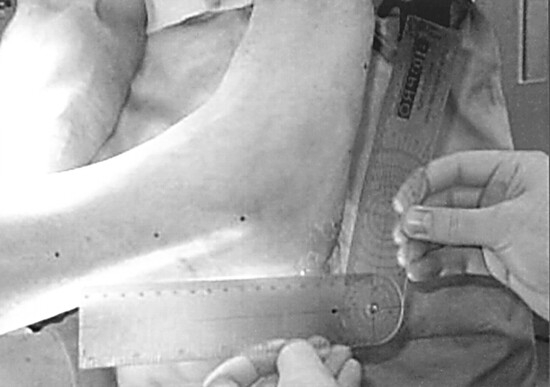
Figure 1.
Technique for measuring preoperative and postoperative dorsiflexion.
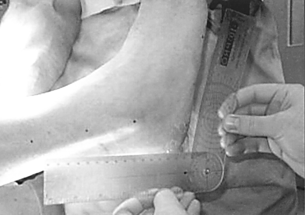
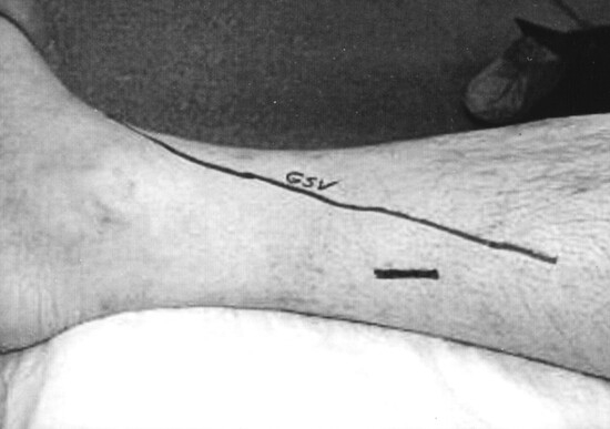
Figure 2.
Identification of vital structures and incision placement.
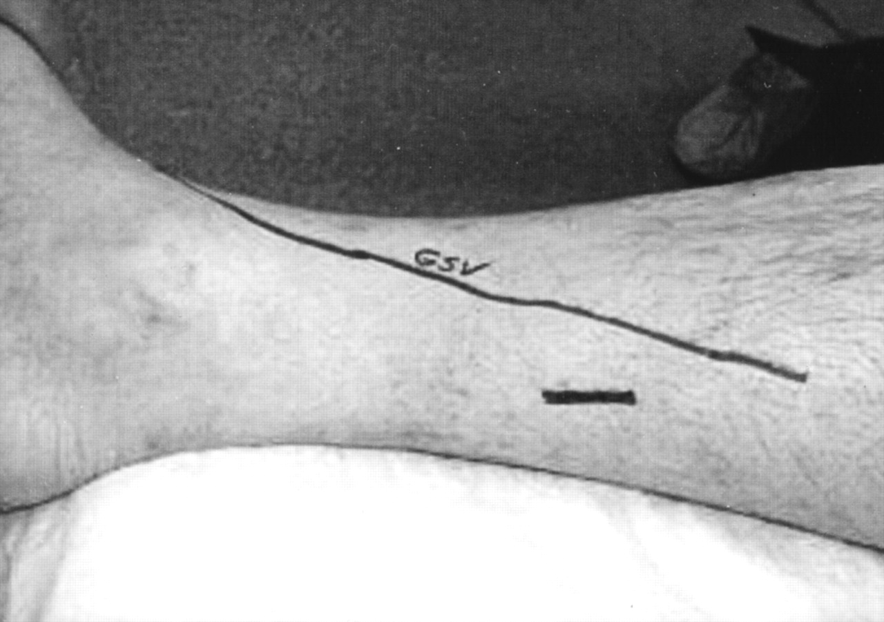
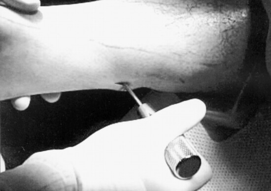
Figure 3.
Insertion of blunt trochar medially.
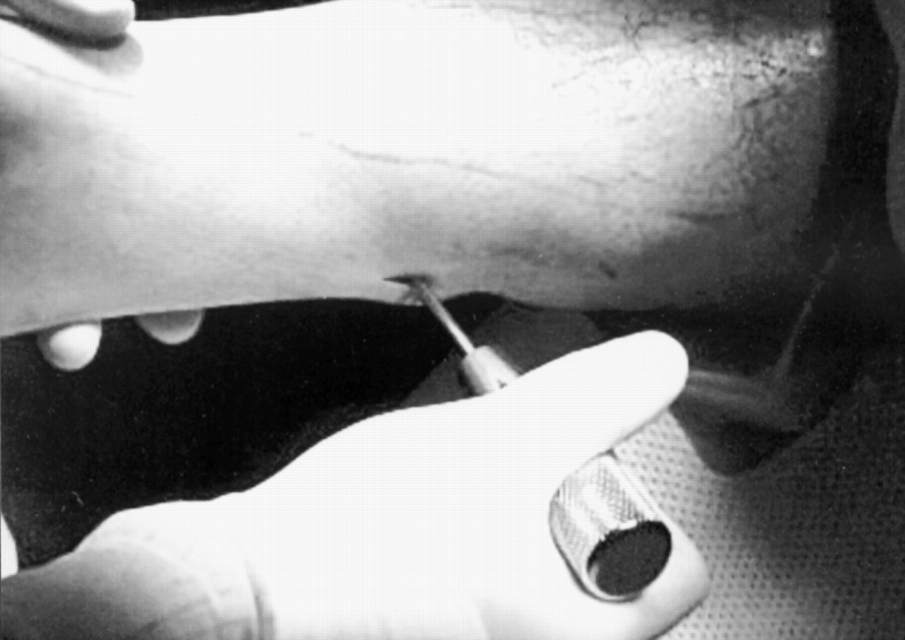
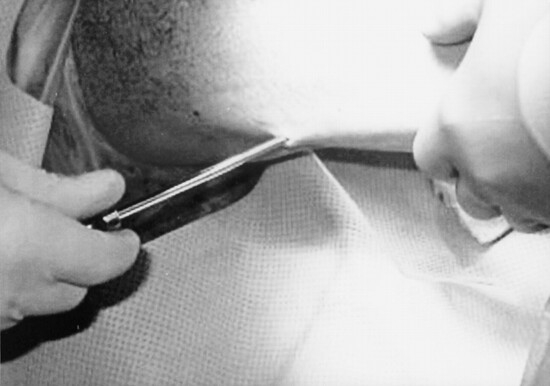
Figure 4.
Insertion of obturator over trochar laterally.
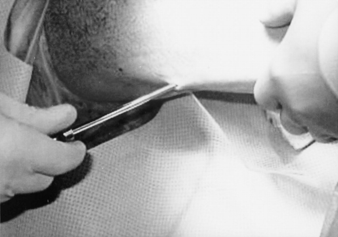
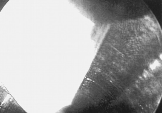
Figure 5.
Endoscopic visualization showing gastrocnemius complex.
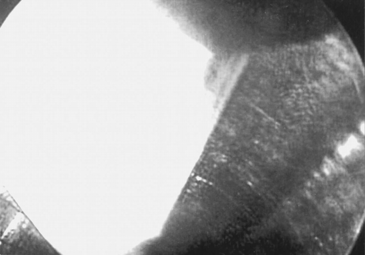
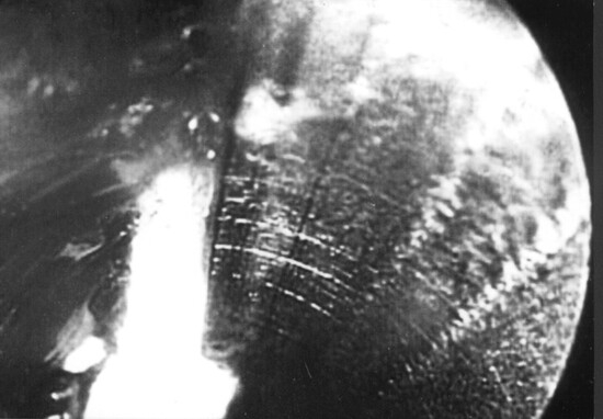
Figure 6.
Endoscopic visualization showing complete release of gastrocnemius complex.
References
- Silfverskiold N: Reduction of the uncrossed two-joint muscles of the leg to one-joint muscles in spastic conditions. Acta Chir Scand56: 315, 1924.
- Root ML: Normal and Abnormal Function of the Foot, p 37, Clinical Biomechanics Corp, Los Angeles, 1977.
- DiGiovanni CW, Kuo R, Tejwani N, et al: Isolated gastrocnemius tightness. J Bone Joint Surg Am84: 962, 2002.
- Subotnick SI: Equinus deformity as it affects the forefoot. JAPA61: 423, 1971.
- Downey MS: “Ankle Equinus,” in Comprehensive Textbook of Foot Surgery, 2nd Ed, ed by ED McGlamry, AS Banks, MS Downey, p 687, Williams & Wilkins, Baltimore, 1992.
- Pinney SJ, Hansen ST, Sangeorzan BJ: The effect on ankle dorsiflexion of gastrocnemius recession. Foot Ankle Int23: 26, 2002.
- Vulpius O, Stoffel A: Orthopadische Operationslehre, p 29, Ferdinand Enke, Stuttgart, 1913.
- Strayer LM: Recession of the gastrocnemius: an operation to relieve spastic contracture of the calf muscles. J Bone Joint Surg Am32: 671, 1950.
- Baker LD: A rational approach to the surgical needs of the cerebral palsy patient. J Bone Joint Surg Am38: 313, 1956.
- Fulp MJ, McGlamry ED: Gastrocnemius tendon recession: tongue in groove procedure to lengthen gastrocnemius tendon. JAPA64: 163, 1974.
- Strayer LM: Gastrocnemius recession: five-year report of cases. J Bone Joint Surg Am40: 1019, 1958.
- Martz CD: Talipes equinus correction in cerebral palsy. Am J Orthop42: 769, 1960.
- Downey MS, Banks AS: Gastrocnemius recession in the treatment of nonspastic ankle equinus: a retrospective study. JAPMA79: 159, 1989.
- Sgarlato TE: Medial gastrocnemius tenotomy to assist body posture balancing. J Foot Ankle Surg37: 546, 1998.
- Tashjian RZ, Appel AJ, Banerjee R, et al: Endoscopic gastrocnemius recession: evaluation in a cadaver model. Foot Ankle Int24: 607, 2003.
- Saxena A: Endoscopic gastrocnemius tenotomy. J Foot Ankle Surg41: 57, 2002.
© 2005 American Podiatric Medical Association