


Abstract
Cutaneous larva migrans is a common skin pathology that occurs in people who have recently visited tropical or subtropical climates. Given the ubiquity of this condition, the podiatric physician may encounter cutaneous larva migrans during clinical practice and should be cognizant of the presenting signs and typical patient history given in these cases. We describe the case of a 62-year-old man who presented with a pruritic, erythematous, serpiginous lesion on the dorsum of his left foot after having vacationed in Florida for several weeks. The patient was treated successfully with oral thiabendazole, 500 mg after meals 4 times daily for 5 days.
Cutaneous larva migrans, first described in 1874 as “creeping eruption,” is a ubiquitous and self-limited skin condition caused by infection with the larvae of mammalian hookworms [1-6]. The skin lesions of cutaneous larva migrans often develop within 2 weeks of a patient’s return from a subtropical or tropical environment, where the patient’s history typically reveals exposure to soil or sand at a beach or to domestic animals [6]. Domestic animals, such as dogs and cats, frequently serve as hosts, although many wild mammals may also be hosts to various species of hookworms [1,2]. The principal nematodes that cause cutaneous larva migrans are Ancylostoma caninum and Ancylostoma braziliense, although Ancylostoma duodenale, Uncinaria stenocephala, Bunostomum phlebotomun, Necator americanus, Gnathostoma species, and Strongyloides species have also been implicated [1-6].
Adult hookworms reside in the intestines of infected mammals, where they produce eggs that are shed through the animals’ feces [1-6]. The nematodes that cause cutaneous larva migrans are ubiquitous; however, cutaneous larva migrans is typically contracted in areas with a warm climate, such as the Caribbean, Africa, South America, Southeast Asia, and the southeastern United States [1,4-6]. Under warm, moist conditions, expelled nematode eggs rapidly develop through several larval forms. First, a rhabditiform larval form develops, followed by a strongyloid larval form, and, finally, an infective filariform larval form [2].
Humans become incidental hosts to these larvae when their skin comes into contact with contaminated soil or sand [1-6]. Hookworm larvae are capable of penetrating unbroken skin, but they most often enter the skin through hair follicles or sweat gland pores [2,6]. The feet are commonly affected [3]. The larvae then begin to migrate, especially at night, through the epidermis and upper dermis at a rate of several centimeters per day [1,2,4,6]. The larvae encounter hyaluronic acid during skin penetration as they migrate between epidermal keratinocytes and through the ground substance of the dermis. Interestingly, it has been shown that infective larvae of the genus Ancylostoma produce secretions with hyaluronidase activity that aids in skin penetration and migration [7]. As the larvae burrow through the skin, a localized yet significant inflammatory reaction is elicited by the antigenicity of the organisms and by their secretion of proteolytic enzymes, such as hyaluronidase [1,6]. In most cases, the migrating larvae produce an intensely pruritic, erythematous, serpiginous lesion that is raised and 2 to 3 mm wide [1,2]. Folliculitis, papular lesions, or painful vesiculobullous lesions may occur along the larval track, and, in uncommon cases, cutaneous larva migrans may manifest only as folliculitis [1,2,5,6].
The larvae almost always remain confined to the upper dermis and rarely penetrate to deeper tissues [1]. The most common complication of cutaneous larva migrans is secondary bacterial infection, although peripheral eosinophilia and allergic reactions may also be encountered [1,2,4-6,8]. A self-limited infection, cutaneous larva migrans usually resolves spontaneously within 6 months if left untreated or, less often, within 1 year [2].
Case Report
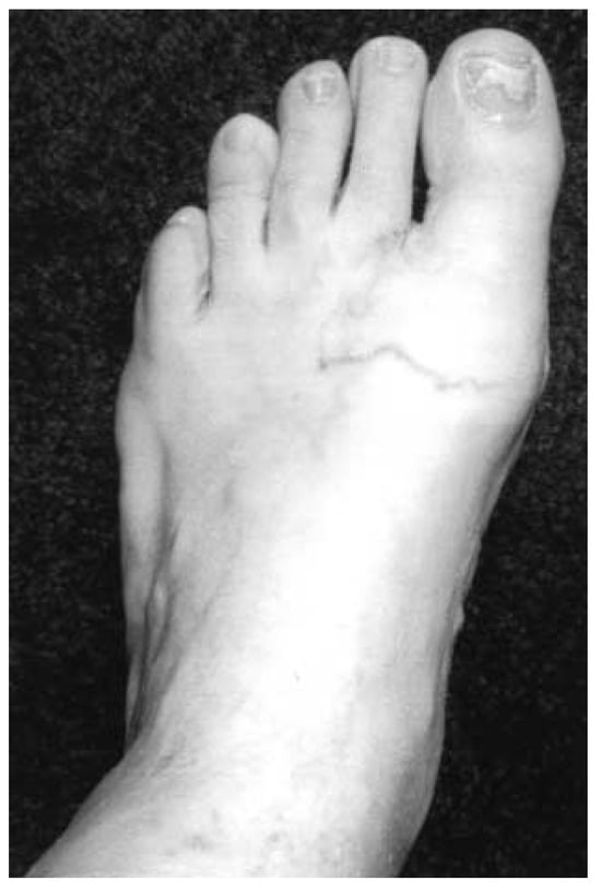
A 62-year-old man presented to his internist’s office with a 1-week history of an erythematous, pruritic lesion on the dorsum of his left foot (Fig. 1). The patient had recently been to Florida, where he had been swimming in a pond, and he was concerned that he might have contracted an infection from the water. The patient was seen in the Podiatry Department outpatient clinic at the Cleveland Clinic Foundation, Westlake, Ohio, later the same day on referral from his internist. On further questioning, the patient related that he had spent several weeks at a friend’s house in Florida, where he had been walking barefoot outside. When asked about the presence of domestic animals at his friend’s house, the patient replied that his friend owns 13 cats. The patient denied nausea, vomiting, fever, and chills.
Figure 1.
Patient’s left foot at initial presentation. Note the small, dark lesion at the medial base of the second digit and the absence of regional cellulitis.
On physical examination, the patient was alert and oriented to person, place, and time. He was in no acute distress and appeared healthy. His neurovascular status was intact. There was an erythematous, papular, and serpiginous lesion extending along the dorsum of the distal aspect of the left foot. This lesion appeared to originate from a 1-mm erythematous, macular lesion located at the dorsal, proximal, and medial aspect of the second digit. There was no surrounding edema, and there was no lower-extremity cellulitis or lymphangitis noted.
On the basis of the patient’s history and clinical presentation, the diagnosis of cutaneous larva migrans was made. The patient was given a prescription for thiabendazole, 500-mg tablets, with instructions to chew one tablet four times daily after meals for 5 days and to take the tablets with fruit juice.
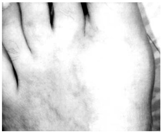
At 1-week follow-up, the patient reported complete resolution of his subjective symptoms. He further noted that the redness of the lesion had markedly diminished within several days of beginning the medication. When asked about oral thiabendazole tolerance, the patient stated that he had experienced no adverse effects. At 3-month follow-up, the patient’s lesion appeared completely healed (Fig. 2); 9 months after initial presentation, the patient remained symptom-free with no relapse.
Figure 2.
Patient’s left foot 3 months after initiation of oral thiabendazole treatment. There is only slight residual hyperpigmentation at the site of the initial skin inflammatory reaction.
Current Recommendations for Treatment
Several treatment options are currently available for cutaneous larva migrans. Oral thiabendazole has traditionally been a mainstay of treatment for many physicians in the United States and elsewhere [6,9]. It may be dosed in multiple ways. Common dosing regimens include 25 to 50 mg/kg once daily for several days, 25 to 50 mg/kg once or twice a day for 2 to 5 days, 50 mg/kg once weekly for a total of four doses, and 500 mg (to be chewed) after meals four times daily with fruit juice for 5 days [2,5,6,9]. Cure rates have been reported to be as high as 89% to 99%, but the drug may be poorly tolerated in some patients, with reported adverse effects of transient dizziness, nausea, vomiting, and headaches [1,5,6,9]. Topical thiabendazole 10% to 15% cream may be prepared by crushing one 500-mg tablet in 5 g of water-soluble cream. This topical formulation must be applied to the lesions three to four times daily for an average of 5 days [1,5,6]. Although use of the topical preparation is associated with only a slightly higher rate of recurrence compared with oral thiabendazole therapy, the topical product has limited value in cases of multiple lesions, and it may be irritating to the skin [2,5].
Oral albendazole has also been used successfully for more than a decade, at 400 to 800 mg daily for 1 to 7 days but generally for at least 3 to 5 days. Albendazole is thought to be better tolerated than oral thiabendazole in many patients, with somewhat milder gastrointestinal upset reported when used in short courses [2,4,6,9]. Although its exact mechanism of action is currently poorly understood, albendazole is effective against numerous nematode species and can produce cessation of larval migration within 24 to 48 hours of administration, corresponding with an early reduction of patient discomfort [2]. As with thiabendazole, cure rates with albendazole are usually very high, approaching 100%; however, reported cure rates in the literature have been as low as 46%, depending on the study population [5,9]. In studies involving people from regions where cutaneous larva migrans is endemic, it is difficult to distinguish between relapse and reinfection.
Oral ivermectin treatment has been shown to be both well tolerated and highly efficacious at a single dose of 12 mg, with a reported cure rate as high as 98% in several large studies [5,9]. In some cases, one or two additional doses may be given at weekly intervals. As is typically seen with oral treatments with other drugs, pruritus resolves after an average of 3 days in patients treated with ivermectin. In a randomized trial comparing ivermectin with albendazole, ivermectin treatment prompted a rapid clinical response and demonstrated a relapse rate of 0%, whereas albendazole treatment showed a rapid initial response but a 54% relapse rate [10]. Furthermore, when used to treat cutaneous larva migrans, no adverse effects have been reported with ivermectin use [5].
The older method of freezing the leading edge of the larval track with liquid nitrogen is associated with skin irritation and patient discomfort, and it often misses the actual larvae, which may be several centimeters beyond the visible track [5,6,9]. Furthermore, the larvae are resistant to low temperatures [2]. For these reasons, cryotherapy is no longer recommended [5,9].
Discussion
Cutaneous larva migrans is a ubiquitous skin condition that affects travelers to tropical and subtropical climates. It is spread to humans through contact with soil that has been contaminated with the filariform larvae of mammalian hookworms. Although generally innocuous in nature, cutaneous larva migrans can be both unsightly for patients and intensely pruritic, with a potential for secondary bacterial skin infections that necessitate prompt treatment. Of the current accepted treatment modalities for cutaneous larva migrans, ivermectin seems to offer the simplest administration and the shortest treatment course, with no reported adverse effects. Because of the high likelihood of pedal involvement, it is important for the podiatric medical community to recognize this condition to facilitate rapid diagnosis and appropriate treatment.
Acknowledgment
Russell G. Clark for assistance with photography.
References
- Davies, H.D.; Sakuls, P.; Keystone, J.S. Creeping eruption. Arch. Dermatol. 1993, 129, 588. [Google Scholar] [CrossRef] [Scilit] [PubMed]
- Albanese, G.; Venturi, C.; Galbiati, G. Treatment of larva migrans cutanea (creeping eruption): a comparison between albendazole and traditional therapy. Int. J. Dermatol. 2001, 40, 67. [Google Scholar] [CrossRef] [Scilit] [PubMed]
- Little, M.D.; Halsey, N.A.; Cline, B.L.; et al. Ancylostoma larva in a muscle fiber of man following cutaneous larva migrans. Am. J. Trop. Med. Hyg. 1983, 32, 1285. [Google Scholar] [CrossRef] [Scilit] [PubMed]
- Biolcati, G.; Alabiso, A. Creeping eruption of larva migrans: a case report in a beach volley athlete. Int. J. Sports Med. 1997, 18, 612. [Google Scholar] [CrossRef] [Scilit] [PubMed]
- Caumes, E. Treatment of cutaneous larva migrans. Clin. Infect. Dis. 2000, 30, 811. [Google Scholar] [CrossRef] [Scilit] [PubMed]
- Silverberg, N.B.; Jackson, R.M.; Laude, T.A.; et al. Picture of the month. Arch. Fam. Med. 1998, 7, 403. [Google Scholar] [CrossRef] [Scilit] [PubMed]
- Hotez, P.J.; Narasimhan, S.; Haggerty, J.; et al. Hyaluronidase from infective Ancylostoma hookworm larvae and its possible function as a virulence factor in tissue invasion and in cutaneous larva migrans. Infect. Immun. 1992, 60, 1018. [Google Scholar] [CrossRef] [Scilit] [PubMed]
- Nakamura-Uchiyama, F.; Yamasaki, E.; Nawa, Y. One confirmed and six suspected cases of cutaneous larva migrans caused by overseas infection with dog hookworm larvae. J. Dermatol. 2002, 29, 104. [Google Scholar] [CrossRef] [Scilit]
- Caumes, E. Treatment of cutaneous larva migrans and Toxocara infection. Fundam. Clin. Pharmacol. 2003, 17, 213. [Google Scholar] [CrossRef] [Scilit] [PubMed]
- Caumes, E.; Carriere, J.; Datry, A.; et al. A randomized trial of ivermectin versus albendazole for the treatment of cutaneous larva migrans. Am. J. Trop. Med. Hyg. 1993, 49, 641. [Google Scholar] [CrossRef] [Scilit] [PubMed]
© 2005 American Podiatric Medical Association