


Abstract
Onychomycosis is a common problem seen in clinical practice. Given the differential diagnosis of dystrophic nails, it is helpful to obtain a definitive diagnosis of dermatophyte infection before initiation of antifungal therapy. Potassium hydroxide preparation and fungal culture, which are typically used in the diagnosis of these infections, often yield false-negative results. Recent studies have suggested that nail plate biopsy with periodic acid–Schiff stain may be a very sensitive technique for the diagnosis of onychomycosis. In this article, we review the literature on the utility of histopathologic analysis in the evaluation of onychomycosis. Many of these studies indicate that biopsy with periodic acid–Schiff is the most sensitive method for diagnosing onychomycosis. We propose that histopathologic examination is indicated if the results of other methods are negative and clinical suspicion is high; therefore, it is a useful complementary technique in the diagnosis of onychomycosis.
Onychomycosis, a fungal infection of the nails, has an estimated prevalence of more than 10% in the general population in countries such as Australia, the United States, and the United Kingdom. It is the most common cause of abnormal nails, but it is clearly not the only cause. Only 50% of dystrophic nails have a fungal etiology. Because the treatment of dermatophyte onychomycosis requires long-term therapy with an oral antifungal medication, with potential adverse effects, it is essential to diagnose the infection correctly. Although onychomycosis caused by nondermatophyte molds is more often resistant to these drugs, these organisms cause few cases of onychomycosis compared with dermatophytes [1].
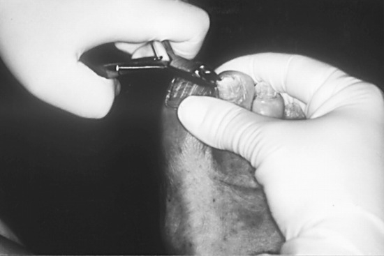
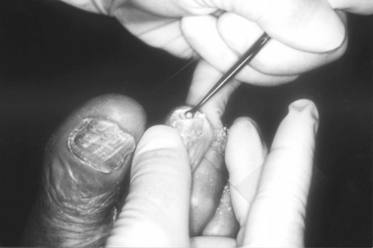
Traditionally, potassium hydroxide (KOH) preparation and fungal culture have been the preferred methods used for the diagnosis of onychomycosis. However, given the inconsistent sensitivity of both of these methods, and the potential delay in results from fungal cultures, histologic evaluation of nail plate specimens using the periodic acid–Schiff method has been considered a complementary method for the diagnosis of mycotic nail infections. Nail plate biopsies with periodic acid–Schiff are also recommended for cases in which clinical suspicion of onychomycosis is strong but negative results are produced from fungal culture and KOH preparation (Figure 1). Recently, several studies [2,3,4,5,6,7,8,9] comparing biopsy/periodic acid–Schiff with the more traditional diagnostic techniques have been published.
Figure 1.
Clipping of the distal nail plate to obtain specimens for periodic acid–Schiff staining.
Traditional Methods: KOH and Culture
Traditional methods used for the diagnosis of onychomycosis are fungal nail culture on Sabouraud dextrose agar and KOH preparation of nail samples. Although these have been standard methods used in clinics, the diagnostic accuracy of the tests varies from 50% to 70%, depending on the methods used to collect and prepare the samples [10].
Direct microscopic examination of KOH-prepared nail samples is the simplest, most inexpensive method used for the diagnosis of mycotic infections. It is often used only to screen for the presence of fungi, but experienced clinicians can differentiate yeast from dermatophytes and dermatophytes from nondermatophytes under the microscope. However, KOH preparations have been reported to have a false-negative rate of 5% to 15%, possibly owing to the low visibility of scantily scattered fungal material in the nail sample [11,12].
Fungal culture is more specific than the KOH preparation in that the causal agent may be grown from the nail specimen. However, false negatives may arise if the nail sample contains only dead or nonviable mycotic organisms, if not enough of the nail was collected, or if the nail sample was clipped distal to the fungal growth [13]. The nail specimen must be crushed thoroughly to enhance fungal growth on the culture medium. This may be another contributing factor to false negatives. In a study by Clayton [14], 2,113 nails were analyzed, and 11% of toenails and 19% of fingernails were demonstrated to be KOH-positive but fungal culture–negative. False-positive cultures also occur secondary to contamination of the growth medium. The normal skin flora contains nonpathogenic fungi that may give a positive fungal culture, and there are laboratory contaminants that can cause disease, especially in the immunocompromised population [12]. Jennings and Rinaldi [15], using data from a multicenter, nationwide, multispecialty survey, investigated the efficacy of in-office dermatophyte test medium and central laboratory cultures used to confirm onychomycosis across samples collected by podiatric, dermatologic, and primary-care physicians. The samples collected by podiatric physicians were either both positive or both negative in 43% and 27% of patients, respectively. Samples harvested by dermatologists were both positive in 37% of patients and both negative in 32%, and samples collected by primary-care physicians were both positive in 28% of patients and both negative in 38%.
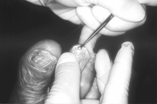
It is important to realize that there are many factors in KOH preparations and fungal cultures that alter their sensitivity and specificity in the diagnosis of onychomycosis. The optimal method of obtaining material, and positive results, for KOH and culture is to curette hyperkeratotic material as proximally under the nail plate as possible, after clipping the distal part of the nail (Figure 2). Although the results demonstrated by the two methods usually correlate, considerable discrepancies still exist between them. A study [16] of 32,000 nails across 83 Australian laboratories showed that 45% of the samples were culture- and KOH-positive. However, 10% to 40% produced only KOH-positive results, and another 10% to 30% produced only culture-positive results.
Figure 2.
Proper method of curetting proximal hyperkeratotic material for potassium hydroxide and culture.
Histologic Evaluation
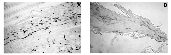
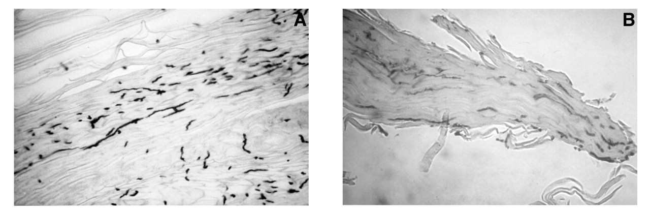
Given the inconsistent sensitivity of KOH preparations and the delay in results from fungal cultures, histologic evaluation of nail plate specimens using the periodic acid–Schiff method has been considered to be a complementary method for the diagnosis of mycotic nail infections (Figure 3). Furthermore, if the nail dystrophy is not due to a fungal infection, the histology could aid in the diagnosis of alternative causes, such as psoriasis or lichen planus. As opposed to culture, however, biopsy/periodic acid–Schiff and KOH preparation do not differentiate between viable and nonviable organisms or provide the species of the pathogen. Recently, several studies [2,3,4,5,6,7,8,9] have been published that compare biopsy/periodic acid–Schiff with the more traditional diagnostic techniques. The results of these studies are summarized in Table 1.
Figure 3.
(A,B). Examples of positive periodic acid–Schiff stains of nail plate biopsy specimens (×40).
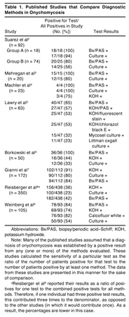
Suarez et al [2] performed two analyses that examined the sensitivity of nail biopsy and fungal culture. In group A, 18 nail samples were preselected from patients with severe onychomycosis. Each sample was divided into two portions; one portion was stained using the periodic acid–Schiff method and the other portion was grown in dermatophyte test medium. All 18 specimens were periodic acid–Schiff–positive; however, only 17 specimens were culture-positive. In group B, 74 nail samples suspected of onychomycosis were stained using the periodic acid–Schiff method and cultured on Sabouraud media using chloramphenicol and cycloheximide. Twenty samples were periodic acid–Schiff–positive; of these, only nine were culture-positive. Conversely, 35 samples were periodic acid–Schiff–negative; of these, five were culture-positive. The authors concluded that routine histopathologic analysis of the nail plate alone is a useful complementary method to fungal culture for diagnosing onychomycosis. This initial study was a favorable indication that biopsy/periodic acid–Schiff might be a useful technique in the evaluation of nail dystrophy.
Mehregan et al [3] investigated nail samples from patients with suspected onychomycosis. Samples were submitted for culture on dermatophyte test medium for 1 month and for biopsy/periodic acid–Schiff. Twelve samples were positive by both methods, three were periodic acid–Schiff–positive and culture-negative, and five were negative by both methods. The authors concluded that in some cases, histology is more sensitive than culture.
Machler et al [4] were the first to analyze three methods. They compared the sensitivity of KOH, biopsy/ periodic acid–Schiff, and culture on Sabouraud agar on 23 nail samples (12 from patients with onychodystrophy of unknown cause and 11 control samples). Of the dystrophic cases, four were demonstrated to be onychomycosis by positive culture and biopsy/periodic acid–Schiff. However, only three of those specimens were KOH-positive. Eight cases of dystrophy were due to psoriasis. In this small study, the authors concluded that biopsy/periodic acid–Schiff was equal to culture and superior to KOH.
Lawry et al [5] examined the sensitivities of various staining and culture techniques used in the diagnosis of onychomycosis on samples taken from 63 patients with clinical findings suggestive of the disease. Nail samples underwent six diagnostic tests. Routine histopathologic examination using periodic acid–Schiff stain (biopsy/periodic acid–Schiff) was 85% sensitive. Sensitivities for KOH dissolution and centrifugation combined with periodic acid–Schiff, fluorescent brightener (Blankophor; AG Scientific, San Diego, California), or chlorazol black E were 57%, 53%, and 53%, respectively. Culture on Sabouraud agar with chloramphenicol and cycloheximide (Mycosel agar; Difco Laboratories, Detroit, Michigan) was 32% sensitive, and on Littman oxgall agar was 23% sensitive. The most sensitive combination of tests, both culture methods plus biopsy/periodic acid–Schiff, was 94% sensitive (not statistically different from the sensitivity of biopsy/periodic acid–Schiff alone [P = 0.26]). They concluded that when onychomycosis is suspected clinically, biopsy/periodic acid–Schiff of the nail is the single most sensitive of the diagnostic tests they evaluated. Because it is quickly performed, biopsy/periodic acid–Schiff is practical for clinical and research purposes. Further study is needed to determine whether sensitivity may be enhanced by combining biopsy/periodic acid–Schiff with cultures obtained using several collection methods (clipping, curetting, and shaving). Such combinations may serve as sensitive and efficient strategies for diagnosing onychomycosis. This study used multiple methods of evaluation and was the first to suggest that combining different methods of evaluation might have a synergistic effect.
Borkowski et al [6] compared the sensitivity of KOH preparations, biopsy/periodic acid–Schiff, and culture techniques for the detection of onychomycosis in 50 cases of clinically suspected onychomycosis. Sixteen cases were KOH-positive, 12 were culture-positive, and 36 were positive by biopsy/periodic acid– Schiff. Analysis showed that biopsy/periodic acid– Schiff was significantly more sensitive than KOH and culture (P = 0.001). There was no significant difference in sensitivity between KOH and culture. The authors concluded that the results suggest that biopsy/ periodic acid–Schiff may be the true “gold standard” for the diagnosis of onychomycosis.
Gianni et al [7] reported a large comparative study that sought to investigate the usefulness of the histologic examination of nail clipping samples in supplementing the standard microscopic and culture techniques for the diagnosis of onychomycosis. They evaluated 172 subjects affected by nail alterations suggestive of onychomycosis. Nail specimens were studied using three different techniques: direct microscopic examination of a 40% KOH-clarified preparation, fungal culture, and histologic examination (biopsy/periodic acid–Schiff). Patients positive for fungal infection were reevaluated using the same techniques after treatment with oral terbinafine, fluconazole, or itraconazole and topical application of bifonazole or ciclopirox for 2 months. Results of direct microscopy were positive in 102 nail specimens (59.3%). The culture test results were positive in 90 cases (52.3%), showing a dermatophyte in 45 samples, a yeast in 23, and a mold in 22. Results of the histologic examination were positive in 94 samples (54.7%). In four cases, it was the only investigation that confirmed the clinical diagnosis of nail mycosis. In most cases, the morphologic aspect of the hyphae or spores also suggested to which group of pathogens (dermatophytes, yeasts, or molds) the fungal elements observed in the histologic sections could be ascribed. They concluded that histologic examination of nail clipping specimens is a relatively inexpensive, rapid, and easily performed procedure and that it is useful to confirm or refute the results of routine microscopy and culture. Moreover, nail histopathologic observation may help in ascribing a pathogenic role for nondermatophyte isolates and in evaluating the effectiveness of antifungal treatment.
Reisberger et al [8] performed the largest comparative study to date. In a prospective study, they compared the traditional mycologic diagnostics using culture medium and KOH preparation with the histopathologic diagnosis of onychomycosis using nail clippings stained with periodic acid–Schiff. Material from 387 nails of 350 patients suspected of having onychomycosis was obtained, and a KOH stain and two fungal cultures (Kimmig agar [Merck, Darmstadt, Germany] with and without cycloheximide) were prepared. In addition, the same specimen was histopathologically examined using periodic acid–Schiff staining. Culture medium and KOH preparation revealed 100 and 156 cases of onychomycosis, respectively, compared with 182 cases by histologic examination. Histologic examination yielded a significantly higher rate of positive results (P < 0.05). Considering the total number of positive results given by at least one of the three methods (total = 438), histologic evaluation was found to give the highest rate of successful recognition of mycotic infection (41.6%). The authors concluded that the histopathologic evaluation of nail clippings stained using periodic acid–Schiff is very quick and easy to perform and will increase the frequency of diagnosing onychomycotic disease above that achieved by culture and KOH preparation alone. However, they noted that because information concerning the vitality of the fungi and accurate identification of the specific pathogen is not available through this investigation alone, mycologic culture remains the indisputable “gold standard” of mycologic diagnostics.
Weinberg et al [9] evaluated 105 patients with suspected onychomycosis using four diagnostic methods: KOH preparation, culture, Calcofluor white preparation, and biopsy/periodic acid–Schiff. Calcofluor white stain binds to cellulose and chitin and fluoresces when exposed to ultraviolet radiation. It is a highly sensitive and specific technique for the detection of fungi. To determine the clinical usefulness and performance characteristics of each test, Calcofluor white was chosen as the gold standard for statistical analysis. In 93 patients, at least one of the four diagnostic methods was positive for the presence of organisms. The following were calculated for each test: sensitivity, specificity, positive predictive value, and negative predictive value. The sensitivities of each of the techniques were as follows: KOH, 80%; biopsy/ periodic acid–Schiff, 92%; and culture, 59%. Both KOH and biopsy/periodic acid–Schiff were more sensitive than culture (P = 0.00002). Biopsy/periodic acid–Schiff was also more sensitive than KOH (P = 0.03). The specificities were as follows: KOH, 72%; biopsy/ periodic acid–Schiff, 72%; and culture, 82%. The positive predictive values calculated for the different techniques were as follows: KOH, 88%; biopsy/periodic acid–Schiff, 89.7%; and culture, 90%. In terms of negative predictive value, the results were as follows: KOH, 58%; biopsy/periodic acid–Schiff, 77%; and culture, 43%. The authors concluded that biopsy/periodic acid–Schiff is the most sensitive method for the diagnosis of onychomycosis. It is also superior to the other methods in its negative predictive value. This study was the first to use a nonstandard technique as a gold standard for evaluation.
Economic and Medical Importance of Proper Diagnosis
In addition to the necessity of proper diagnosis for patient care, there are also economic issues relating to the diagnosis of onychomycosis. Mehregan and Gee [13] addressed the issue of cost-effectiveness in a study analyzing the cost of treating onychomycosis with itraconazole or terbinafine. The study analyzed nail samples from 688 patients who were suspected of having onychomycosis based on clinical observations. The presence of disease in the samples was determined by histopathologic periodic acid–Schiff staining. Four hundred forty-four samples were reported to be disease-positive.
Using a cost-analysis framework that included the average costs of monitoring blood values during the extrapolated treatment period and mean medication costs for a normal therapeutic regimen, a total of $540 for medication and $50 for laboratory costs per case was calculated. Expanding the unit cost of $590 to include the 688 patients in the study, a total expenditure of $405,920 would be generated.
The total cost of testing all of the patient samples with periodic acid–Schiff would be $34,400. Diagnosis of positive disease using clinical observation and periodic acid–Schiff staining resulted in only 444 patients being diagnosed as having disease. The total cost of testing all of the patients and treating only those with positive periodic acid–Schiff results generated a total cost of $269,360. This represents a 44% decrease in the theorized overall expenditures.
One aspect of these treatment regimens that was neglected in the researchers’ cost analysis is the cost of adverse effects experienced by the patients, which would be generated by physician visits and subsequent laboratory work, days lost from work, and so on. Any patient who takes these medications would be at risk of adverse effects such as drug rash, gastrointestinal abnormalities, drug-drug interactions, and hepatotoxicity [10]. Using this study as an example, 244 people would be at risk as a result of unnecessary treatment. Taking this into account, an overall savings of 44% may be underestimated [13].
Conclusions
The misdiagnosis of onychomycosis in any patient leads to a delay in the proper treatment of the underlying condition causing the nail abnormalities. Proper diagnosis not only allows for more efficient expenditure of funds and services but also is paramount for the timely alleviation of the condition. Culture and KOH, the traditional methods of diagnosis, have several advantages. They are easily performed by most clinicians and are inexpensive. In addition, culture offers accurate identification of the specific causative pathogen in onychomycosis. Biopsy/periodic acid–Schiff seems to be highly useful as a complementary method for proper diagnosis. The one limitation of this method is that it does not provide the exact species of the causative agent. This method can be useful when there is strong clinical suspicion of onychomycosis but negative results are produced from fungal culture and KOH preparation.
References
- GHANNOUM, M.A.; HAJJEH, R.A.; SCHER, R.; ET, A.L. A large-scale North American study of fungal isolates from nails: the frequency of onychomycosis, fungal distribution, and antifungal susceptibility patterns. J Am Acad Dermatol 2000, 43, 641. [Google Scholar] [CrossRef] [Scilit] [PubMed]
- SUAREZ, S.M.; SILVERS, D.N.; SCHER, R.K.; et al. Histologic evaluation of nail clippings for diagnosing onychomycosis. Arch Dermatol 1991, 127, 1517. [Google Scholar] [CrossRef] [Scilit] [PubMed]
- MEHREGAN, D.A.; MEHREGAN, D.R.; RINKER, A. Onychomycosis. Cutis 1997, 59, 247. [Google Scholar] [PubMed]
- MACHLER, B.C.; KIRSNER, R.S.; ELGART, G.W. Routine histologic examination for the diagnosis of onychomycosis: an evaluation of sensitivity and specificity. Cutis 1998, 61, 217. [Google Scholar] [PubMed]
- LAWRY, M.A.; HANEKE, E.; STROBECK, K.; et al. Methods for diagnosing onychomycosis: a comparative study and review of the literature. Arch Dermatol 2000, 136, 1112. [Google Scholar] [CrossRef] [Scilit] [PubMed]
- BORKOWSKI, P.; WILLIAMS, M.; HOLEWINSKI, J.; et al. Onychomycosis: an analysis of 50 cases and a comparison of diagnostic techniques. JAPMA 2001, 91, 351. [Google Scholar] [CrossRef] [Scilit] [PubMed]
- GIANNI, C.; MORELLI, V.; CERRI, A.; et al. Usefulness of histological examination for the diagnosis of onychomycosis. Dermatology 2001, 202, 283. [Google Scholar] [CrossRef] [Scilit] [PubMed]
- REISBERGER, E.M.; ABELS, C.; LANDTHALER, M.; et al. Histopathological diagnosis of onychomycosis by periodic acid–Schiff–stained nail clippings. Br J Dermatol 2003, 148, 749. [Google Scholar] [CrossRef] [Scilit] [PubMed]
- WEINBERG, J.M.; KOESTENBLATT, E.K.; TUTRONE, W.D.; et al. Comparison of diagnostic methods in the evaluation of onychomycosis. J Am Acad Dermatol 2003, 49, 193. [Google Scholar] [CrossRef] [Scilit] [PubMed]
- LEYDEN, J. Pharmacokinetics and pharmacology of terbinafine and itraconazole. J Am Acad Dermatol 1998, 38, 542. [Google Scholar] [CrossRef] [Scilit] [PubMed]
- ELEWSKI, B.E. Large scale epidemiological study of causal agents of onychomycosis: mycological findings from the multicenter onychomycosis study of terbinafine. Arch Dermatol 1997, 133, 1317. [Google Scholar] [CrossRef] [Scilit] [PubMed]
- WEITZMAN, I.; SUMMERBELL, R.C. The dermatophytes. Clin Microbiol Rev 1995, 8, 240. [Google Scholar] [CrossRef] [PubMed]
- MEHREGAN, D.C.; GEE, S.L. The cost effectiveness of testing for onychomycosis versus empiric treatment of onychodystrophies with oral antifungal agents. Cutis 1999, 64, 407. [Google Scholar] [PubMed]
- CLAYTON, Y.M. Clinical and mycological diagnostic aspects of onychomycosis and dermatomycoses. Clin Exp Dermatol 1992, 17 (suppl 1), 37. [Google Scholar] [CrossRef] [Scilit] [PubMed]
- JENNINGS, M.B.; RINALDI, M.G. Confirmation of dermatophytes in nail specimens using in-office dermatophyte test medium cultures: insights from a multispecialty survey. JAPMA 2003, 93, 195. [Google Scholar] [CrossRef] [Scilit] [PubMed]
- ELLIS, D.H. Diagnosis of onychomycosis made simple. J Am Acad Dermatol 1999, 40, S3. [Google Scholar] [CrossRef] [Scilit] [PubMed]
© 2005 American Podiatric Medical Association