


Abstract
The Keller procedure has been used during the past century for the treatment of first metatarsophalangeal joint pathology. Many modifications to the procedure have been made, including interposition of the joint capsule into the first metatarsophalangeal joint space. Capsular interposition is often the most difficult step in performing the Keller bunionectomy. This article describes a new, simplified technique for capsular interposition with the use of a dorsal capsular flap and soft-tissue anchors.
Keywords:
N/A In 1904, William L. Keller described the procedure that bears his name in the New York Medical Journal.[1] Through the years, the indications for the procedure have not changed. These indications include a symptomatic, nonsalvageable first metatarsophalangeal joint in an elderly, sedentary patient that features degenerative changes, joint subluxation, hallux abductus, hallux limitus, an increased first inter-metatarsal angle, or abnormal proximal or distal articular set angles.[2,3]
However, during the past century, many modifications to the Keller procedure have been made. Interpositioning of the first metatarsophalangeal joint capsule into the void created by the resection of the proximal phalangeal base of the hallux is one modification that has been used by many surgeons. The interposed capsule acts as a biologic spacer about which fibrosis develops.[2] This fibrosis then maintains the distance between the first metatarsal head and the stump of the proximal phalanx, preventing shortening of the hallux.
Many techniques for capsular interpositioning have been described in the literature, from bunching of the capsule into the first metatarsophalangeal joint void to a box stitch or purse-string suturing of linear capsulotomies.[4,5] Two additional techniques that have been used are 1) U-shaped medial capsular flaps across the metatarsal head sutured dorsally and plantarly and 2) folding the medial flap across the metatarsophalangeal joint and then suturing it to the periosteum of the proximal phalanx.[2,6] Kelikian[7] described the interposition of dorsal and plantar flaps. Walter et al[3] used a J-shaped medial capsular flap sutured to the lateral capsule of the first metatarsophalangeal joint.
In the elderly patient, often the lateral capsule and plantar soft tissue is thin and the suture knots do not hold. Also, it is difficult to maneuver the suture needle in the small space available at the metatarsophalangeal joint. Mroczek and Miller[8] stated that “the most difficult aspect of the procedure involves anchoring the capsular flap into the joint.” Presented here is a simplified technique of capsular interpositioning for the Keller bunionectomy with the use of soft-tissue anchors.
Technique
The first metatarsophalangeal joint is approached through a standard linear dorsomedial skin incision, and the dissection is carried down to the level of the joint capsule. Next, a proximally based U-shaped capsulotomy is made over the dorsomedial aspect of the first metatarsophalangeal joint, extending distally to the proximal one-third of the proximal phalanx (Fig. 1). The capsule is reflected proximally, exposing the base of the proximal phalanx and the head of the first metatarsal. The medial eminence of the first metatarsal and the base of the proximal phalanx are freed of all soft tissue and are resected in the usual manner. Extensor hallucis longus tendon lengthening may be performed at this time.
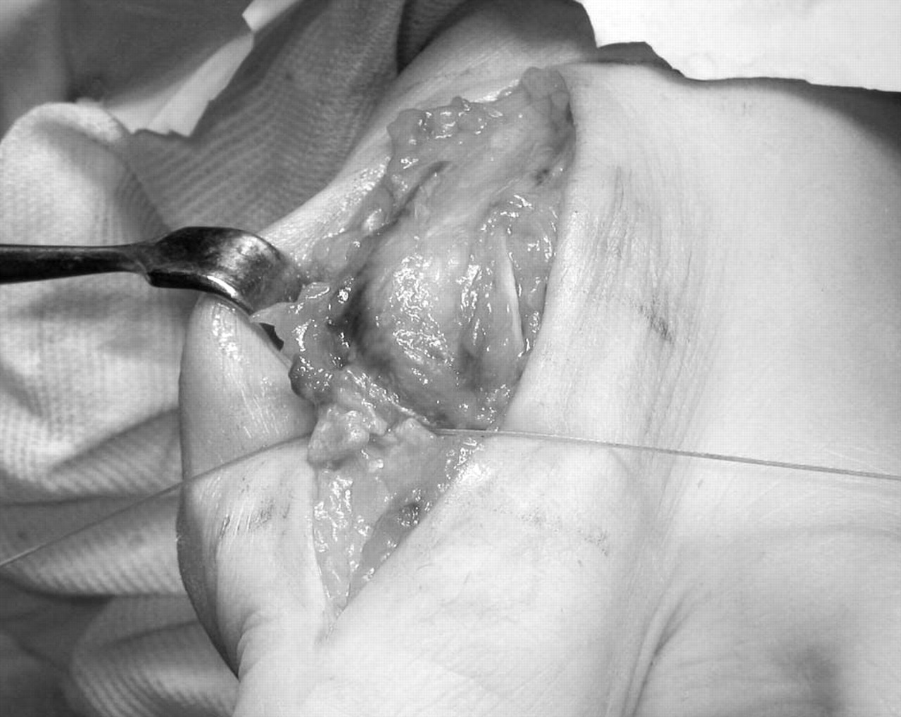
After the appropriate resection of bone is completed, the capsule is interposed into the metatarsophalangeal joint space from dorsal to plantar around the metatarsal head (Fig. 2). The capsule is secured in place with the use of two soft-tissue anchors (Mini Bio-Anchors; Arthrex Inc, Naples, Florida). One anchor is placed plantar laterally and the second plantar medially into the first metatarsal head, and they are sutured using the attached suture material to the distal lateral and distal medial aspects of the capsule, respectively (Fig. 3). With the help of an assistant, the surgeon can tie the sutures simultaneously or independently of each other, completely covering the metatarsal head with the capsule (Fig. 4). A Kirschner wire passed through the interposed capsule from the hallux into the first metatarsal head may then be used for stabilization of the hallux. The proximal lateral and proximal medial aspects of the capsular flap are sutured to the adjacent capsule or periosteum using absorbable suture material. The wound is then closed and a dressing is applied in the usual fashion.
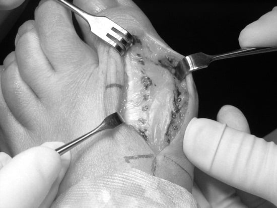
Figure 1.
The dorsomedial U-shaped capsulotomy is outlined with a skin marker.
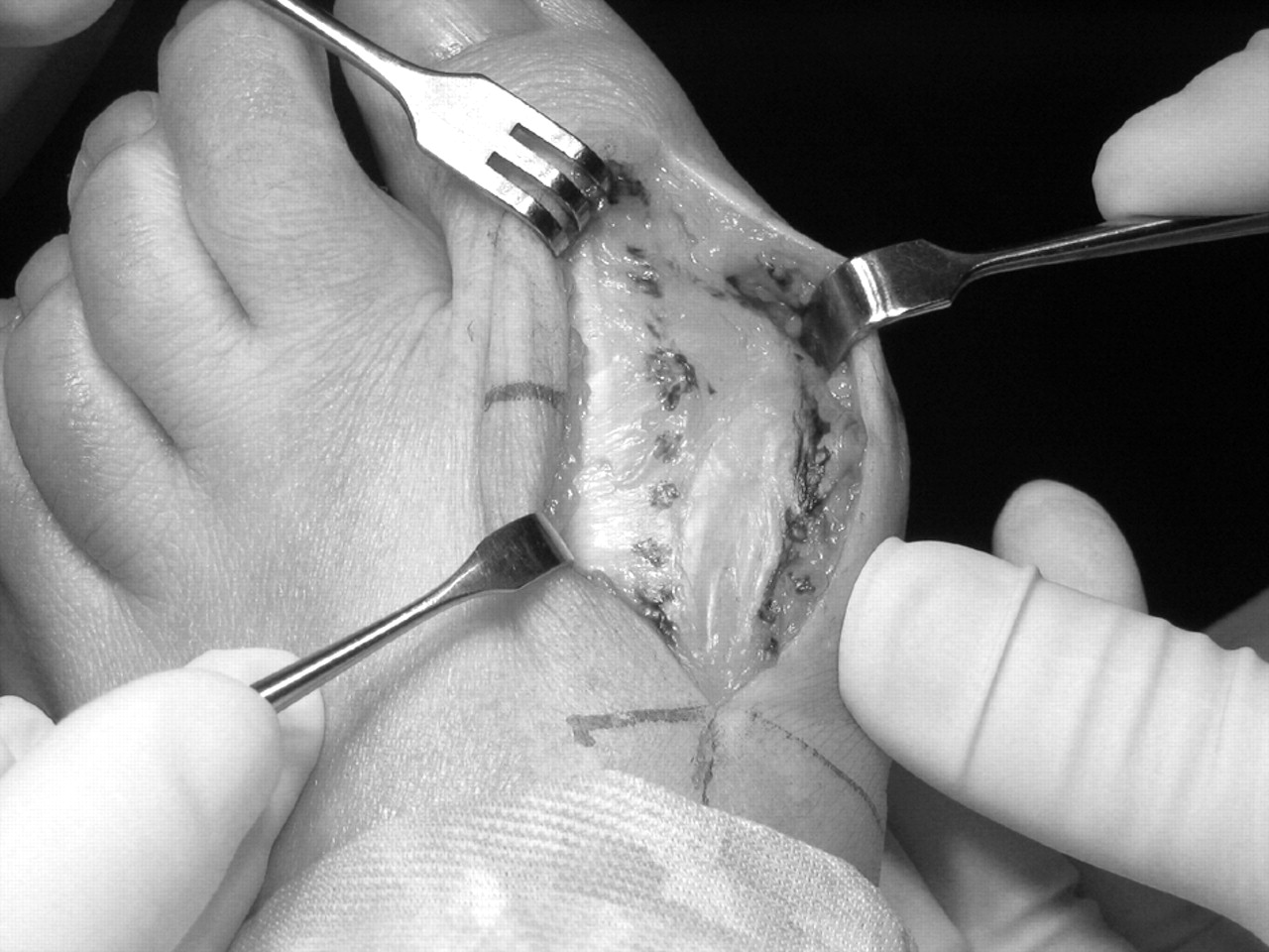
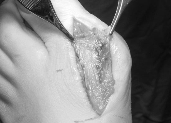
Figure 2.
The capsular flap is pulled distally around the first metatarsal head.
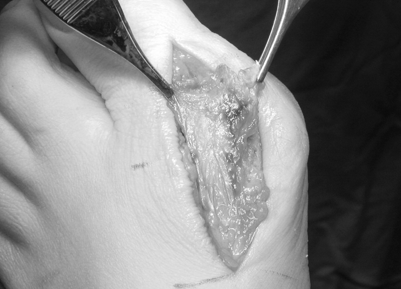
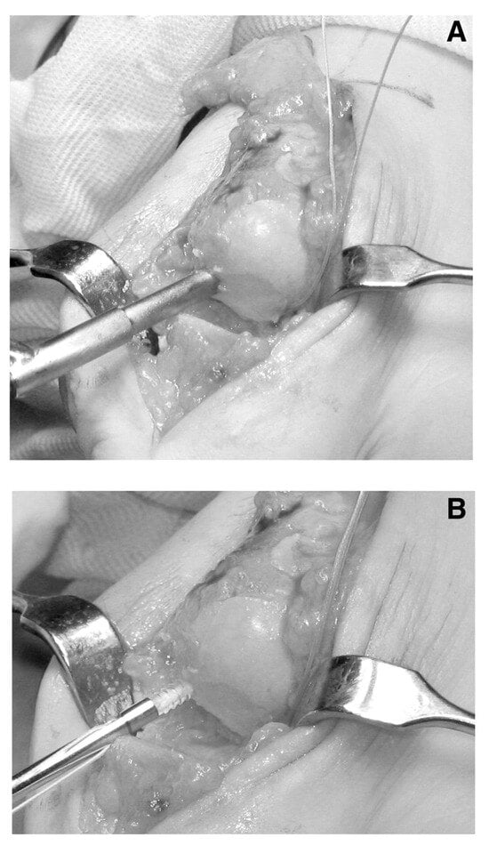
Figure 3.
A, A drill hole is created in the plantar medial aspect of the first metatarsal head to accommodate the soft-tissue anchor. The sutures to the lateral anchor, which has been inserted, can be seen. B, Insertion of the soft-tissue anchor.
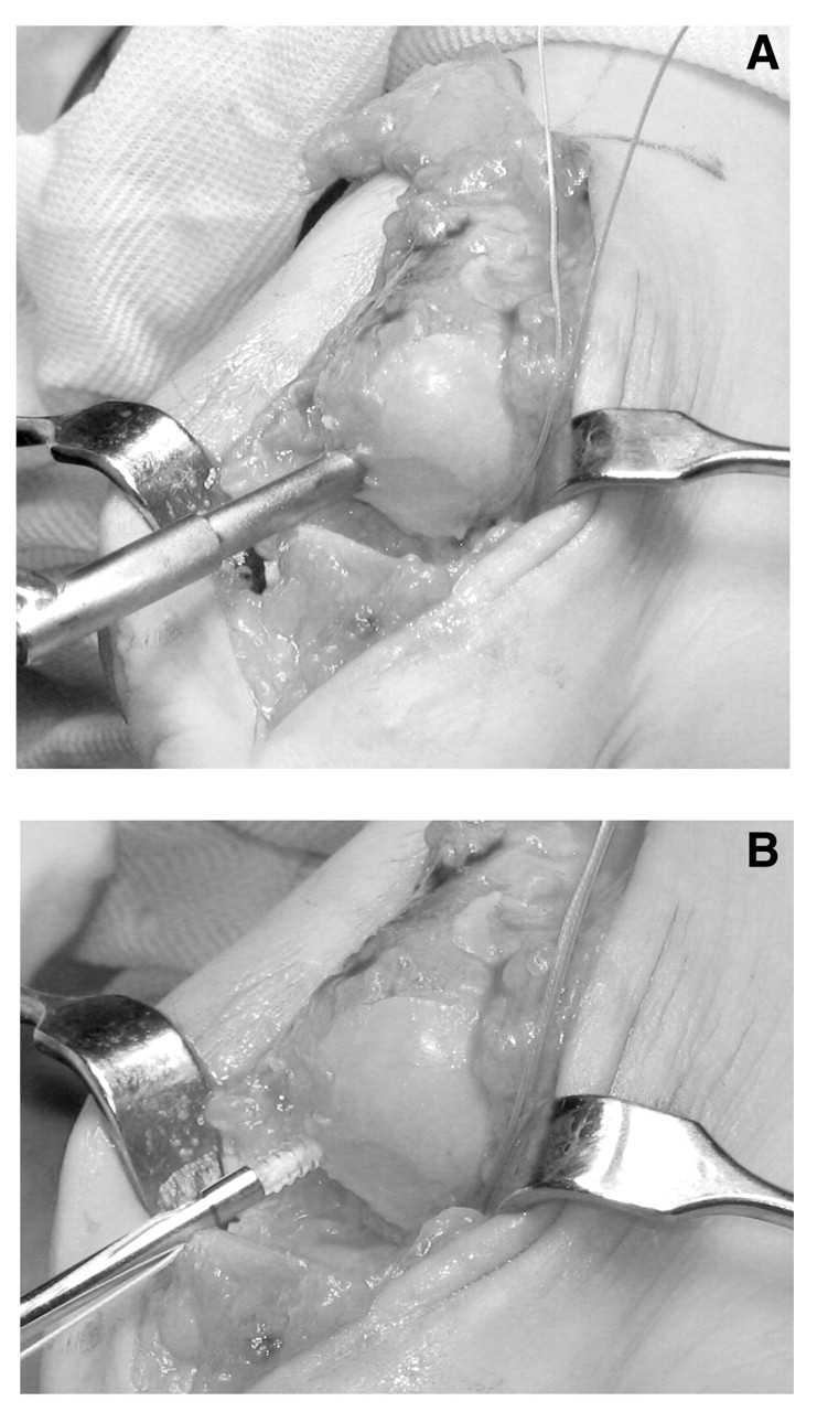
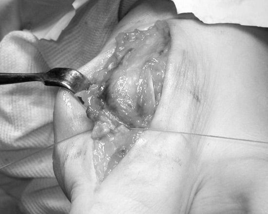
Figure 4.
The suture ends are tied down and the capsular flap is interposed into the void created at the first metatarsophalangeal joint.
Conclusion
The use of soft-tissue anchors when performing the Keller bunionectomy provides the surgeon with an easy method for interposing the capsule into the first metatarsophalangeal joint. The U-shaped capsulotomy described with this technique provides excellent exposure to the first metatarsal head and the proximal phalanx as well as coverage over the metatarsal head. The soft-tissue anchors sit deep to the articular surface of the metatarsal head, and their size provides enough space for the use of two or even three anchors if desired. They ensure that the interposed capsular flap will remain in the proper position without suture knot slippage or pullout from surrounding soft tissue, thereby allowing for fibrosis to occur and to maintain the space between the metatarsal head and the stump of the proximal phalanx, which prevents shortening of the hallux.
References
- Keller WL: Surgical treatment of bunions and hallux valgus. .N Y Med J80::741. ,1904. .
- Ganley JV, Lynch FR, Darrigan RD: Keller bunionectomy with fascia and tendon graft. .JAPMA76::602. ,1986. .
- Walter MJ, Mostone E, Buxbaum F, et al: The Keller-Austin bunionectomy. .J Foot Surg26::400. ,1987. .
- McGlamry ED, Kitting RW, Butlin WE: Keller bunionectomy and hallux valgus correction: an appraisal and current modifications sixty-six years later. .JAPA60::161. ,1970. .
- Richardson EG, Donley BG: “Disorders of Hallux,” in Campbell’s Operative Orthopaedics, 9th Ed, ed by ST Canale, p 1647, CV Mosby, St Louis. ,1998. .
- Mann RA, Coughlin MJ: “Adult Hallux Valgus,” in Surgery of the Foot and Ankle, 7th Ed, ed by MJ Coughlin, RA Mann, p 221, CV Mosby, St Louis. ,1999. .
- Kelikian H: “Arthroplastic Resections,” in Hallux Valgus, Allied Deformities of the Forefoot and Metatarsalgia, p 215, WB Saunders, Philadelphia. ,1965. .
- Mroczek KJ, Miller SD: The modified oblique Keller procedure: a technique for dorsal approach interposition arthroplasty sparing the flexor tendons. .Foot Ankle Int24::521. ,2003. .
© 2005 American Podiatric Medical Association