


Abstract
Pes cavus is a complex deformity with various components. The etiology is multifaceted, which can result in changes to the rearfoot, forefoot, or both areas. Proper patient evaluation is crucial to understanding the deformity and its management. Various soft-tissue and osseous procedures may be used in the reconstruction of the symptomatic cavus foot deformity. This article discusses the clinical and radiographic findings of pes cavus, as well as the various etiologies of this challenging foot deformity and the surgical principles of its correction.
Pes cavus often appears in childhood as a complex foot deformity. Its etiology is multifaceted, and although idiopathic pes cavus may seem to be the culprit, the initial manifestation is often caused by a progressive neurologic disorder. Understanding the true etiology behind each cavus foot is important. Treatment measures and outcomes are often influenced by the pathogenesis of the deformity. Untreated pes cavus can be disabling and can result in significant difficulties for the patient and the surgeon.
The deformity is often accompanied by various muscular imbalances. Such imbalances may stem from an intrinsic or extrinsic muscular defect caused by an upper or lower motor neuron lesion. Understanding the pathomechanics of such deformities is crucial for formulating a good treatment plan. The process begins with a thorough history and an extensive physical examination, including a detailed neurologic work-up.
A comprehensive clinical examination, in conjunction with a good radiographic assessment, can aid the surgeon’s procedure selection and optimize patient outcomes. No two cavus feet are alike. Patients should be evaluated and treated on the basis of their unique characteristics.
Etiology
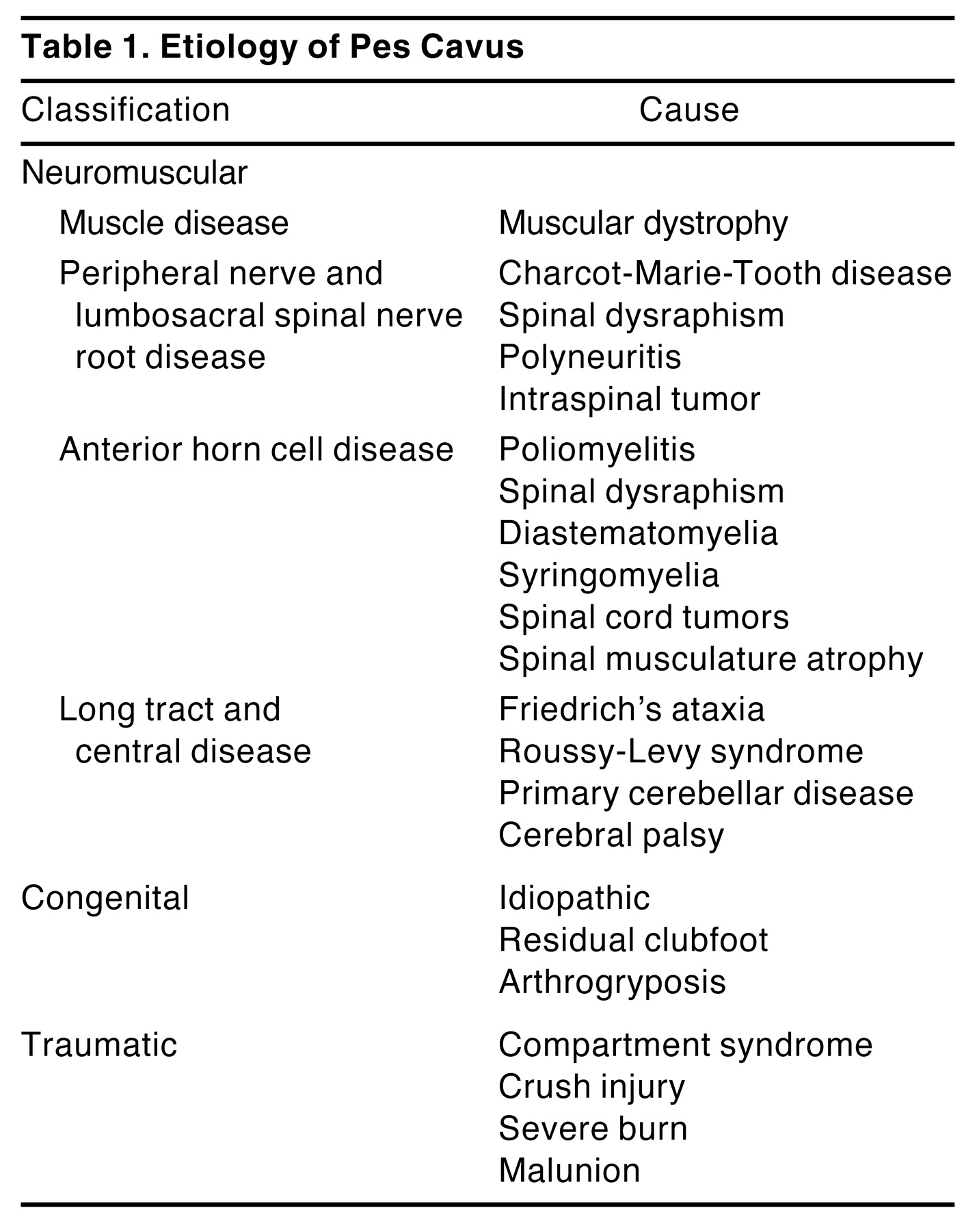
Pes cavus is the general term used to describe a high-arched foot (Figure 1). The deformity consists of several different components, including varying degrees of rearfoot varus, forefoot equinus, metatarsus adductus, and digital deformities. Pes cavus is largely caused by muscle imbalance. In the classic article by Brewerton et al [1], 77 cases of pes cavus were reviewed, and 51 (66%) were found to have clinical evidence of a neurologic origin. When electromyographic and nerve conduction studies were used, an additional 7 cases (9%) were found to have neuromuscular involvement, totaling 75% of the 77 cases demonstrating a neurologic etiology. Ibrahim [2] classified the etiologies of pes cavus as neuromuscular, congenital, and traumatic (Table 1). The most important reason to identify the etiology of the cavus foot is to establish whether the deformity is progressive or static, as this will affect the treatment plan.
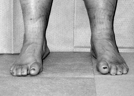
Figure 1.
Bilateral idiopathic pes cavus.
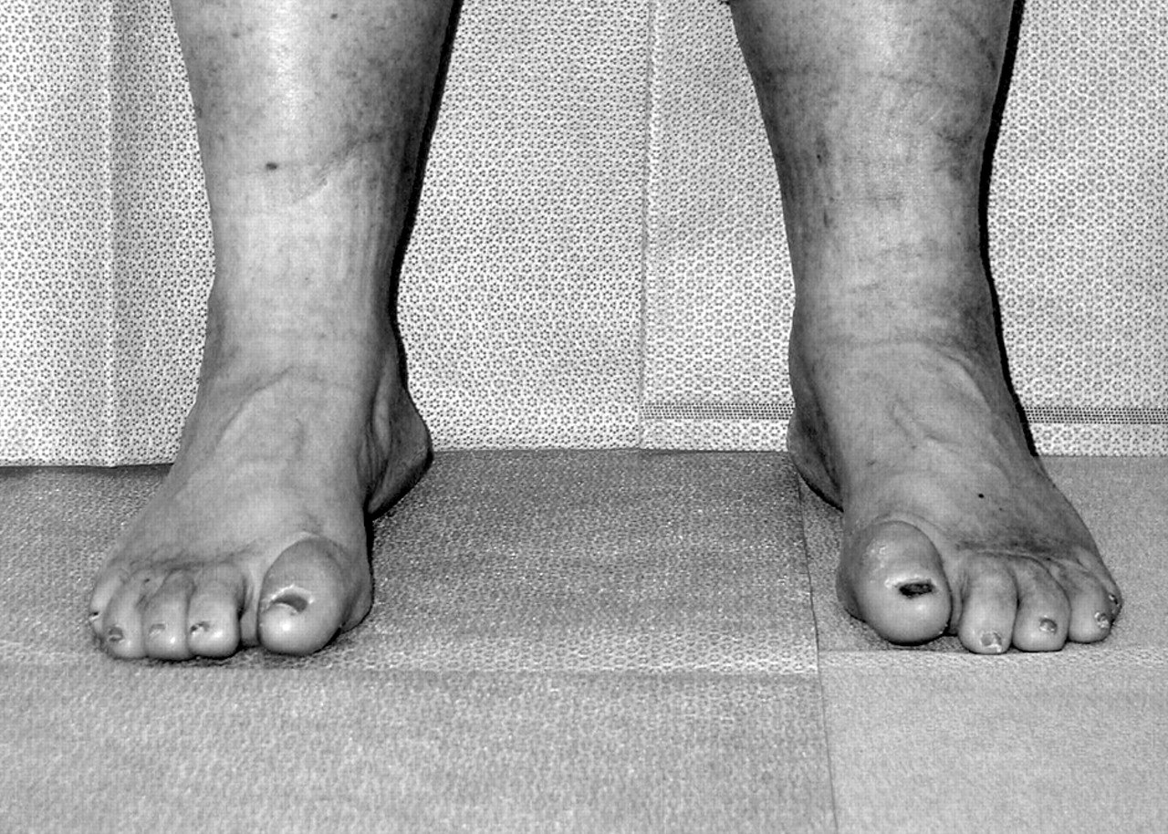
Table 1.
Etiology of Pes Cavus.
Muscle imbalance is the fundamental force behind the cavus foot. Mann and Missirian [3] hypothesized the cause of pes cavus in Charcot-Marie-Tooth disease using an agonist/antagonist theory. When the peroneus brevis muscle atrophies and weakens, it no longer plantarflexes and everts the foot. The opposing tibialis posterior tendon pulls the foot into plantarflexion at the subtalar and ankle joints and inverts the calcaneus. With weakening of the tibialis anterior tendon, the peroneus longus muscle pulls the first ray and forefoot into plantarflexion and eversion. When the forefoot valgus deformity is rigid, weight-bearing forces a compensatory rearfoot varus to allow the forefoot to become plantigrade.
Digital deformities occur as the intrinsic stabilizers of the toes weaken. Contractures at the metatarsophalangeal joints develop because of the overpowering pull of the long extensor muscles. Furthermore, the long toe flexors produce a flexor contracture at the level of the interphalangeal joints, resulting in a claw toe deformity. A weakened tibialis anterior tendon results in a longer, harder contraction of the toe extensors during gait to assist in ankle joint dorsiflexion. This compensation exacerbates the claw toe deformity and creates retrograde pressure at the metatarsophalangeal joint, thus increasing the plantarflexory force of the forefoot. With plantarflexion of the metatarsals and atrophy of the intrinsic muscles, the plantar fascia and ligaments shorten, which exacerbates the cavus foot deformity.
Cavus deformities caused by poliomyelitis have greater tibialis anterior strength, with significant weakness of the gastrocnemius soleus complex. This creates less forefoot plantarflexion but produces a marked increase in calcaneal inclination and rearfoot cavus [4].
Each pes cavus case is different, even if they have the same etiology. They differ in shape, reducibility, function, and degree of neurologic involvement. Each foot must be carefully examined to understand the specific deforming forces and to create an individualized approach to treatment.
Clinical Examination
Evaluation for pes cavus begins with a complete family history. Because up to 75% of all cavus deformities are of neurologic origin, every effort should be made to establish the etiology [1]. Identifying the etiology yields information regarding the progressive or nonprogressive nature of the deformity. This distinction is important because it affects surgical decision making. Idiopathic pes cavus is a nonprogressive deformity with varying amounts of neuromuscular pathology [4,5]. A neurologic consultation is often recommended. Neurologic testing should rule out any spinal lesions and cerebellar dysfunction and should include lower-extremity sensory and motor nerve conduction studies with electromyographic evaluation [4,6].
Patients often complain of difficulty with footwear and painful callus formation beneath the metatarsal heads. Patients with neuropathy may present with or have a history of plantar ulcerations [7,8]. Rearfoot varus and compensated forefoot valgus predispose patients to lateral ankle trauma and recurrent sprains, resulting in chronic ankle pain as the chief complaint [9].
The goals of the clinical evaluation are to define the deformity and to determine which components, if any, are flexible. The foot should be examined nonweightbearing and weightbearing, as well as during normal gait. After muscle strength is evaluated, three main anatomical areas are examined: 1) the rearfoot and ankle, 2) the midfoot, and 3) the distal foot and toes.
Ankle joint range of motion is often limited by a pseudoequinus deformity [10]. When the forefoot plantarflexes on the rearfoot or with isolated rearfoot varus, the ankle joint has to dorsiflex for the foot to be plantigrade. This compensation may use all of the available ankle joint dorsiflexion, resulting in talar neck impingement with the anterior tibial plafond. This impingement often results in an osseous-type equinus, which is frequently associated with anterior ankle pain and contributes to high forefoot pressures during the midstance and propulsive phases of gait [11].
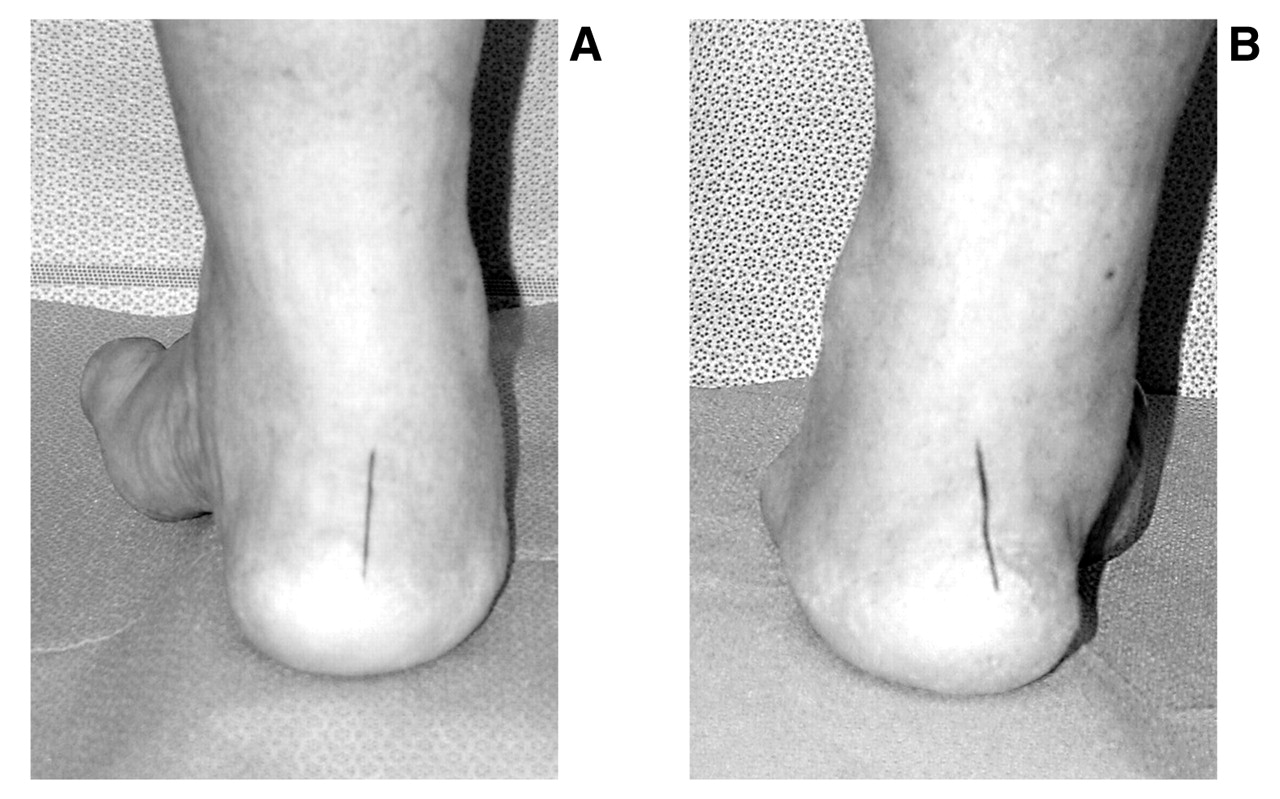
Subtalar joint range of motion should be evaluated for reducibility of the calcaneus to the neutral position. The position of the calcaneus relative to the floor may also be assessed using a Coleman lateral block test (Figure 2) [12]. Using a 1-inch block, the first ray is allowed to hang over the edge of the block, and the heel and the lateral foot remain on the block. If the calcaneal varus corrects, rearfoot deformity is reducible and is a result of subtalar compensation for a forefoot valgus or a plantarflexed first ray. If the varus deformity does not correct, the deformity is considered fixed and typically involves the subtalar joint or calcaneus.
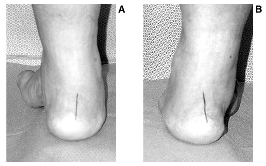
Figure 2.
A, Reduction of calcaneal varus using a lateral Coleman block test; B, no change in calcaneal varus using the same test.
The flexibility of the midfoot is evaluated at the level of the midtarsal joint. Deformity of the midfoot is primarily in the sagittal plane. A soft-tissue contracture of the plantar fascia and intrinsic musculature may be flexible early in the deformity. Reducibility or rigidity of the midfoot and rearfoot do not necessarily occur together, and these areas should be evaluated separately.
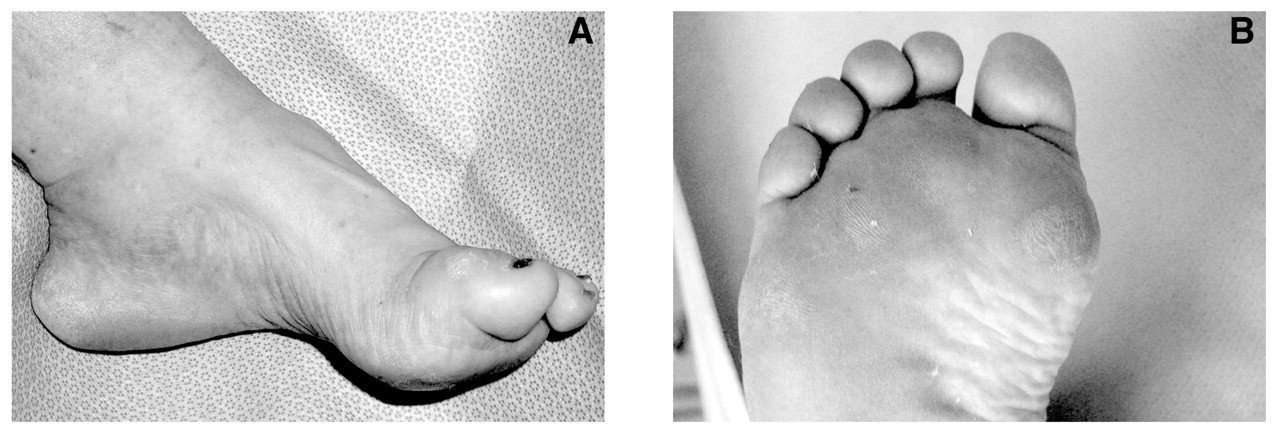
Forefoot-to-rearfoot position is observed with the subtalar joint in the neutral position if possible. The metatarsals may be plantarflexed equally as a unit, or the first ray may be plantarflexed alone (Figure 3). A rigid forefoot valgus with a flexible subtalar joint is a classic mechanism for rearfoot varus commonly seen in cavus foot deformities. The transverse plane position of the metatarsals is also observed. Metatarsus adductus may be present, with varying degrees of reducibility. The toes are often rigidly extended at the metatarsophalangeal joints and flexed at the interphalangeal joints owing to muscle imbalances between the long extensor muscles and the stabilizing intrinsic muscles. Digital deformities are often accentuated in the swing phase of gait because the extensor digitorum longus and extensor hallucis longus muscles fire longer and harder than usual to compensate for a weak tibialis anterior tendon.
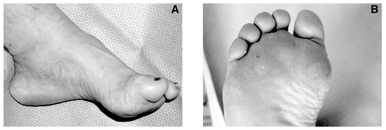
Figure 3.
A, A clinically plantarflexed first ray; B, a callus production pattern associated with a plantarflexed first ray (note the absence of pressure-induced lesions under the central metatarsal heads).
Radiographic Examination
Weightbearing radiographs of the foot and ankle should be evaluated. The lateral talar–first metatarsal angle (Meary’s angle), the calcaneal inclination angle, and the parallelism of the metatarsals are evaluated on the lateral projection (Figure 4). Meary’s angle helps define the apex of the deformity and quantify the sagittal plane component [13]. A calcaneal inclination angle greater than 30° has been suggested to indicate a “cavus deformity with excessive heel varus” [4]. The longitudinal axes of the metatarsals are compared and may show involvement of one or more rigidly plantarflexed metatarsals contributing to a forefoot valgus.
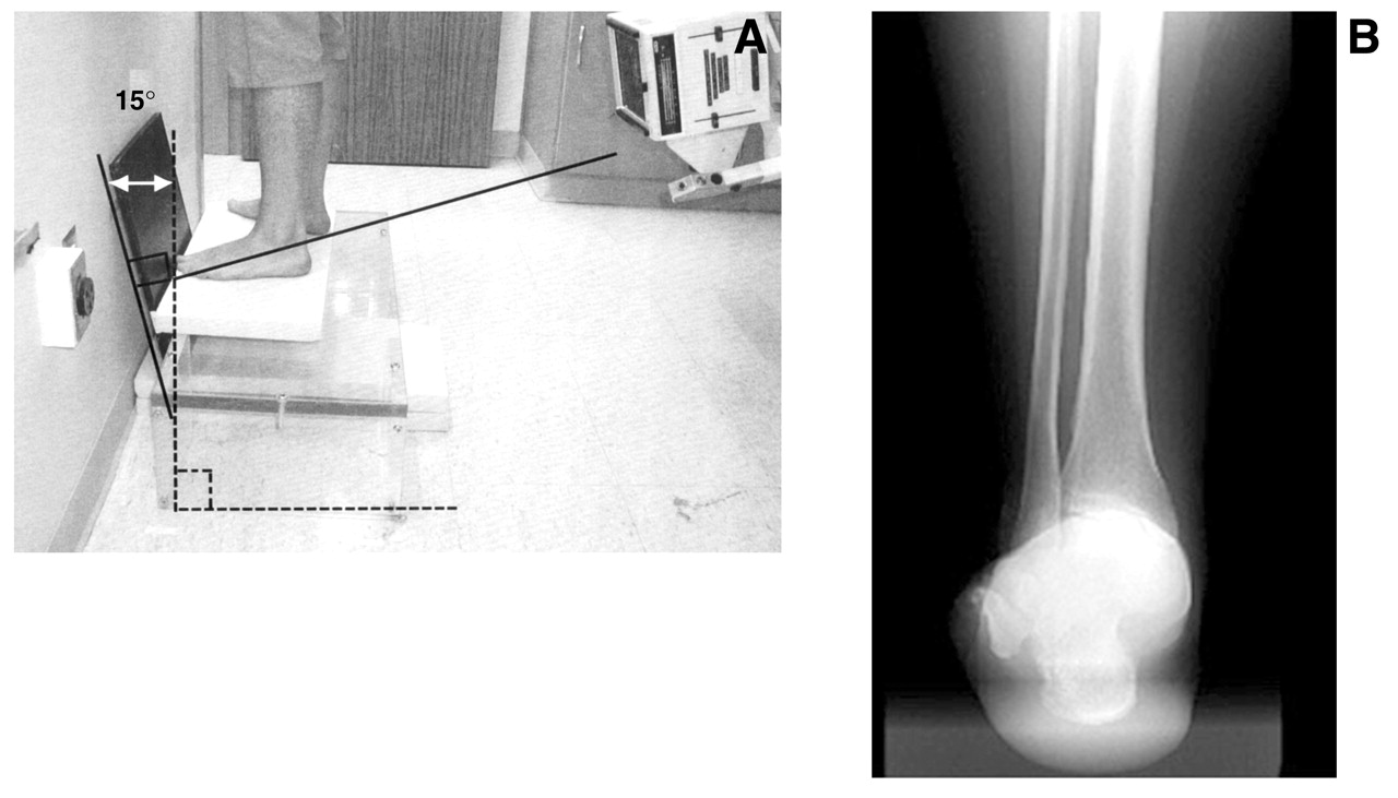
Anteroposterior projections of the foot may show transverse plane deformities of the metatarsus or lesser tarsus (Figure 5). However, significant overlap of the talus on the calcaneus as well as the lesser tarsus sometimes leads to radiographic “whiteout” and makes evaluation of these areas difficult. We routinely obtain long leg axial and rearfoot alignment views to help identify the level of deformity and to rule out a suprastructural skeletal deformity [14]. The long leg calcaneal axial radiograph uses a 14 × 17-inch film, and the x-ray projection is aimed 45° cephalad centered at the subtalar joint (Figure 6). This allows visualization of the subtalar joint and aids in identifying any structural and calcaneal-to-tibial deformities. The rearfoot alignment (modified Cobey view) radiograph uses an elevated platform and a 14 × 17-inch film positioned 15° from vertical with a posterior x-ray projection perpendicular to the film and centered at the ankle joint (Figure 7) [15]. This view evaluates the ankle joint and identifies any calcaneal-to-tibial deformities. The rearfoot alignment view represents a radiographic resting calcaneal stance position.
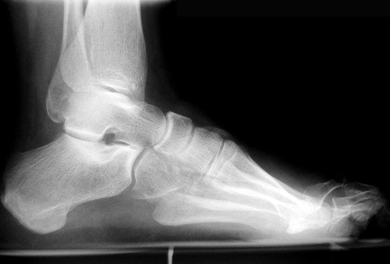
Figure 4.
Lateral radiograph showing classic findings of pes cavus: an increased talar–first metatarsal angle (Meary’s angle), an increased calcaneal inclination angle, a plantarflexed first ray relative to the lesser metatarsals, and digital contractures.
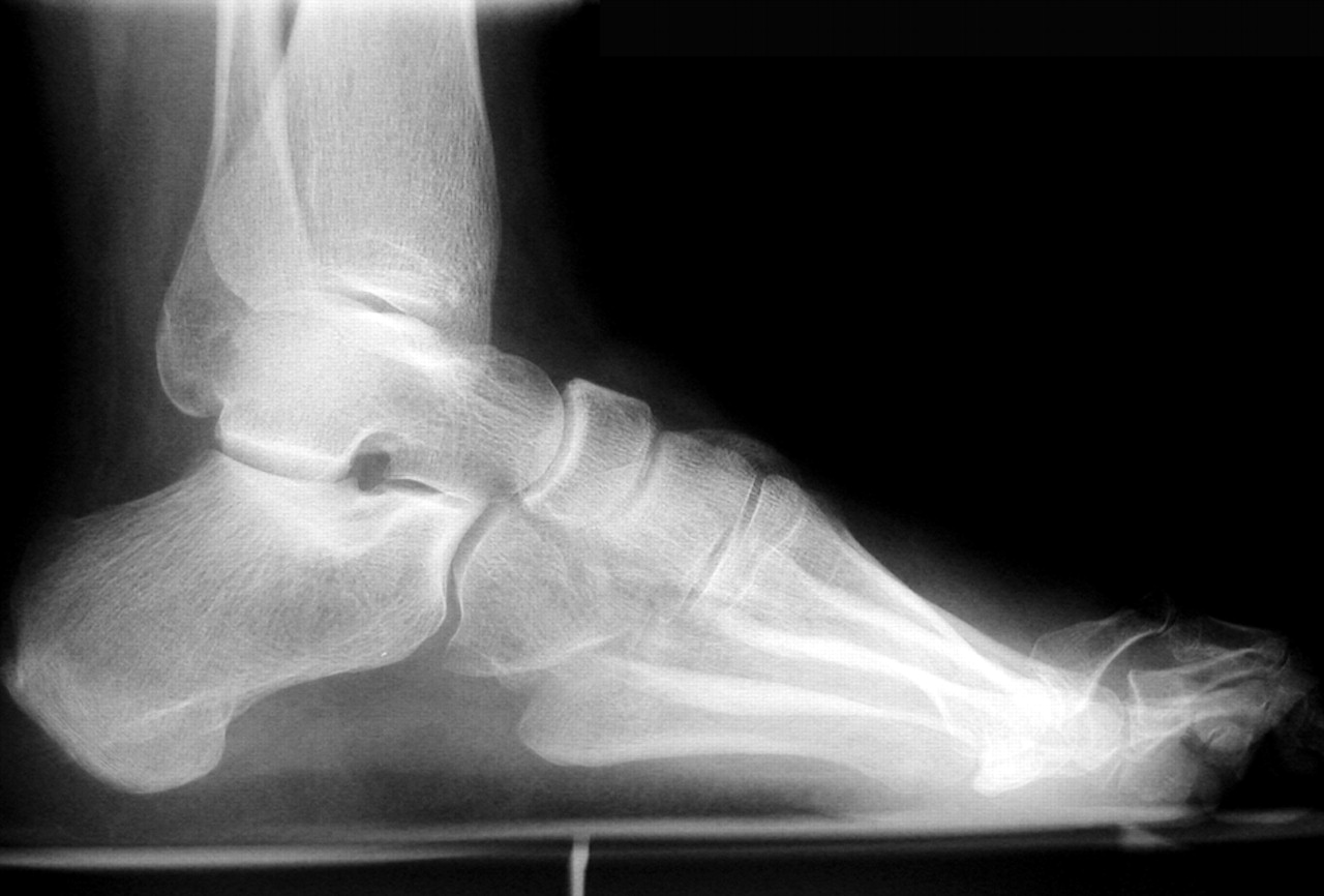
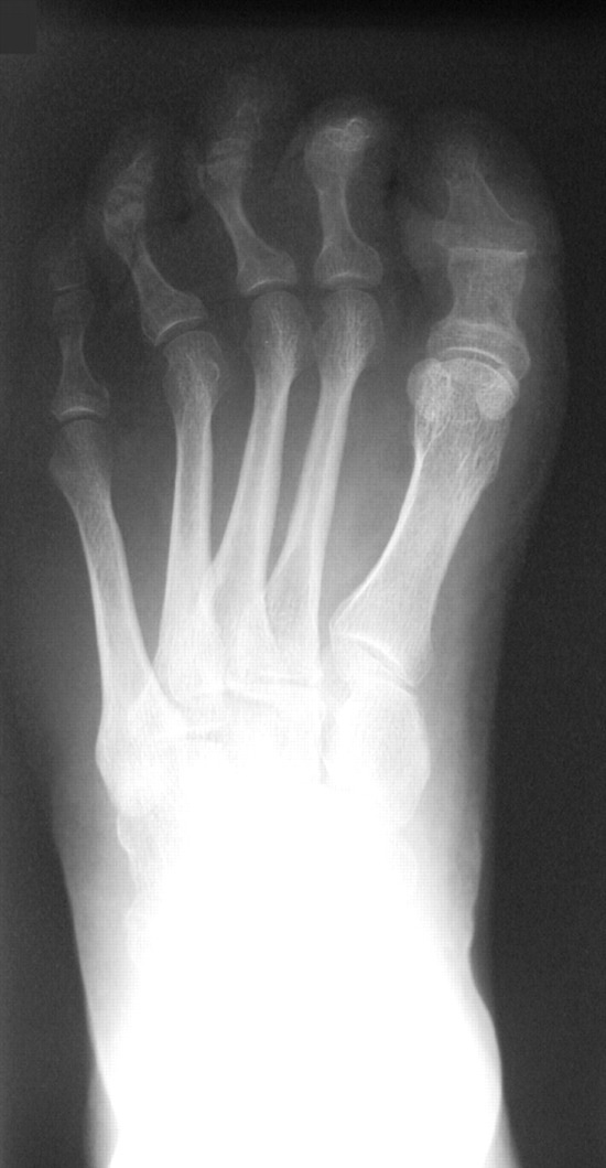
Figure 5.
Dorsoplantar radiograph showing mild forefoot adduction associated with cavus foot deformity.
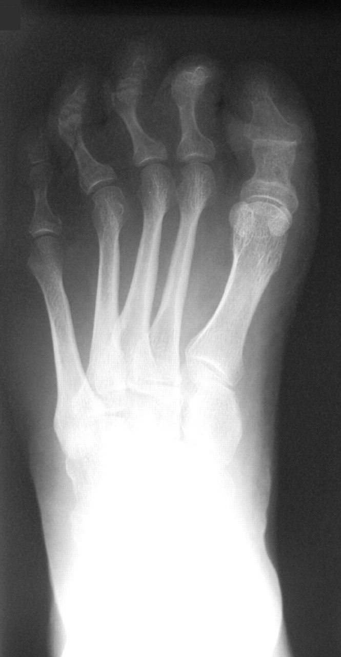
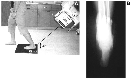
Figure 6.
A, Patient positioning for the long leg calcaneal axial radiograph; B, mild calcaneal varus in relation to the tibia.
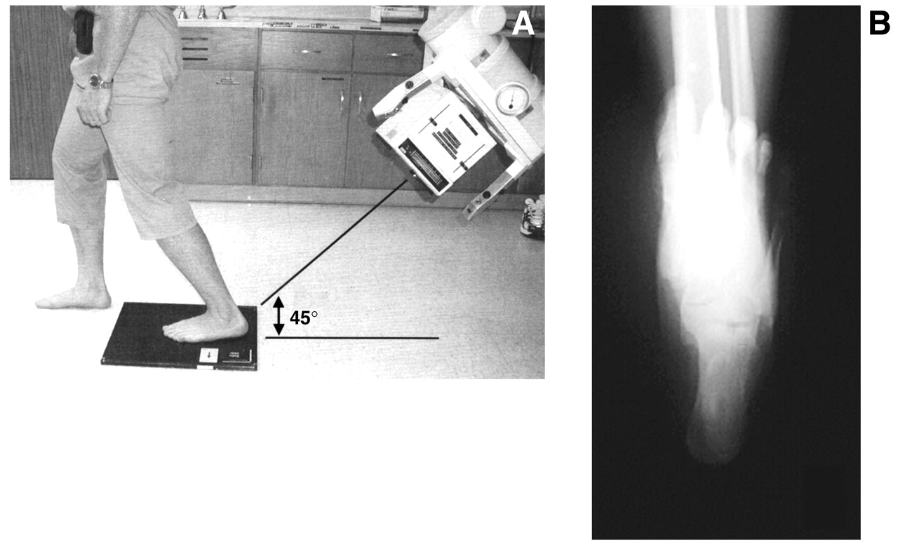
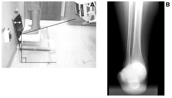
Figure 7.
A, Patient positioning for the rearfoot alignment (modified Cobey view) radiograph; B, calcaneal varus in relation to the ankle joint and tibia.
The ankle is evaluated for alignment and degenerative joint changes. The anterior ankle joint may show talotibial abutment and the presence of an osseous-type pseudoequinus. Patients with chronic lateral ankle instability may show an increased talocrural angle or evidence of talar dome osteochondral lesions [16]. Magnetic resonance imaging may be needed to fully evaluate suspicious lesions as well as any pathology of the lateral collateral ligaments and peroneal tendons that is common in chronic ankle instability [17].
Treatment
The goal of treatment is to achieve a foot that functions as a painless, mobile, plantigrade unit with good muscular balance. Conservative care in cavus foot management often fails owing to the progressive nature of the disorder and the degree of deformity that most patients have at the time of diagnosis [18]. In the presence of a nonprogressive idiopathic cavus foot, balanced padding, orthoses, metatarsal bars, and shoe modifications can be beneficial. However, when conservative measures fail and painful sub-metatarsal calluses and ankle instability persist, surgical intervention is often indicated.
Most foot deformities are secondary to intrinsic factors that cause abnormal foot mechanics or a variability among anatomical structures. Cavus deformity, however, should be considered a manifestation of a neuromuscular disorder with muscle imbalance until proven otherwise [18]. At least two-thirds of patients who seek treatment for symptomatic cavus foot deformities have an underlying neurologic defect, with more than half of these patients having Charcot-Marie-Tooth disease [1]. The prognosis of surgical reconstruction is greatly affected not only by the procedure selection itself but also by the progression of the patient’s neuromuscular status [11]. Therefore, treatment of the underlying neurologic abnormality must precede treatment of the foot.
Once the etiology has been established and surgical intervention has been decided on, proper procedure selection is extremely important. Several attempts to classify cavus foot deformities have been made [11,19,20]. However, no algorithm or classification scheme has been developed to help one choose a procedure or a combination of procedures that would address the unique deformity of a particular patient. No one procedure works for all cavus feet. The patient’s age, the flexibility of the deformity, the magnitude and nature of the deforming forces, and the anticipated foot demands must all be taken into account when considering treatment options [8]. Evaluation for proper procedure selection begins before surgery and continues throughout surgery. Some decisions may be delayed until a second operative procedure is performed on the basis of the outcome of the first [21]. The selection process begins and is influenced by understanding whether the cavus foot deformity being treated is progressive or static. As previously mentioned, a good neurologic examination and the help of a neurologist before surgery can aid in this decision-making process. Unfortunately, there are no known treatments or cures for many of the neurologic manifestations that produce a cavus foot. Established muscle weakness and imbalances are not reversible, even with successful arrest of the neurologic deterioration [18]. Therefore, the sequela of cavus foot often progresses.
A combination of soft-tissue and bony procedures may be necessary to satisfactorily correct a cavus foot deformity. Mosca [18] described three sound principles of cavus foot management: 1) correct all of the segmental deformities while preserving joint motion, 2) balance the remaining muscle forces, and 3) leave reasonable treatment options available for possible recurrence of deformity and pain. Using the preoperative assessment tools previously discussed, one can better identify the apex of the cavus foot deformity and can properly select the procedure or combination of procedures that will best serve the patient. Once the apex of the deformity has been identified, the surgeon should begin with soft-tissue release. Osseous procedures used to correct secondary bone deformities may be better determined after soft-tissue release has been performed.
Soft-Tissue Release
Soft-tissue release, popularized by Steindler [22,23], is designed to reduce the contracture of the plantar aponeurosis that is typical in most cavus foot deformities. This contracture leads to forefoot plantarflexion with an associated calcaneal varus. It is generally believed that soft-tissue release is better suited for the juvenile patient with less rigid deformities. However, as skeletal maturity evolves, soft-tissue release is typically considered as an adjunct to osseous procedures [11,24]. Several soft-tissue release procedures have been described [19,22,23,25,26]. The Steindler stripping maneuver, however, has been favored in recent years when contractures of the aponeurosis and plantar musculature are evident. The plantar fascia and the abductor hallucis, flexor digitorum brevis, and abductor digiti quinti muscles are stripped from the calcaneus when performing this procedure (Figure 8). The long plantar ligament may be palpated laterally and incised if it is believed to contribute to the deformity. The Steindler stripping maneuver will not correct rigid or fixed deformities.
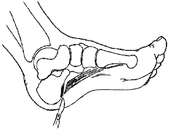
Figure 8.
The Steindler stripping procedure performed through a medial approach.
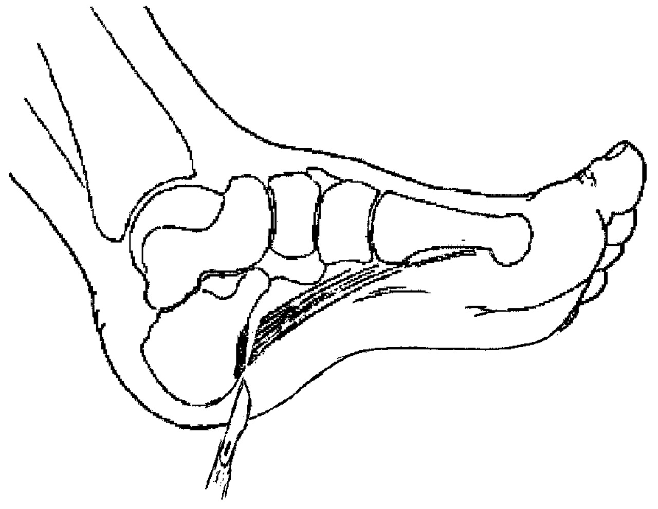
Neurovascular injury, tendon laceration, partial skin sloughs, and overcorrection have been reported with use of the Steindler stripping maneuver [19,27]. Attention to detail and proper surgical technique will help minimize complications.
Tendon Transfers
Various other soft-tissue procedures have been advocated in the treatment of cavus foot deformities. Tendon transfers help counterbalance muscular abnormalities associated with cavus disorders. Balancing deforming muscular forces by selecting the appropriate tendon and transferring it under the correct tension remains one of the most challenging aspects of cavus foot reconstruction [18]. Although transferring tendons alone may not permanently correct a cavus foot, removal of the deforming force is key in controlling a progressive deformity [11].
Several principles must be followed to achieve a successful tendon transfer. Understanding the deforming forces is critical. Alleviating one deforming force only by creating another should be avoided. Joints on which a transferred tendon should act should demonstrate adequate range of motion and be free of arthrosis. Furthermore, the axis of motion of these joints should be as close to perpendicular to the pull of the tendon as possible [11].
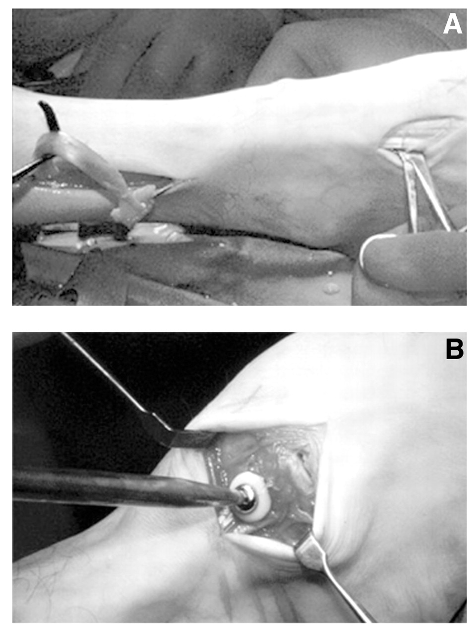
Posterior tibial tendon transfer through the interosseous membrane to the dorsum of the foot remains a valuable tool in eliminating its contribution to cavus foot deformity and provides dorsiflexory assistance when a weak anterior muscle group leads to a footdrop deformity, as seen in Charcot-Marie-Tooth disease (Figure 9) [28–32]. Stabilization of the rearfoot may be necessary owing to loss of a primary supinator, resulting in a severe pes valgus deformity through progressive subtalar joint pronation [11]. In the case of anterolateral muscle weakness, this transfer has been used with good results. Watkins et al [33], in 1954, described 29 patients who underwent a posterior tibial tendon transfer. They described good-to-excellent results in 24 patients. However, only one patient demonstrated peroneal tendon weakness before surgery. In 1961, Lipscomb and Sanchez [34] reported results in ten patients who had undergone transfer of the posterior tibial tendon for the treatment of post-traumatic common peroneal nerve palsy. All but one of the patients was judged to have a satisfactory result. These patients, however, had all undergone a simultaneous or previous triple arthrodesis. The authors believed it was important to stabilize the joints for the transfer to function maximally. In the presence of anterolateral muscle weakness, rearfoot stabilization can help prevent complications such as pes valgus and can enhance the overall pulling power of the transfer. This stabilization may be accomplished by isolated talonavicular, subtalar joint, or triple arthrodesis. Stabilization may not be necessary if the calcaneus cannot be everted past perpendicular [11].
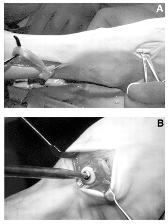
Figure 9.
Posterior tibial tendon transfer for neurogenic pes cavus and anterior muscle weakness. The posterior tibialis tendon is transferred through the interosseous membrane and is passed distally to the dorsal incision (A) and anchored to the dorsolateral cuneiform (B). (Courtesy of the Foot and Ankle Institute of the Western Pennsylvania Hospital, Pittsburgh).
Other types of tendon transfers have been advocated for the treatment of specific disorders when managing cavus foot deformities. The Girdlestone-Taylor transfer of the flexor digitorum longus tendon can be used in the surgical treatment of claw toe deformities [35]. The Jones tenosuspension may be used if there is an anterior muscle imbalance that allows for the extensor hallucis to hyperextend the metatarsophalangeal joint, causing a secondary hallux malleus [36]. The Hibbs suspension is often used in conjunction with the Jones procedure, in which the extensor digitorum longus tendon is transferred through a drill hole in the third cuneiform [37]. The Hibbs suspension helps load the midtarsal joint in dorsiflexion without significantly altering dorsiflexion of the ankle joint. The cavus foot deformity may be reduced if it is flexible, and this procedure is typically limited to flexible anterior cavus foot deformities.
The split anterior tibial tendon transfer is an effective adjunctive procedure in the treatment of a flexible cavus foot. Originally described in 1974 [38], it helps prepare the foot for weightbearing with a pronatory influence across the subtalar and midtarsal joint axis [11]. The lateral half of the tibialis anterior tendon is sectioned and anastomosed to the peroneus tertius tendon near the base of the fifth metatarsal. By transferring half of the tendon laterally, a greater dorsiflexory force is created that is perpendicular to the ankle joint. The split anterior tibial tendon transfer procedure is recommended only when there are weak extensor digitorum and extensor hallucis longus muscles but a strong tibialis anterior tendon. The procedure is rarely used alone. Other soft-tissue and osseous procedures may be used effectively with this procedure.
Osteotomies
A plantarflexed first metatarsal may be identified as the culprit in an anterior cavus foot deformity. This can be ascertained through clinical testing, as described previously here. A plantarflexed first metatarsal may be corrected by a dorsal closing wedge osteotomy at its base. This same type of closing osteotomy may also be performed in the first cuneiform with similar success [39]. Multiple basilar dorsal closing wedge osteotomies may be performed at the lesser metatarsals if clinically warranted and if a fixed anterior cavus foot deformity persists. More often, a fixed anterior cavus foot deformity is best managed through an osteotomy in the midtarsal joints.
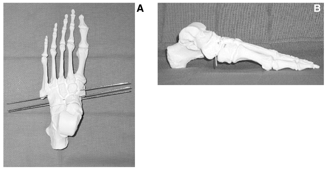
Several midfoot osteotomies have been developed to reduce a fixed anterior cavus foot deformity. The dorsal closing wedge osteotomy described by Cole [40], favored by us and surgeons at the Foot and Ankle Institute of the Western Pennsylvania Hospital, Pittsburgh, corrects forefoot equinus by means of an osteotomy through the naviculocuneiform joints and the cuboid laterally (Figure 10). Although this procedure is technically challenging, we simplified it by using fluoroscopy-assisted guide pins and demonstrated great success with use of the osteotomy when treating fixed anterior cavus foot deformities (Figure 11). We reviewed 11 feet in 8 patients who underwent the Cole osteotomy. A 100% fusion rate was achieved, with all patients at final follow-up being independently active and able to wear conventional shoes. A questionnaire was administered to patients at an average of 21 months after surgery; 80% of those who responded (four of five patients [seven of eight feet]) would recommend this surgery to others [41].
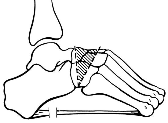
Figure 10.
The Cole procedure for midfoot osteotomy, which is often combined with other soft-tissue releases, such as the Steindler stripping procedure.
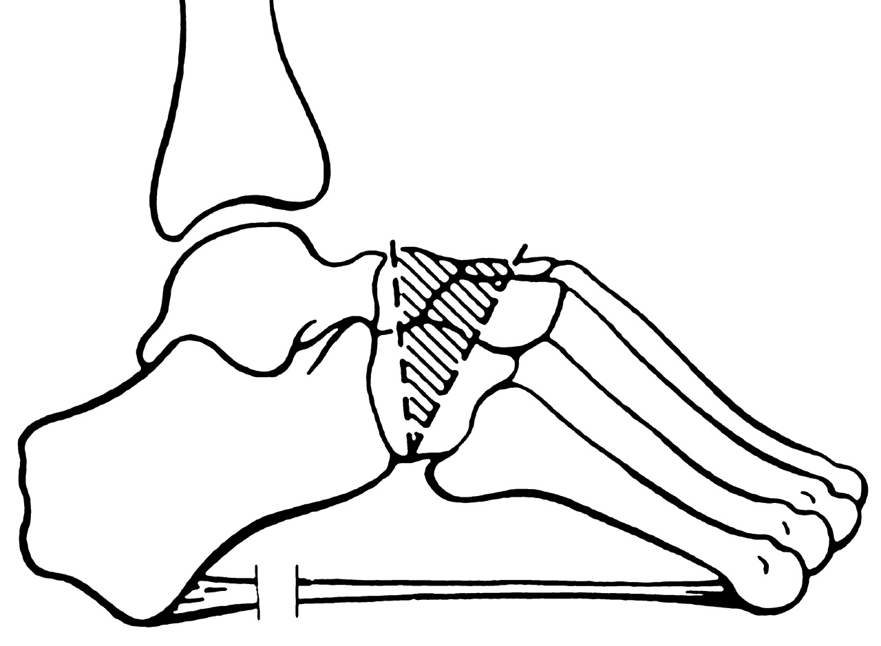
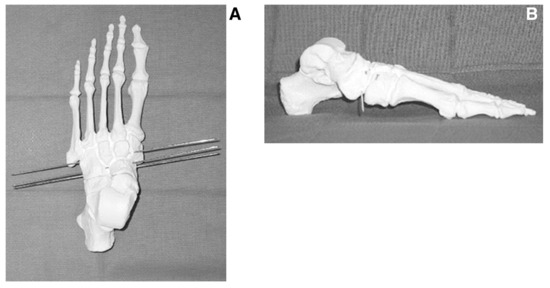
Figure 11.
Dorsoplantar (A) and lateral (B) views of the foot model demonstrating guide pin placement for performing a Cole midfoot osteotomy.
Few authors have published results primarily of the Cole osteotomy. The original article by Saunders [42] reviewed 102 feet in 86 patients with minimum follow-up of 2 years. Only 49% had good-to-excellent results. Leal et al [43] reported on eight feet without commenting on any results or complications. Levitt et al [44] reported a 30% pseudarthrosis rate with use of the Cole osteotomy. The Cole osteotomy can offer excellent reduction in fixed anterior cavus foot deformities but is technically challenging and must be performed with great care. An overzealous wedge resection can lead to a rocker-bottom foot deformity and continued difficulties with ambulation. Intraoperative imaging is paramount in visualizing the correct alignment of the osteotomy and in ensuring that no inadvertent intra-articular damage is done to the midtarsal joints.
Other types of osteotomies have been described for the correction of forefoot equinus. Japas [45] described a V-shaped osteotomy with the apex at the middle cuneiform and medial border just proximal to the first metatarsocuneiform joint and the lateral border proximal to the fifth metatarsocuboid joint. More recently, Jahss [46] described a truncated wedge osteotomy and arthrodesis through the tarsometatarsal joints. Although both may be technically complex, they achieve correction similar to that of the Cole osteotomy.
When the rearfoot position does not change with the lateral block test (indicating a relatively fixed rearfoot deformity), calcaneal osteotomies should be considered [8]. Dwyer [47] popularized a closing lateral wedge osteotomy of the calcaneus to correct the varus component of the rearfoot (Figure 12). This osteotomy may also allow for correction of an increased calcaneal pitch by superiorly displacing the posterior fragment. In addition to a lateral closing wedge, lateral translation and a medially based opening wedge osteotomy has been advocated for the treatment of rigid rearfoot varus. Gould [48] described a variation of this, as did Samilson and Dillon [49], who used a crescentic calcaneal osteotomy to achieve a more normal rearfoot position.
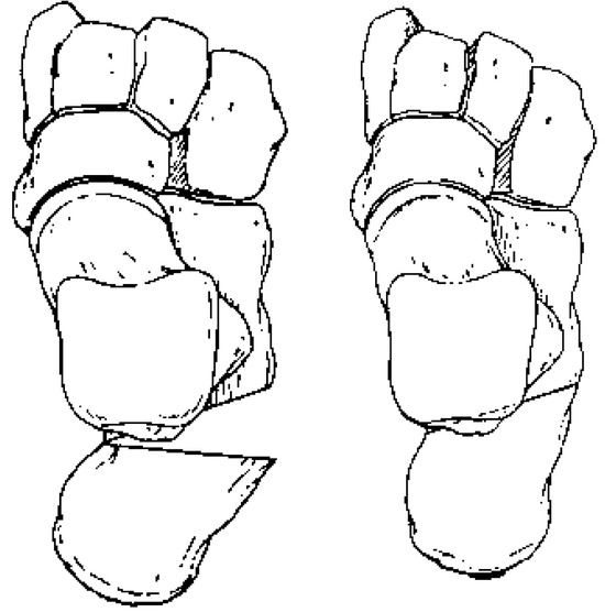
Figure 12.
The Dwyer closing calcaneal osteotomy for structural calcaneal varus.
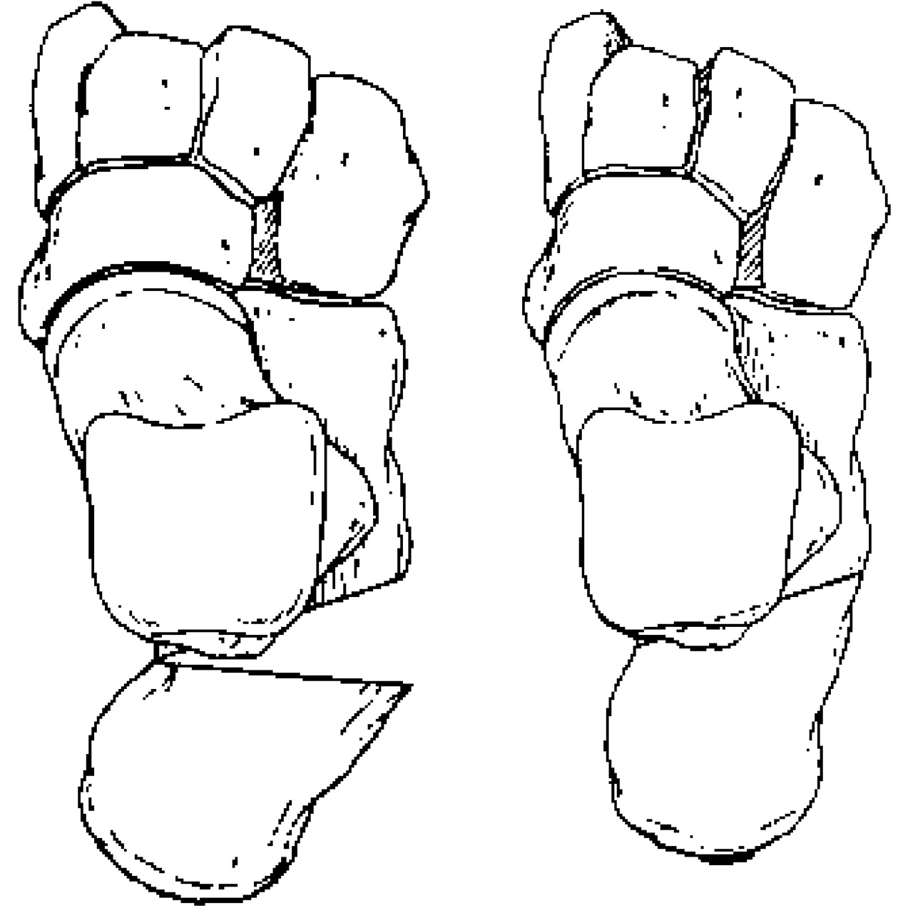
Combined with the Steindler stripping procedure, the Dwyer osteotomy has been advocated in the treatment of pediatric cavus foot in children older than 3 years [50]. The best results are obtained in the idiopathic cavus foot. Recurrence of deformity is more frequent in children with neuromuscular disease. The work of Levitt et al [44] supports these findings by casting doubt on the long-term effectiveness of calcaneal osteotomies in the treatment of pes cavus due to progressive neurologic conditions. They advocated triple arthrodesis as a salvage procedure when other procedures have failed or when stabilization is essential to prevent progression of the deformity with further neurologic deterioration.
Arthrodesis
Arthrodesis procedures of the tarsal and midtarsal regions are frequently used to stabilize and correct severe rigid cavus foot deformities. Triple arthrodesis has been the most widely accepted procedure in the management of pes cavus due to Charcot-Marie-Tooth disease or other progressive neurologic disorders. The stage at which this stabilization procedure should be performed is controversial. In patients with a severe rigid cavus foot deformity and markedly limited range of motion of the subtalar joint, triple arthrodesis is the best surgical option [51]. Correction of rearfoot varus is achieved through subtalar joint resection, whereas the remaining cavus foot deformity can be corrected through resection of the transverse tarsal joint. A conventional triple arthrodesis with planal resection is typically sufficient for deformities of a lesser degree. Levitt et al [44] advocated the Lambrinudi approach, which facilitates greater correction for more severe deformities.
These procedures may be combined with osteotomies, tendon transfers, and digital procedures. Stabilization of the foot by subtalar or midtarsal arthrodesis is a complex procedure that involves multiple joints, with appropriate realignment of each. Such realignment relies on proper joint preparation, with adequate osseous resection for correction. Proposed resections are typically ascertained from preoperative radiographs. However, one should be cautious when using such analysis. Defining a three-dimensional problem from a two-dimensional radiograph can be misleading [11]. Ankle joint stability should also be evaluated before any rearfoot stabilization procedure. If instability exists, then the ankle should be addressed at the time of surgery.
Arthrodesis procedures are not without complications. Most complications of triple arthrodesis in patients with Charcot-Marie-Tooth disease parallel and occur at the same frequency as those in patients with other diagnoses [52]. Undercorrection occurred in 45% of patients and overcorrection in 15% in a study by Wukich and Bowen [53]. Wetmore and Drennan [54] reported a 33% incidence of residual postoperative deformity, with undercorrection being the most common. Nonunions in triple arthrodesis most commonly occur at the talonavicular joint and were reported in approximately 7% of patients in the series by Wetmore and Drennan and 15% of patients reported by Wukich and Bowen. Degenerative arthritis after triple arthrodesis has also been widely reported in the literature. Angus and Cowell [55] reported on 80 feet after triple arthrodesis for a variety of deformities. Degenerative arthritis occurred in 31 of 80 ankles and in 43 of 80 midfeet.
Conclusion
Pes cavus is a complicated foot deformity of multi-faceted etiology and varying severity. Once the diagnosis of pes cavus has been made, understanding its etiology is crucial. Pes cavus should be managed as a sequela of a neurologic manifestation until proven otherwise. Proper clinical and radiographic testing should be implemented before any treatment to maximize correction and to halt further potential progression. No two cavus feet are alike, and they should be evaluated and treated on an individual basis. Careful surgical procedure selection should be based on the age of the patient, the characteristics of the deformity, and the anticipated foot demands. The various treatment options mentioned in this article serve as a basis for cavus foot reconstruction and should not be regarded as a panacea for all cavus feet. A significant learning curve should be anticipated before proficiency is attained in the surgical treatment of cavus foot deformities, which should be managed only by surgeons with extensive training in this type of foot pathology.
References
- Brewerton D, Sandifer P, Sweetnam D: “Idiopathic” pes cavus: an investigation into its etiology.BMJ2:659,1968.
- Ibrahim K: “Pes Cavus,” in Surgery of the Musculoskeletal System, ed by CM Evarts, p 4015, Churchill Livingstone, New York,1990.
- Mann RA, Missirian J: Pathophysiology of Charcot-Marie-Tooth disease.Clin Orthop234:221,1988.
- Wapner KL: “Pes Cavus,” in Foot and Ankle Disorders, Vol 2, ed by M Meyerson, p 919, WB Saunders, Philadelphia,2000.
- Amiot R, Coulter T, Nute M, et al: Surgical treatment of adult idiopathic cavus foot.J Bone Joint Surg Am85:1400,2003.
- Whittaker VB: Pes cavus: the value of electrodiagnosis.J R Army Med Corps109:234,1963.
- Jahss MH: Evaluation of the cavus foot for orthopedic treatment.Clin Orthop181:52,1983.
- Holmes JR, Hansen ST Jr: Foot and ankle manifestations of Charcot-Marie-Tooth disease.Foot Ankle14:476,1993.
- Sammarco GJ, Taylor R: Cavovarus foot treated with combined calcaneus and metatarsal osteotomies.Foot Ankle Int22:19,2001.
- Whitney AK, Green DR: Pseudoequinus.JAPA72:365,1982.
- Smith TF, Pitts T, Green DR: “Pes Cavus,” in Comprehensive Textbook of Foot Surgery, 2nd Ed, Vol 1, ed by ED McGlamry, AS Banks, MS Downey, p 742, Williams & Wilkins, Baltimore,1992.
- Coleman SS, Chestnut WJ: A simple test for hindfoot flexibility in the cavovarus foot.Clin Orthop123:60,1977.
- Tachdjian MO: “Pes Cavus,” in Pediatric Orthopedics, Vol 2, ed by MO Tachdjian, p 1378, WB Saunders, Philadelphia,1972.
- Paley D: The correction of complex foot deformities using Ilizarov’s distraction osteotomies.Clin Orthop293:97,1993.
- Marks RM: “Radiographic Evaluation of the Foot and Ankle,” in Foot and Ankle Disorders, Vol 1, ed by M Meyerson, p 83, WB Saunders, Philadelphia,2000.
- Karlsson J, Lansinger O: Lateral instability of the ankle joint.Clin Orthop276:253,1992.
- Kerr R: MRI of soft tissue disorders of the ankle.Clin Podiatr Med Surg19:285,2002.
- Mosca V: The cavus foot.J Pediatr Orthop21:423,2001.
- Paulos L, Coleman SS, Samuelson KM: Pes cavovarus.J Bone Joint Surg Am62:942,1980.
- Alexander I, Johnson K: Assessment and management of pes cavus in Charcot-Marie-Tooth disease.Clin Orthop246:273,1989.
- Smith TF, Castellano B: “Rigid/Flexible Cavus Foot Deformities,” in Reconstructive Surgery of the Foot and Leg: Update ’87, ed by ED McGlamry, p 68, The Podiatry Institute, Tucker, GA,1987.
- Steindler A: Operative treatment of pes cavus.Surg Gynecol Obstet24:612,1917.
- Steindler A: Stripping of the os calcis.Am J Orthop Surg2:8,1920.
- Jay RM: “Cavus Deformity,” in Pediatric Foot and Ankle Surgery, ed by RM Jay, p 211, WB Saunders, Philadelphia,1999.
- Rugh JT: The plantar fascia: a study of its anatomy and its pathology in talipes cavus: a new operation for its correction.Am J Surg14:335,1932.
- Thomas W: On the treatment of talipes cavus.Birmingham Med Rev34:1,1917.
- Sherman FC, Westin GW: Plantar release in the correction of deformities of the foot in childhood.J Bone Joint Surg Am63:1382,1981.
- Jacobs JE, Carr CR: Progressive muscular atrophy of the peroneal type: orthopedic management and end result study.J Bone Joint Surg Am32:27,1950.
- Shapiro F, Brensan MJ: Orthopedic management of childhood neuromuscular disease: part II. Peripheral neuropathies, Friedreich’s ataxia, and arthrogryposis multiplex congenital.J Bone Joint Surg Am64:949,1982.
- Erying EJ, Earl WC, Brockmyer F: Posterior tibial tendon transfers in neuromuscular conditions other than anterior poliomyelitis.Arch Phys Med Rehabil55:124,1974.
- Turner JW, Cooper RR: Anterior transfer of the tibialis posterior through the interosseous membrane.Clin Orthop83:241,1972.
- Ober FR: Tendon transposition in the lower extremity.N Engl J Med209:52,1933.
- Watkins MB, Jones JB, Ryder CT, et al: Transplantation of the posterior tibial tendon.J Bone Joint Surg Am36:1181,1954.
- Lipscomb PR, Sanchez JJ: Anterior transplantation of the posterior tibial tendon for persistent palsy of the common peroneal nerve.J Bone Joint Surg Am45:60,1961.
- Girdlestone GR: Physiotherapy for hand and foot.Chartered Soc Physiother32:167,1947.
- Jones R: An operation for paralytic calcaneocavus.Am J Orthop Surg4:371,1908.
- Hibbs RA: An operation for “clawtoe.”. JAMA73:1583,1919.
- Hoffer MM, Reising JA, Garrett AM: The split anterior tibial tendon transfer in the treatment of spastic varus hindfoot of childhood.Orthop Clin North Am5:31,1974.
- Fowler SB, Brooks AL, Parish TF: The cavovarus foot.J Bone Joint Surg Am51:757,1959.
- Cole WH: The treatment of claw foot.J Bone Joint Surg22:895,1940.
- Tullis B, Mendicino RW, Catanzariti AR, et al: The Cole midfoot osteotomy: a retrospective review of 11 procedures in 8 patients.J Foot Ankle Surg43:160,2004.
- Saunders JT: Etiology and treatment of clawfoot.Arch Surg30:2,1935.
- Leal LO, Bosta SO, Feller DP: Anterior tarsal resection (Cole osteotomy).J Foot Surg27:259,1988.
- Levitt RL, Canale ST, Cook AJ, et al: The role of foot surgery in progressive neuromuscular disorders in children.J Bone Joint Surg Am55:1396,1973.
- Japas LM: Surgical treatment of pes cavus by tarsal V-osteotomy: a preliminary report.J Bone Joint Surg Am50:927,1968.
- Jahss MH: Tarsometatarsal truncated-wedge arthrodesis for pes cavus and equinovarus deformity of the forepart of the foot.J Bone Joint Surg Am62:713,1980.
- Dwyer FC: The present status of the problem of pes cavus.Clin Orthop106:254,1975.
- Gould N: Surgery in advanced Charcot-Marie-Tooth disease.Foot Ankle4:267,1984.
- Samilson RL, Dillon W: Cavus, cavovarus and calcaneocavus: an update.Clin Orthop177:125,1983.
- Dekel S, Weissman SL: Osteotomy of the calcaneus and concomitant plantar stripping in children with talipes cavovarus.J Bone Joint Surg Br55:802,1973.
- Alexander IJ, Johnson KA: Assessment and management of pes cavus in Charcot-Marie-Tooth disease.Clin Orthop246:273,1989.
- Roy DR, Al-Sayyad MJ: Complications of surgery of the foot and ankle in hereditary neurological disorders.Clin Orthop1:181,2001.
- Wukich DK, Bowen JR: A long term study of triple arthrodesis for correction of pes cavovarus in Charcot-Marie-Tooth disease.J Pediatr Orthop9:433,1989.
- Wetmore RS, Drennan JC: Long-term results of triple arthrodesis in Charcot-Marie-Tooth disease.J Bone Joint Surg Am71:417,1989.
- Angus PD, Cowell HR: Triple arthrodesis: a critical long-term review.J Bone Joint Surg Br68:260,1986.
© 2005 American Podiatric Medical Association