


Abstract
Forty patients (12 men and 28 women) treated with isolated subtalar joint arthrodesis were retrospectively reviewed. The average patient age was 50 years (range, 21–76 years). Preoperative diagnoses included posterior tibial tendon dysfunction, post-traumatic arthritis, nontraumatic arthritis, and subtalar joint middle facet coalition. The average follow-up was 15 months (range, 12–74 months). Subjective postoperative questionnaire results were classified as satisfied (n = 32), satisfied but with reservations (n = 4), or dissatisfied (n = 4). Eighty-three percent of the patients (n = 33) stated that they would undergo the procedure again. Minor complications (those that resolved with nonoperative treatment) occurred in 55% of the patients. However, the major complication rate was only 12.5%. This study showed no statistical correlation between the preoperative diagnosis and the postoperative outcome. Our results also suggested that the prevalence of complications is slightly higher than in previous reports. Isolated subtalar joint arthrodesis is an effective treatment for pain and deformity of the rearfoot.
Isolated subtalar joint arthrodesis has been recommended for post-traumatic arthropathy, neurologic disorders, primary osteoarthritis, talocalcaneal coalition, posterior tibial tendon insufficiency, and inflammatory arthritis. The goals of this procedure are to eliminate pain, restore stability, and realign the rearfoot. High patient satisfaction and low complication rates have been reported with this procedure [1–9]. Isolated subtalar joint arthrodesis has been recommended as an alternative to triple arthrodesis for pathology confined to the subtalar joint. Isolated subtalar joint arthrodesis preserves approximately 50% of the midtarsal joint motion preserved with triple arthrodesis [7,10,11].
Although isolated subtalar joint arthrodesis is well documented, most studies neglect to evaluate how the preoperative diagnosis influences postoperative outcomes and complications. The purpose of this study was to identify the complications and patient satisfaction associated with isolated subtalar joint arthrodesis. In addition, we attempted to correlate these complications to the preoperative diagnosis.
Methods
Forty consecutive patients who underwent isolated subtalar joint arthrodesis between January 1, 1994, and December 31, 2000, were retrospectively reviewed at the Western Pennsylvania Hospital, Pittsburgh. Isolated subtalar joint arthrodeses were performed in patients with a preoperative diagnosis of adult-acquired flatfoot secondary to posterior tibial tendon dysfunction, tarsal coalition, and primary and post-traumatic subtalar joint osteoarthritis. Patients were excluded from this study if any additional arthrodesis procedures were concomitantly performed. Medical records and radiographs were evaluated. The collected clinical data included sex, age, preoperative diagnosis, occupation, history of systemic illness, previous surgery, type and duration of nonoperative care, concomitant procedures, type of fixation, type of bone graft, time to osseous union, follow-up time, time to full weightbearing, smoking history, and postoperative complications. Nonoperative therapy included modification of activities or occupational status, nonsteroidal anti-inflammatory medications, immobilization, orthoses, ankle-foot orthoses, various types of braces, physical therapy, and arthrocentesis with corticosteroids. All of the patients in this study were not adequately helped by nonoperative care.
The diagnostic tools used to evaluate the subtalar joint included standard foot and ankle radiographs, advanced imaging (computed tomography and magnetic resonance imaging), and diagnostic intra-articular injections. Preoperative and postoperative weightbearing anteroposterior and lateral foot and ankle radiographs and calcaneal axial radiographs were evaluated for degenerative joint disease, tarsal coalition, and osseous union of the subtalar joint. Osseous union after subtalar joint arthrodesis was recorded when no subtalar joint motion was detected on clinical examination and when evidence of trabeculation across the arthrodesis site was observed on plain film radiographs (Figure 1). Patients who did not meet these criteria were evaluated with the use of computed tomography. A delayed union was defined as a successful fusion 6 to 9 months after surgery with the same aforementioned clinical and radiographic union characteristics. Radiographic angles were not analyzed owing to the variety of preoperative foot pathologies. In addition, a postoperative subjective questionnaire was used at the last follow-up visit to determine patient satisfaction. Patients were questioned about their level of pain, cosmesis, functional capacities, use of walking aids, ability to wear shoes, and willingness to undergo the surgery again (Table 1).
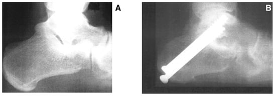
Figure 1.
Preoperative (A) and postoperative (B) radiographs demonstrating successful subtalar joint arthrodesis.
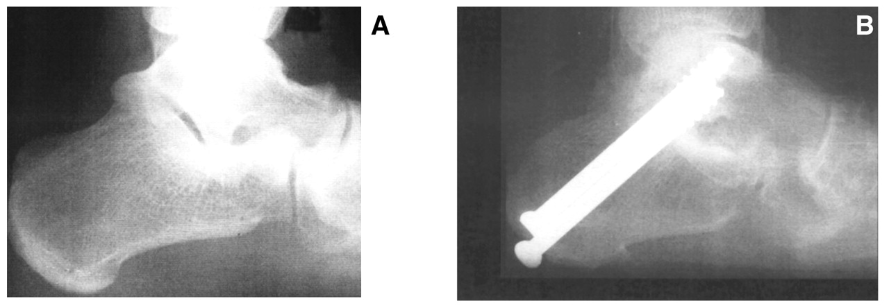
Table 1.
Patient Satisfaction Questionnaire.
This study evaluated minor and major postoperative complications. Minor complications were defined as transient postoperative symptoms that resolved with nonoperative treatment. Major complications were defined as continued postoperative symptoms that were resistant to nonoperative treatment or that required revisional surgery. These types of complications were compared with the types of preoperative diagnoses.
The procedure was performed with the patient in the supine position, with a bump under the ipsilateral hip to gain access to the lateral rearfoot. General or spinal anesthesia was administered, and a thigh tourniquet was used for hemostasis. An incision was made beginning at the tip of the fibula and extending over the sinus tarsi toward the fourth metatarsal base, with care taken to identify all neurovascular structures. A communicating branch from the intermediate dorsal cutaneous nerve to the sural nerve was sometimes encountered. The peroneal tendon sheath was incised just distal to the tip of the fibula, and the tendons were retracted plantarly. A deep fascial incision was made beginning just inferior to the lateral malleolus and extending distally just inferior to the extensor digitorum brevis muscle belly. A vertical deep fascia–periosteal incision was made along the sinus tarsi and extending inferiorly to meet the horizontal incision. All further dissection was subperiosteal.
The soft-tissue contents of the sinus tarsi were evacuated, which permitted visualization of the subtalar joint middle and posterior facets and allowed access to the underlying cortical bone for decortication. The subtalar joint was mobilized, and a lamina spreader was placed in the sinus tarsi region. The articular cartilage was debrided from the posterior and middle facets. Emphasis was placed on developing a healthy cancellous substrate conducive to primary arthrodesis while maintaining joint contour and talar height. Limited debridement techniques were used, which permitted positional realignment without large deficits and prevented lateral fibular impingement from excessive loss of talar height. In addition, maintaining a portion of the subchondral plate enhanced the stiffness of the fixation construct.
The surgical site was irrigated of all cartilaginous debris, and the subchondral surface was prepared with fenestration and scaling. The subtalar joint was then positioned, and provisional fixation was obtained using large cannulated guide pins. This maneuver was performed under image intensification. Large-diameter cancellous screws were then delivered over the guide pins. Placement was confirmed by intraoperative imaging. The dorsal aspect of the calcaneus and the undersurface of the talus in the sinus tarsi were then decorticated. Bone graft was packed into the sinus tarsi to augment the primary arthrodesis site.
Postoperative management included 6 to 9 weeks in a nonweightbearing short-leg cast. Patients were then advanced to weightbearing casts, rocker-bottom braces, and then standard footwear. Progression to weightbearing depended on radiographic evidence of consolidation at the arthrodesis site.
Descriptive and inferential statistical procedures were used to analyze the resulting data. For each variable collected, the numbers and percentages or means and SDs were calculated. To analyze predictors of complications, a χ2 analysis was performed. A statistically significant difference was defined as P ≤ .05.
Results
The 12 men (30%) and 28 women (70%) included in the study had an average age of 50 years (range, 21–76 years). Preoperative diagnoses included posterior tibial tendon dysfunction (n = 19), post-traumatic arthritis (n = 11), nontraumatic arthritis (n = 6), and subtalar joint middle facet coalition (n = 4). Seventeen patients were smokers (42.5%) and 23 were nonsmokers (57.5%). One patient underwent revisional subtalar joint arthrodesis for a previous nonunion. One patient had a medical history of psoriatic arthritis. The average follow-up was 15 months (range, 12–74 months), and the mean time to full weightbearing was 14 weeks (range, 7–60 weeks).
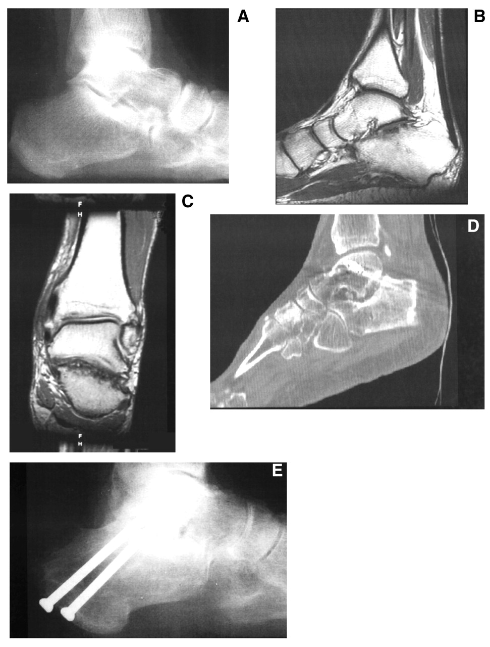
Radiographic and clinical assessment demonstrated that all but four feet went on to primary union, for an overall union rate of 90%. The overall time to union ranged from 7 to 45 weeks, with an average of 12.6 weeks. Of the four patients with nonunions, two underwent revisional arthrodesis and went on to successful union, one was lost to follow-up, and one refused any further surgical intervention and at the time of follow-up was symptomatic, wore a brace, and was receiving disability benefits (Figure 2). Three of the four nonunions were in patients who smoked. There was a 17.6% nonunion rate in the smoking group and a 4.6% nonunion rate in the nonsmoking group. Delayed unions occurred in only three patients, none of whom were augmented with a bone graft.
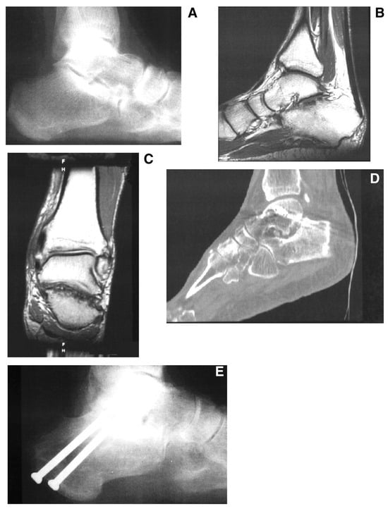
Figure 2.
Case study of a 40-year-old man with nontraumatic arthritis of the subtalar joint. A preoperative lateral radiograph (A) and proton-density magnetic resonance images (B and C) demonstrate severe arthritic changes. A computed tomographic scan (D) and a lateral radiograph (E) 1 year after surgery confirm a nonunion.
Bone grafts were used in 18 patients (45%) to enhance primary union, to provide and maintain structural realignment, or to fill a defect. The bone grafts that were implanted included autogenous anterior iliac crest grafts (n = 4), autogenous tibial strut grafts (n = 3), allogenic cancellous grafts (n = 5), and autogenous cancellous grafts (n = 6). Seven patients received a structural bone graft. Ancillary surgical procedures included tendo Achillis lengthening (n = 12), flexor digitorum longus tendon transfer (n = 13), direct repair of the tibialis posterior tendon (n = 2), subtalar joint middle facet coalition resection (n = 4), and excision of an os tibiale externum (n = 2). Large cannulated (7.0- or 7.3-mm) screws were used for internal fixation in all patients. Thirty-three patients had screws oriented from the plantar proximal calcaneal tuberosity dorsal distally into the talus, whereas in seven patients the screws were directed from the dorsal neck of the talus plantar posteriorly into the calcaneus.
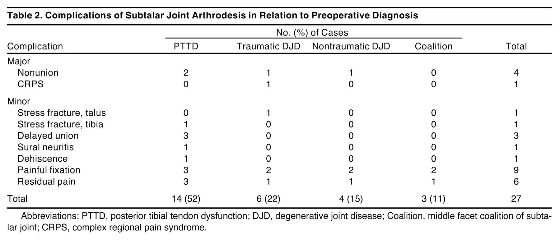
The overall complication rate for an isolated subtalar joint fusion was 37.5% (15 of 40 patients). Nine of these patients had both minor and major complications. The minor complication rate was 55% and included nine patients with painful internal fixations, six with residual postoperative pain, one with sural neuritis, three with delayed unions, one with a stress fracture of the talus, one with a stress fracture at the distal tibial metaphysis (the harvest site of a bone graft), and one with wound dehiscence. The major complication rate was 12.5% and included one patient with complex regional pain syndrome and four with nonunions.
The nine patients with painful internal fixation had the screws removed, which provided immediate relief of symptoms. All other minor complications went on to resolve uneventfully with the use of conservative treatment consisting of extended periods of nonweightbearing, anti-inflammatory medications, corticosteroid injections, and local wound care. The patient with complex regional pain syndrome received inadequate relief from all treatment options and was referred to a pain management center. One patient with residual postoperative pain related continued mild-to-moderate pain with activity that was controlled by using anti-inflammatory medications.
Patients were grouped based on the preoperative diagnosis: posterior tibial tendon dysfunction, post-traumatic arthritis, nontraumatic arthritis, and middle facet talocalcaneal coalition. Twelve (63%) of the 19 patients with posterior tibial tendon dysfunction had minor complications, including painful internal fixation (n = 3), sural neuritis (n = 1), delayed union (n = 3), stress fracture at the tibial graft site (n = 1), wound dehiscence (n = 1), and residual postoperative pain (n = 3). Two (10.5%) of the 19 patients with posterior tibial tendon dysfunction had major complications, both of which were nonunions. Four (36%) of the 11 patients with post-traumatic arthritis had minor complications, including painful internal fixation (n = 2), stress fracture of the talus (n = 1), and residual postoperative pain (n = 1). Two (18%) of the 11 patients with post-traumatic arthritis had major complications, including complex regional pain syndrome (n = 1) and nonunion (n = 1). Three (75%) of the four patients with a middle facet talocalcaneal coalition experienced minor complications, including residual postoperative pain (n = 1) and painful internal fixation (n = 2). There were no nonunions or major complications in the coalition group. Three (50%) of the six patients with nontraumatic arthritis had minor complications, including painful internal fixation (n = 2) and residual postoperative pain (n = 1). One nonunion (16.7%) was the only major complication recorded in this group (Table 2).
Table 2.
Complications of Subtalar Joint Arthrodesis in Relation to Preoperative Diagnosis.
All 40 patients completed a subjective questionnaire at the last follow-up visit. Fifteen percent of the patients had pain with limitation of activities. Sixty-five percent of the patients were satisfied with the resultant appearance of the foot. The ability to function on uneven terrain was at least somewhat difficult in 65% of the patients. Ninety-five percent of the patients were able to wear either normal or athletic footwear. Thirty-two patients (80%) were satisfied with the procedure, four (10%) were satisfied but with reservations, and four (10%) were dissatisfied with the results of their surgery. Thirty-three patients (82.5%) responded that they would undergo the procedure again, and seven (17.5%) stated that they would not (Table 1).
A statistical analysis was performed to compare all of the previously mentioned data. No statistical significance was found owing to the small sample size. However, we discovered a trend that showed that patients who smoked had a greater chance of nonunion.
Discussion
There were 28 women (70%) and 12 men (30%) in this review. The high female-to-male ratio was similar to that of other isolated subtalar joint arthrodesis series that had a large number of patients with posterior tibial tendon dysfunction [5]. Several studies [2,12–15] report the opposite, which can be attributed to the larger number of post-traumatic etiologies in these series. The average patient age of 50 years was comparable to that of Mangone et al [6], whose average age was 53 years. However, our patient population was approximately a decade older than the populations of previous studies [7,13] whose preoperative diagnoses included mostly traumatic etiologies.
The average follow-up was 15 months, which is short compared with that of other studies [5,7,8]. Because of the short follow-up, radiographic evaluation of angular measures and adjacent joint arthritis were not analyzed. Adjacent joint arthrosis has been a reported long-term follow-up finding after rearfoot arthrodesis procedures [7,16].
Proponents of isolated subtalar joint arthrodesis agree that this procedure increases stress on the adjacent joints but preserves most of the motion at the midtarsal joint. Thus the incidence of clinically significant arthrosis at the ankle joint may be less compared with that of triple arthrodesis [11,15]. Talonavicular joint motion has been shown to decrease 13% to 45% in vitro and 40% in vivo after isolated subtalar joint arthrodesis [7,10]. Mild-to-moderate degenerative changes in the ankle joint (36%) and midtarsal joint (41%) have also been demonstrated after subtalar joint arthrodesis [7]. It is well known that neither slight varus nor excessive valgus is well tolerated after rearfoot surgical procedures [7,10,17–20]. We believe that positional realignment of the subtalar joint is critical and may be the most significant factor relative to outcome.
There were 36 feet that went on to primary union; therefore, our overall union rate was 90%. The union rates reported in the literature vary from 84% to 100% [5–8,13]. The overall time to union averaged 12.6 weeks, which is comparable to that of other studies [7]. The mean time to full weightbearing was 14 weeks, which is similar to that of previous reports of approximately 12 weeks until unprotected weightbearing [6].
Smoking has been clearly shown to impede bone healing [1,13,21]. The results of our retrospective study demonstrate that three of four nonunions occurred in smokers. Also, one of the three delayed unions in our study occurred in a smoker. Cobb et al [22] reported that the relative risk of nonunion was increased 3.75 times for the active smoker undergoing ankle joint arthrodesis. Bednarz et al [1] reported that 4 of 28 isolated subtalar joint arthrodeses resulted in nonunions, all of which were in smokers. Another study [13] reviewed 184 isolated subtalar joint arthrodeses and reported that 73% of 30 nonunions occurred in smokers. Haverstock and Mandracchia [21] recommend that patients who smoke seek medical treatment for nicotine addiction before elective foot and ankle surgery owing to the risk of delayed healing and nonunion.
Bone grafts were used in 45% of our patients to enhance fusion of the arthrodesis site. All seven patients treated with structural bone grafts (autogenous anterior iliac crest grafts and autogenous tibial strut grafts) went on to primary fusion. This finding differs from that of a study by Easley et al [13], which showed that only 83% of the structural autogenous grafting cases went on to union. In this same study, 40% of patients who received structural allograft went on to nonunion. In contrast, none of our patients had a structural allograft used. The use of structural allogenic bone graft in joint arthrodesis is a relative contraindication [23]. Nonstructural autogenous bone grafting in subtalar joint arthrodesis was used in a study [8] of 45 fusions, with only one patient experiencing nonunion. Kitaoka and Patzer [5] stated that bone grafting is unnecessary when fusing the subtalar joint in patients with posterior tibial tendon dysfunction.
Our study revealed an overall complication rate of 37.5%. Some of the isolated subtalar joint arthrodesis literature indicates high union rates and low complication rates [5,6]. However, these studies were concentrated only on correcting flatfoot deformities. In contrast, Easley et al [13] reported markedly higher complication rates for the correction of a variety of preoperative diagnoses. Our study reflected similar findings.
Our major complications (12.5%) were complex regional pain syndrome and nonunion. Complex regional pain syndrome occurred in one patient who had a subtalar joint fusion secondary to a traumatic degeneration of the joint. Similarly, Amendola and Lammens [12] reported complex regional pain syndrome as a complication after subtalar joint arthrodesis following a calcaneal fracture. Delayed unions occurred in three patients with a preoperative diagnosis of posterior tibial tendon dysfunction. To our knowledge, delayed unions have been unreported in the literature, and few studies have mentioned malunion as a postoperative complication [7]. No malunions were identified in our study. Four nonunions occurred in our series, and, as with the study by Easley et al [13], we found that neither the preoperative diagnosis nor the complexity of the fusion (bone grafting or revisional arthrodesis) had any relationship to the nonunion rate. Like previous reports, our study showed that smoking greatly increased the risk of nonunion in subtalar joint fusion.
Minor complications (55%) included painful internal fixation, sural neuritis, wound dehiscence, stress fractures, and residual postoperative pain. Although stress fractures occurred in two patients, after surgery they healed uneventfully. These minor complications are frequently encountered in any foot and ankle surgical procedure, and they were treated successfully with conservative care. Although defined as a minor complication, painful internal fixation can lead to additional interventions, such as operative or advanced imaging. Similar to our results, Easley et al [13] and Carr et al [2] found painful fixation or heel pad irritation in approximately 20% of their patients. Therefore, we modified the screw placement to avoid the weightbearing area of the plantar calcaneus. Residual postoperative pain was common in our study and in many others [5,7,13]. We theorize that this may be the result of an unrecognized suprastructural deformity, and we currently use a radiographic protocol to evaluate rearfoot alignment to prevent this oversight.
Most articles that discuss subtalar joint arthrodesis use a modified version of the American Orthopaedic Foot and Ankle Society ankle-hindfoot scoring system [5,7,13]. Easley et al [13] found that 84% of their patients returned to their usual daily activities after arthrodesis. Mangone et al [6] reported that 82% of their patients would have the procedure performed again. Kitaoka and Patzer [5] discovered that some of their patients had ankle pain when walking on uneven surfaces. The results of our subjective questionnaire are difficult to compare; however, most of our patients were satisfied with their results and would undergo the procedure again.
Subtalar joint pathology is variable and includes post-traumatic talocalcaneal arthritis, calcaneal fracture, talocalcaneal arthritis, nontraumatic arthritis, talocalcaneal coalition, and posterior tibial tendon dysfunction. Many studies have reviewed the outcomes of isolated subtalar joint fusions, but most studies fail to mention the significance of the preoperative etiology in determining the outcome. As with studies by Mann et al [7] and Russotti et al [8], our study included a variety of preoperative diagnoses.
Isolated subtalar joint arthrodesis has been advocated for traumatic rearfoot arthrosis [24–28]. Easley et al [13] reviewed 174 subtalar joint fusions, of which 138 (79%) were secondary to a traumatic event. They found that most of these traumatic cases went on to successful arthrodesis. Similarly, our patients with traumatic etiologies had mostly favorable results.
Kitaoka and Patzer [5] looked specifically at subtalar joint arthrodesis in 21 patients with posterior tibial tendon dysfunction. Our 19 patients with flatfoot deformities had more complications compared with their results. We theorize that the increased complication rate may be due to the lack of staging flatfoot deformities in both studies.
Easley et al [13] looked at subtalar joint coalitions and found that only 75% went on to union. However, our study showed that all patients with coalitions had primary union, with no major complications. Although we have not had success with subtalar coalition resection, some authors [29,30] favor this method over primary arthrodesis.
Our results show that complication rates vary depending on the preoperative subtalar joint etiology. Unfortunately, this study was limited by small sample sizes for each etiology. Therefore, we could not statistically correlate the preoperative diagnosis to the type or rate of postoperative complications.
Conclusion
Isolated subtalar joint arthrodesis with or without an adjunctive soft-tissue procedure is an effective treatment for pain and deformity of the rearfoot. The results of our study suggest that complications are not uncommon after isolated subtalar joint arthrodesis, and they have a greater prevalence than reported in some previous studies. Isolated subtalar joint arthrodesis should be considered as a salvage procedure for painful rearfoot pathology.
Acknowledgments
Michael Gallina, DPM, for his assistance with this research.
References
- Bednarz PA, Beals TC, Manoli A II: Subtalar distraction bone block fusion: an assessment of outcome.Foot Ankle Int18:785,1997.
- Carr JB, Hansen ST, Benirschke SK: Subtalar distraction bone block fusion for late complications of os calcis fractures.Foot Ankle9:81,1988.
- Cracchiolo A III, Pearson S, Kitaoka H, et al: Hindfoot arthrodesis in adults utilizing a dowel graft technique.Clin Orthop257:193,1990.
- Dahm DL, Kitaoka HB: Subtalar arthrodesis with internal compression for post-traumatic arthritis.J Bone Joint Surg Br80:134,1998.
- Kitaoka HB, Patzer GL: Subtalar arthrodesis for posterior tibial tendon dysfunction and pes planus.Clin Orthop345:187,1997.
- Mangone PG, Fleming LL, Fleming SS, et al: Treatment of acquired adult planovalgus deformities with subtalar fusion.Clin Orthop341:106,1997.
- Mann RA, Beaman DN, Horton GA: Isolated subtalar arthrodesis.Foot Ankle Int19:511,1998.
- Russotti GM, Cass JR, Johnson KA: Isolated talocalcaneal arthrodesis: a technique using moldable bone graft.J Bone Joint Surg Am70:1472,1988.
- Sammarco GJ, Tablante EB: Subtalar arthrodesis.Clin Orthop349:73,1998.
- Astion DJ, Deland JT, Otis JC, et al: Motion of the hindfoot after simulated arthrodesis.J Bone Joint Surg Am79:241,1997.
- Mann RA, Baumgarten M: Subtalar fusion for isolated subtalar disorders: preliminary report.Clin Orthop226:260,1988.
- Amendola A, Lammens P: Subtalar arthrodesis using interposition iliac crest bone graft after calcaneal fracture.Foot Ankle Int17:608,1996.
- Easley ME, Trnka HJ, Schon LC, et al: Isolated subtalar arthrodesis.J Bone Joint Surg Am82:613,2000.
- Flemister AS Jr, Infante AF, Sanders RW, et al: Subtalar arthrodesis for complications of intra-articular calcaneal fractures.Foot Ankle Int21:392,2000.
- Noble J, McQuillan WM: Early posterior subtalar fusion in the treatment of fractures of the os calcis.J Bone Joint Surg Br61:90,1979.
- Graves SC, Mann RA, Graves KO: Triple arthrodesis in older adults: results after long-term follow-up.J Bone Joint Surg Am75:355,1993.
- Jacobs AM, Oloff L, Visser HJ: Calcaneal osteotomy in the management of flexible and nonflexible flatfoot deformity: a preliminary report.J Foot Surg20:57,1981.
- Fitzgibbons TC: Valgus tilting of the ankle joint after subtalar (hindfoot) fusion: complication or natural progression of valgus hindfoot deformity?. Orthopedics19:415,1996.
- Elfman H: The transverse tarsal joint and its control.Clin Orthop16:41,1960.
- Catanzariti AR, Lee MS, Mendicino RW: Posterior calcaneal displacement osteotomy for adult acquired flatfoot.J Foot Ankle Surg39:2,2000.
- Haverstock BD, Mandracchia VJ: Cigarette smoking and bone healing: implications in foot and ankle surgery.J Foot Ankle Surg37:69,1998.
- Cobb TK, Gabrielsen TA, Campbell DC II, et al: Cigarette smoking and nonunion after ankle arthrodesis.Foot Ankle Int15:64,1994.
- Czitrom A: “Indications and Uses of Morselized and Small-Segmented Allograft Bone in General Orthopaedics,” in Allografts in Orthopedic Practice, 4th Ed, ed by A Gross, p 119, Williams & Wilkins, Baltimore,1992.
- Burton DC, Olney BW, Horton GA: Late results of subtalar distraction fusion.Foot Ankle Int19:197,1998.
- Gallie W: Subastragalar arthrodesis in fractures of the os calcis.J Bone Joint Surg Am25:731,1943.
- Johansson JE, Harrison J, Greenwood FA: Subtalar arthrodesis for adult traumatic arthritis.Foot Ankle2:294,1982.
- Myerson M, Quill GE Jr: Late complications of fractures of the calcaneus.J Bone Joint Surg Am75:331,1993.
- Reich R: End results in fractures of the calcaneus.JAMA99:1909,1932.
- Scranton PE Jr: Treatment of symptomatic talocalcaneal coalition.J Bone Joint Surg Am69:533,1987.
- Olney BW, Asher MA: Excision of symptomatic coalition of the middle facet of the talocalcaneal joint.J Bone Joint Surg Am69:539,1987.
© 2005 American Podiatric Medical Association