


Abstract
Triple arthrodesis is indicated for pain, deformity, and instability of the tritarsal complex. Although osseous consolidation is important to outcome, success depends primarily on appropriate realignment. A poorly positioned triple arthrodesis may result in continued pain and disability, gait disturbances, excessive stress in adjacent joints, and footwear problems. We present a protocol for intraoperative realignment of the triple arthrodesis guided by image intensification. This protocol for alignment evaluation was followed in nine patients who underwent triple arthrodesis for a variety of pathologies.
Triple arthrodesis is indicated in pathologies such as posterior tibial tendon dysfunction stages III and IV, posterior tibial tendon dysfunction stage II with severe concomitant rearfoot instability, rigid flatfoot deformities (eg, tarsal coalition), and post-traumatic rearfoot arthrosis [1,2,3,4]. The goals of the procedure include deformity correction with appropriate realignment to protect the ankle joint, restoration of rearfoot stability, sound arthrodesis, and improvement of symptoms. Significant complications, such as malunion, loss of correction, continued instability, gait disturbances, and adjacent joint degeneration, can result from inadequate realignment. Thus realignment is the most critical factor in acceptable outcomes for the triple arthrodesis.
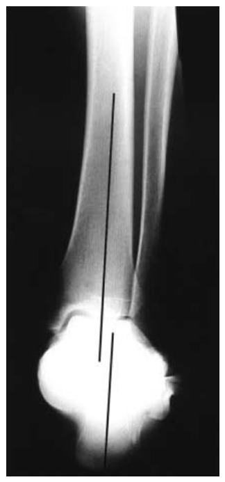
Intraoperative positioning should result in a perpendicular relationship between the forefoot and rearfoot in the frontal plane and a parallel relationship in the transverse plane. The rearfoot should be realigned parallel to the lower leg in the frontal plane (Figure 1). Achieving these positional relationships will result in a stable and plantigrade foot that will protect the ankle and lead to minimal gait disturbances.
Figure 1.
Rearfoot alignment radiograph demonstrating the normal relationship of the calcaneus to the distal one-third of the tibia.
We used a protocol for evaluating the alignment achieved by the triple arthrodesis. Forefoot-to-rearfoot alignment is assessed using standard anteroposterior and lateral radiographs. Rearfoot position is evaluated using rearfoot alignment radiographs. This protocol for alignment evaluation was followed for nine patients who underwent triple arthrodesis.
Materials and Methods
We reviewed nine triple arthrodeses in nine patients at the Foot and Ankle Institute of Western Pennsylvania Hospital, Pittsburgh, between January 1, 2000, and December 31, 2002. Postoperative follow-up occurred at 1 year.
Surgical Technique
Standard lateral and medial incisional approaches were used. Joint resection involved limited debridement techniques to preserve the contour of the subchondral plate. The subchondral plate was then developed into a healthy cancellous substrate to promote osseous consolidation. A structural autogenous bone graft from the iliac crest was used in the subtalar joint to realign severe frontal plane deformities in several patients. Subtalar joint fixation was achieved using two 7.3-mm screws. The screws were inserted using a plantar-to-dorsal approach. The talonavicular joint was fixated using two crossed 4.0- or 4.5-mm screws, and the calcaneocuboid joint was fixated using two crossed 4.5-mm screws or one axially directed 7.3-mm screw.
Intraoperative Alignment
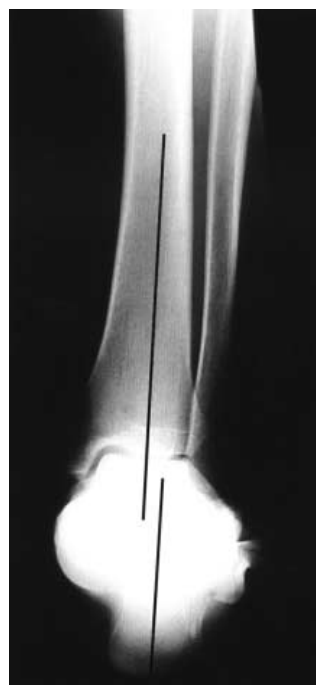
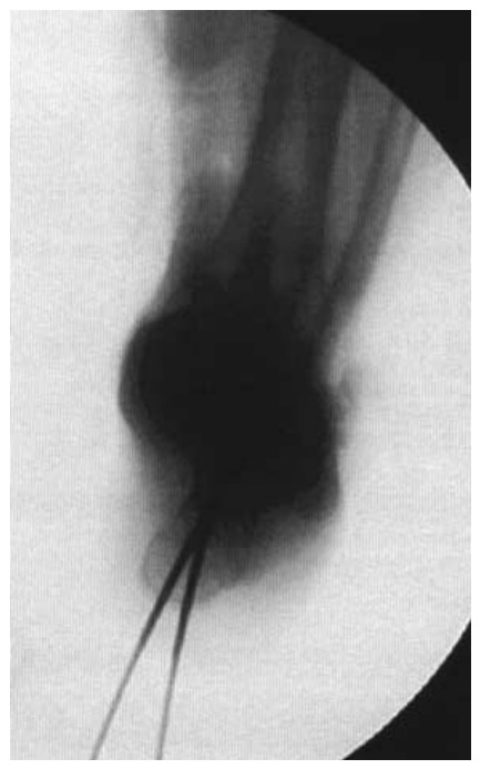
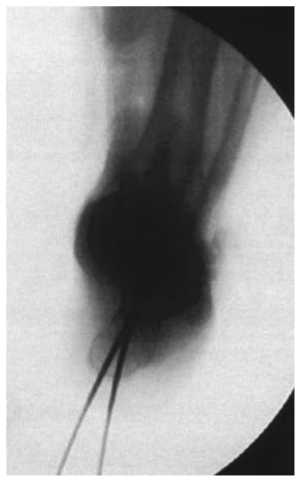
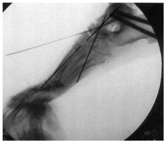
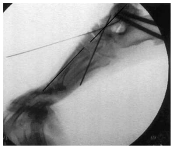
Fluoroscopy was used to confirm and adjust realignment of the rearfoot to the leg and the forefoot to the rearfoot. The position of the subtalar joint was analyzed first. The calcaneal axial view was used to ensure that the long axis of the calcaneus was parallel to the mid-diaphyseal line of the distal tibia (Figure 2). Provisional fixation was achieved using guidewires for a 7.3-mm cannulated screw. Fixation was performed over the guidewires after radiographic confirmation of position. The forefoot was then positioned parallel to the rearfoot in the frontal and transverse planes (Figure 3). Realignment was confirmed by evaluating the talar–first metatarsal angle on the anteroposterior and lateral views.
Figure 2.
Intraoperative fluoroscopic assessment of the rearfoot alignment view before final screw fixation. The calcaneus is aligned parallel to the distal one-third of the tibia and slightly lateral to it.
Figure 3.
Intraoperative fluoroscopic assessment of the forefoot-to-rearfoot relationship before final screw fixation. Note the parallel relationship.
Preoperative and Postoperative Radiographic Evaluation
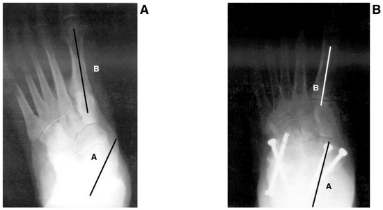
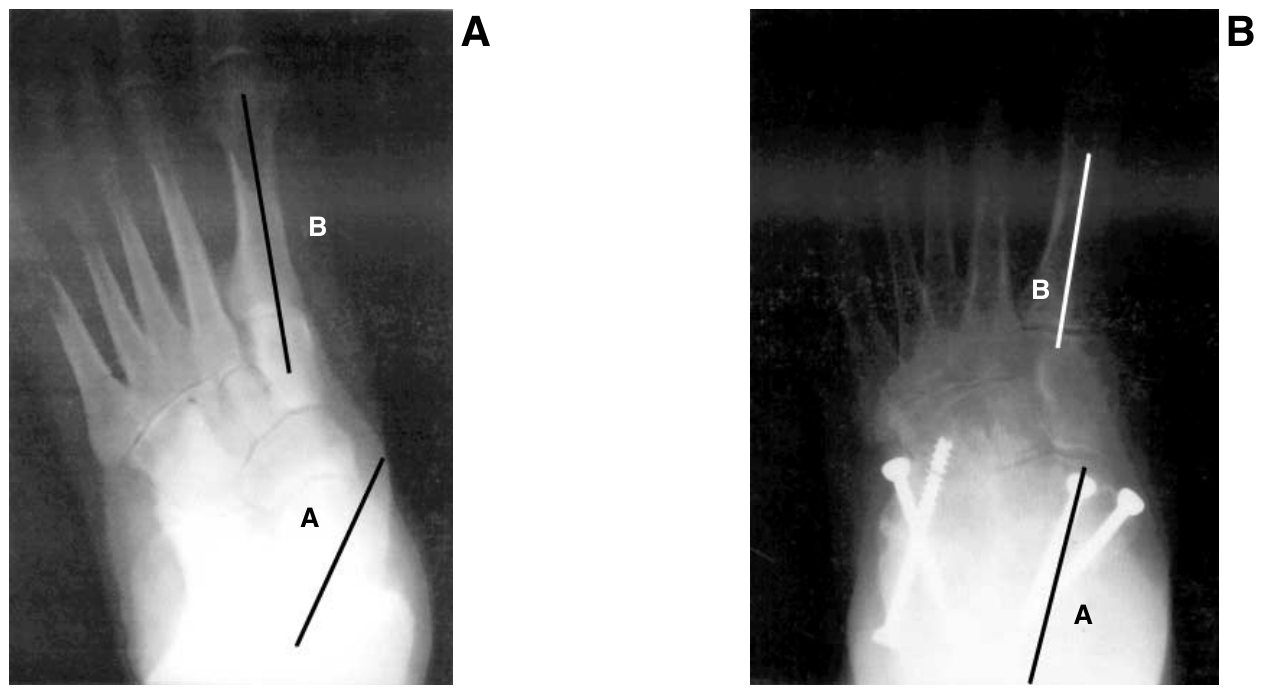
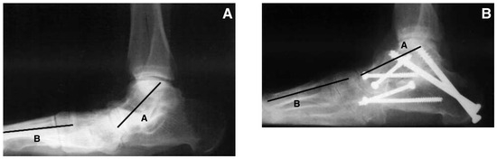
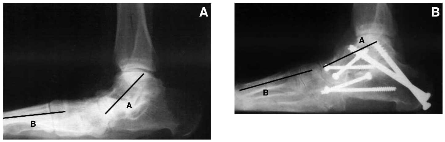
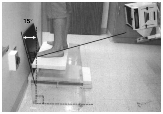
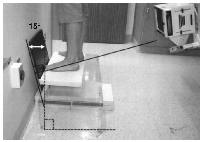
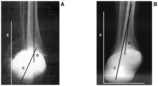
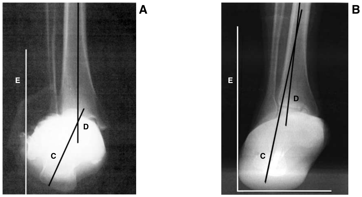
The patients were evaluated for forefoot-to-rearfoot position using standard anteroposterior (transverse plane alignment) and lateral (frontal plane alignment) radiographs (Figure 4 and Figure 5). The anteroposterior talar–first metatarsal angle was used for the transverse plane assessment (0° = a parallel forefoot-to-rearfoot position), and the lateral talar–first metatarsal angle was used for the frontal plane assessment (0° = a perpendicular relationship between the plane of the forefoot and the vertical bisection of the rearfoot). Preoperative and postoperative evaluations of leg-to-ground, rearfoot-to-leg, and rearfoot-to-ground relationships in the frontal plane were performed using rearfoot alignment radiographs (Figure 6 and Figure 7). All angular relationships were measured using the X-Caliper digital measuring device (Eisenlohr Technologies, Davis, California).
Figure 4.
Preoperative (A) and postoperative (B) radiographic transverse plane assessment. AB indicates the anteroposterior talar–first metatarsal angle.
Figure 5.
Preoperative (A) and postoperative (B) radiographic frontal plane assessment. AB indicates the lateral talar–first metatarsal angle.
Figure 6.
Patient positioning for the rearfoot alignment radiograph.
Figure 7.
Preoperative (A) and postoperative (B) radiographic assessment of the rearfoot alignment views. C indicates the calcaneal bisection; D, the distal tibial bisection; E, the ground bisection; DE, the leg-to-ground angle; CD, the rearfoot-to-leg angle; and CE, the rearfoot-to-ground angle.
Results
The change in alignment of the forefoot to the rearfoot in the transverse plane (as measured by the anteroposterior talar–first metatarsal angle) and the change in alignment of the forefoot to the rearfoot in the frontal plane (as measured by the lateral talar–first metatarsal angle) seem to be similar. The data actually show that the forefoot is more closely realigned with the rearfoot in the frontal plane than in the transverse plane (an average change of 13.7° for the lateral talar–first metatarsal angle compared with 10.8° for the anteroposterior talar–first metatarsal angle).
The average change in heel position relative to the leg was 13.7° and relative to the ground was 11.4° for rearfoot realignment. The data show that rearfoot realignment to within 5° of rectus was achieved in five of the nine patients.
Discussion
Triple arthrodesis is a predictable procedure regarding improvement of symptoms. A successful long-term outcome, characterized by a return to usual footwear and a functional ambulatory status with no significant gait abnormalities, depends primarily on appropriate alignment.
Fluoroscopy was used to confirm and adjust the intraoperative alignment of the rearfoot to the leg in the frontal plane and the forefoot to the rearfoot in the transverse and frontal planes. Realignment of these segments was evaluated 1 year after surgery with standard foot and rearfoot alignment radiographs. Realignment of the forefoot to the rearfoot in the frontal plane (talar–first metatarsal angle, lateral foot radiograph) was more consistently attained compared with realignment in the transverse plane (talar–first metatarsal angle, anteroposterior foot radiograph).
The intraoperative goal is a parallel rearfoot-to-leg relationship. Acceptable rearfoot-to-leg positions were achieved in five of the nine patients. An abnormal relationship of the leg to the ground will result in a deviated or nonperpendicular relationship of the heel to the ground. Knowledge of an abnormal leg-to-ground orientation will allow for an adjustment to the heel-to-leg relationship in the operating room, with the final result being a heel-to-ground position that is closer to perpendicular. Thus it is important to evaluate all three relationships.
References
- Bennett GL, Graham CE, Mauldin DM: Triple arthrodesis in adults. .Foot Ankle Int12::138. ,1991. .
- Bednarz PA, Monroe MT, Manoli A: Triple arthrodesis in adults using rigid internal fixation: an assessment of outcome. .Foot Ankle Int20::356. ,1999. .
- Horton GA, Olney BW: Triple arthrodesis with lateral column lengthening for treatment of severe planovalgus deformity. .Foot Ankle Int16::395. ,1995. .
- Fortin PT, Walling AK: Triple arthrodesis. .Clin Orthop365::91. ,1999. .
Additional References
- Graves SC, Mann RA, Graves KO: Triple arthrodesis in older adults. .J Bone Joint Surg Am75::355. ,1993. .
- Hutchinson B: Current concepts in triple arthrodesis. .Clin Podiatr Med Surg17::1. ,2000. .
- Jones CK, Nunley JA: Osteonecrosis of the lateral aspect of the talar dome after triple arthrodesis. .J Bone Joint Surg Am81::1165. ,1999. .
- Kissel CG, Hulst TJ, Blacklidge DK, et al: Nonresection triple arthrodesis: a retrospective analysis. .J Foot Ankle Surg37::490. ,1998. .
- Maenpaa H, Lehto MU, Belt EA: What went wrong in triple arthrodesis? an analysis of failures in 21 patients. .Clin Orthop391::218. ,2001. .
- Pell RF, Myerson MS, Schon LC: Clinical outcome after primary triple arthrodesis. .J Bone Joint Surg Am82::47. ,2000. .
- Reade B, Atlas G, Distazio J, et al: Making triple arthrodesis easier and more reliable. .J Foot Ankle Surg38::242. ,1999. .
- Song SJ, Lee S, O’Malley MJ, et al: Deltoid ligament strain after correction of acquired flatfoot deformity by triple arthrodesis. .Foot Ankle Int21::573. ,2000. .
© 2005 American Podiatric Medical Association