


Abstract
This article describes a proximal phalangeal osteotomy for the correction of abductus deformity of the hallux. The modification uses a Z-osteotomy, which retains the length of the hallux, corrects the abductus component, and provides excellent rigid internal fixation. This procedure may be used only in mild-to-moderate cases. (J Am Podiatr Med Assoc 94(4): 404–408, 2004)
An integral component of hallux abducto valgus is an abnormal position of the hallux that may be caused by increased or abnormal hallux abductus interphalangeal or distal articular set angles. During the past century, many articles have been published that discuss reduction or correction of the abductus deformity of the hallux.
The brief article by Akin[1] published in 1925 was the first to describe a cuneiform-type osteotomy of the proximal phalanx to correct hallux abductus. Various modifications have been described since publication of Akin’s article that encompass numerous techniques, including oblique and cylindric osteotomies.[2–10] Still other publications describe fusion of the hallux interphalangeal joint11 and the first metatarsophalangeal joint,[12] which may indirectly correct an abnormal hallux abductus component. However, most of these techniques inherently shorten the hallux.
A variety of methods for fixation of proximal phalangeal osteotomies have been described, including staples,[2] sutures,[6,13] screws,[11,14] cerclage wires,[3,10,15] and Kirschner wires.[15] Screw fixation at a perpendicular angle has proved to be superior to the other methods of fixation of osteotomies under normal conditions.[16,17]
The technique described here is superior to all other procedures involving a mild-to-moderate deformity by maintaining the hallucal length and providing excellent rigid internal fixation. This procedure reduces the risk of delayed-union and nonunion bone healing through the perpendicular orientation of fixation. By eliminating percutaneous Kirschner wires, the possibility of a pin-tract infection is also removed.
One disadvantage of this procedure is the relatively high manual dexterity that is required to perform it. Although the use of Kirschner wires as guides assists in this task, a wedge must be created. The wedge of bone in some circumstances is created simply from the width of the saw blade.
Technique
This procedure is usually performed concurrently with bunion repair. However, it may be performed as a sole and separate procedure. In cases in which the procedure involves the first metatarsophalangeal joint, it is generally performed as a secondary procedure.
A dorsal incision is made or extended distally to the hallux interphalangeal joint. Dissection is carried through the subcutaneous tissue according to the surgeon’s preference. The extensor hallucis longus tendon is retracted laterally. The periosteum of the proximal phalanx is incised and reflected. Care is taken to preserve the insertion of the extensor hallucis brevis tendon as much as possible. A 0.028 Kirschner wire is inserted into the dorsal aspect of the proximal phalanx at its proximal end near the insertion of the extensor hallucis brevis tendon. The Kirschner wire is used as an apex for osteotomy. The placement is perpendicular in the transverse and frontal planes. Osteotomies are performed using the Kirschner wire as a guide (Fig. 1). A second Kirschner wire may be placed in the most distal aspect of the proximal phalanx to act as a guide. The osteotomies are made at 90° to one another on the dorsum of the proximal phalanx. The osteotomies are not made through and through until completion of the outline of the Z-osteotomy. The proposed pie wedge osteotomy is marked and cut using a bone saw (Fig. 2). The Kirschner wires are removed. Through-and-through osteotomies are now completed. The pie wedge–shaped bone is completely cut through and through and removed (Fig. 3). If shortening is desired, then further osteotomies are performed as described by Hodor and Hess.[18] The bone is reapproximated (Fig. 4) and held in place using a bone clamp. The foot is loaded to assess correction. Further feathering to reduce the angle may be performed as indicated. Two 1.5-mm cortical screws are placed perpendicular to the osteotomy site using AO technique. Owing to the displacement, gapping occurs at the proximal lateral aspect. Bone may be packed into the defect created by displacement of the hallux (Fig. 5).
As deemed appropriate, redundant bone on the medial aspect of the hallux interphalangeal joint may also be removed, including removal of the medial condyle on the distal phalanx (Figs. 6 and 7). If this is desired, then the incision is extended to the medial nail fold, which will assist in accessing the medial aspect of the hallux interphalangeal joint. Closure is performed using standard technique. Postoperative care is determined by the adjunct bunion procedure, if performed.
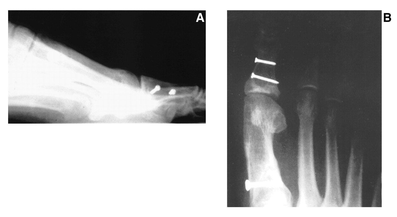
Radiographs provide preoperative (Fig. 8) and 1-week (Fig. 9) and 6-month (Fig. 10) postoperative assessments. The radiographs involved a proximal first metatarsal osteotomy, bunionectomy, and Z-osteotomy of the proximal phalanx on the right foot. The radiographs clearly demonstrate the Z-osteotomy and its application.
Discussion
The advantages of this technique include reduction of the abnormal hallux abductal position, maintenance of hallucal length (if desired), elimination of the possibility of pintract infection, and provision of excellent rigid internal fixation. The traditional Akin procedure provides correction of the hallux abductus component, but it also shortens the hallux and does not provide a means for compressive internal fixation that is provided by perpendicularly placed screws. Screw fixation at a perpendicular orientation also reduces the risk of delayed union and nonunion.
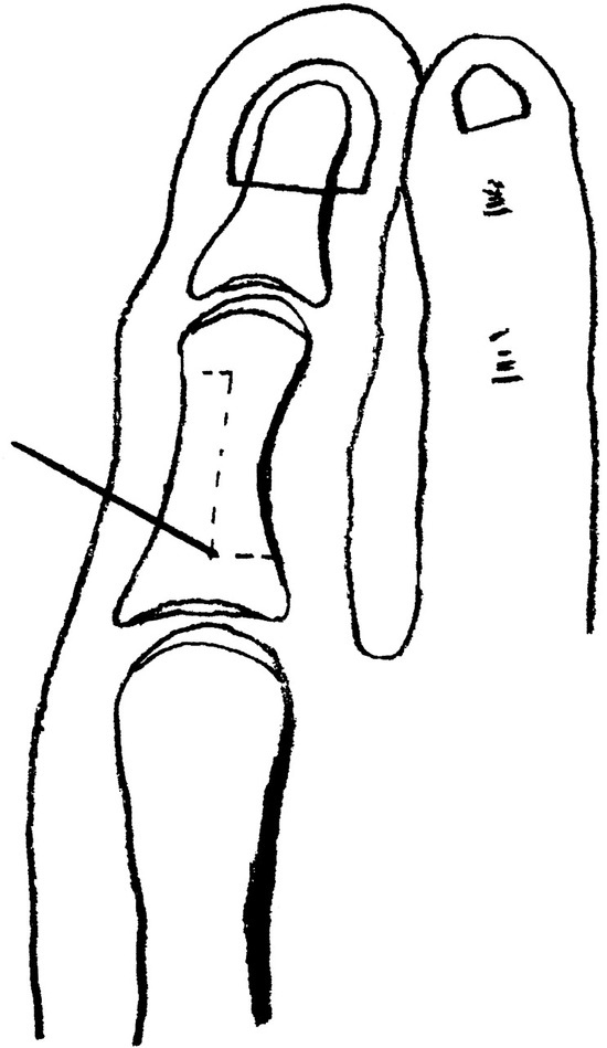
Figure 1.
With a 0.028 Kirschner wire in place, the Z-osteotomy is outlined.
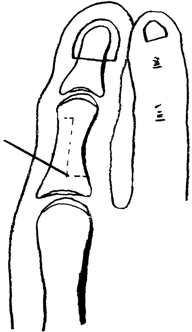
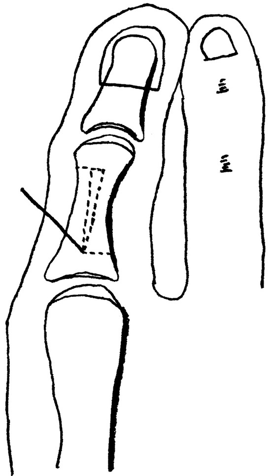
Figure 2.
The pie wedge osteotomy is outlined.
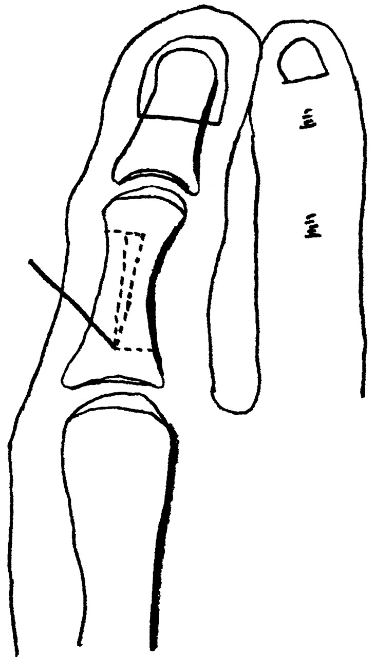
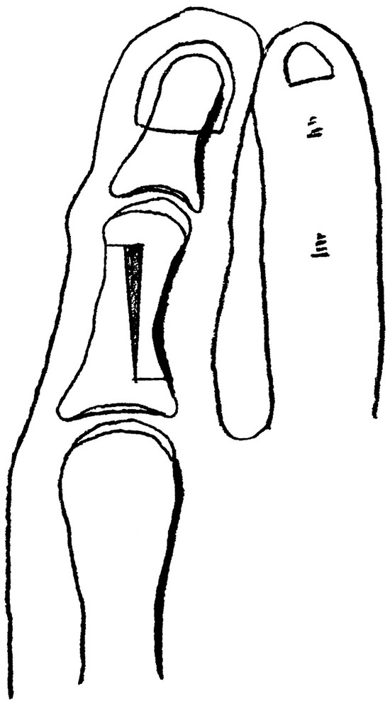
Figure 3.
View after the osteotomy is performed. The pie wedge–shaped bone has been removed.
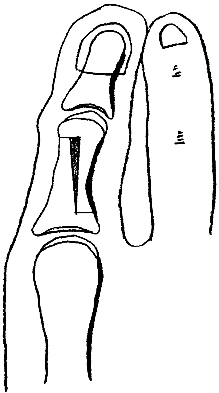
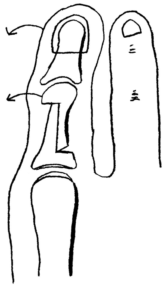
Figure 4.
The hallux is manipulated medially, which reduces the osteotomy on the proximal phalanx. Note the gapping that occurs at the proximal lateral aspect of the proximal phalanx.
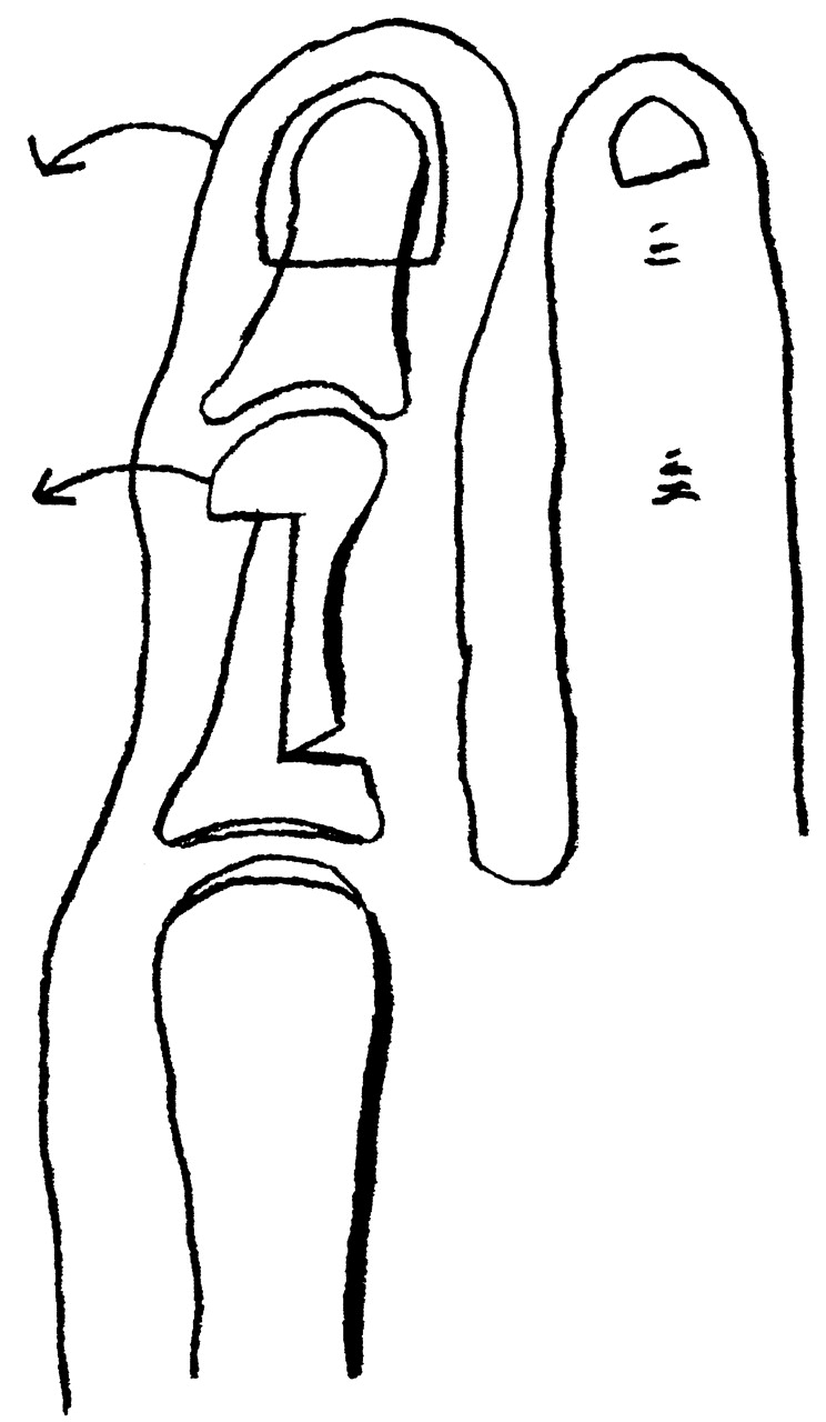
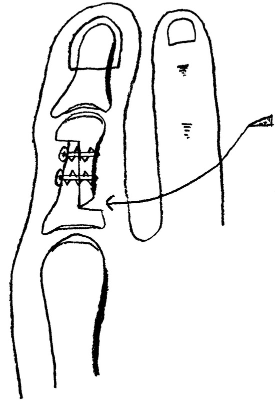
Figure 5.
Two 1.5-mm screws are in place. Bone is packed into the gap at the proximal lateral aspect of the proximal phalanx.
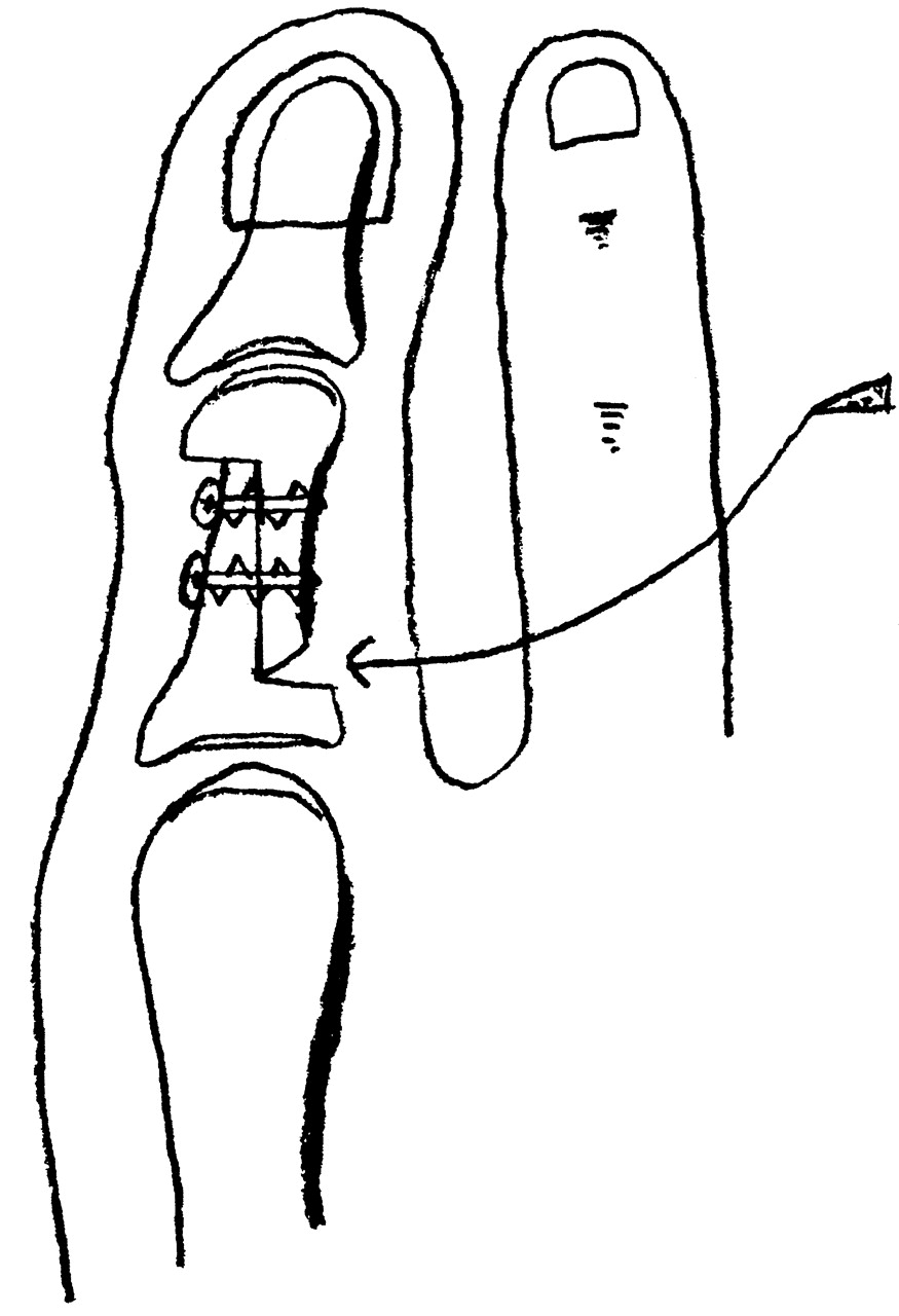
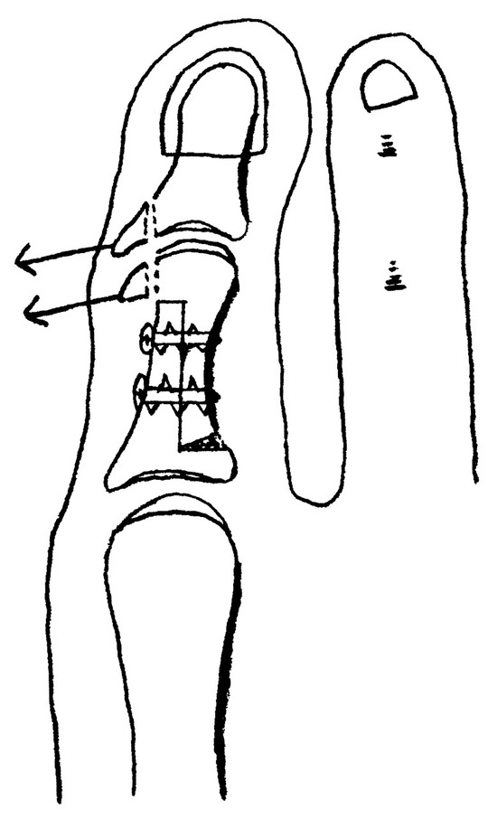
Figure 6.
Redundant bone on the medial aspect of the head of the proximal phalanx and medial condyle of the base of the distal phalanx is removed.
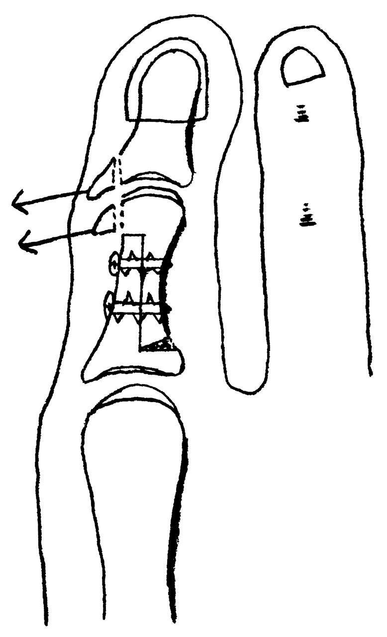
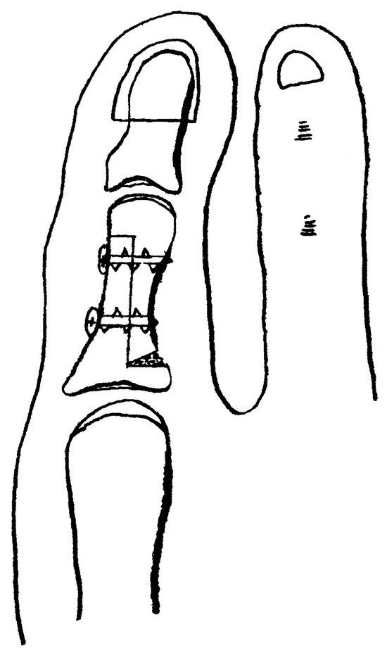
Figure 7.
Final result with two 1.5-mm screws in place.
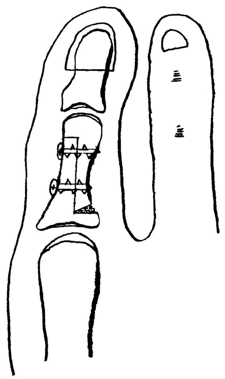
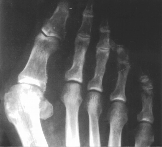
Figure 8.
Preoperative anteroposterior radiographic view.
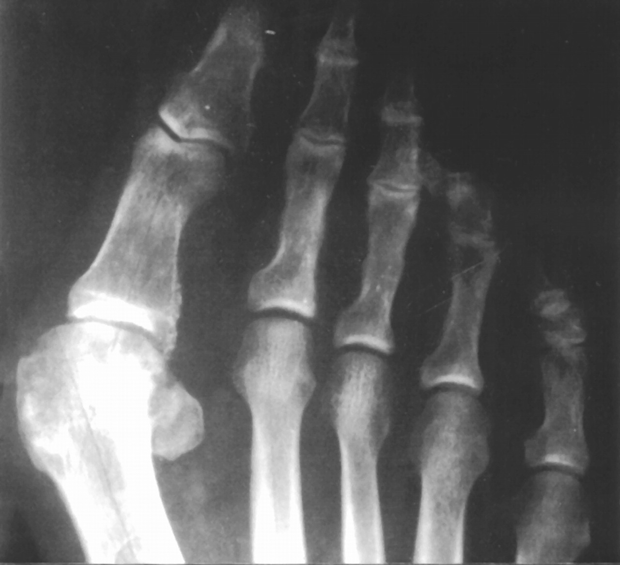
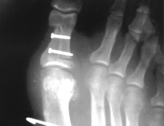
Figure 9.
One-week postoperative anteroposterior radiographic view.
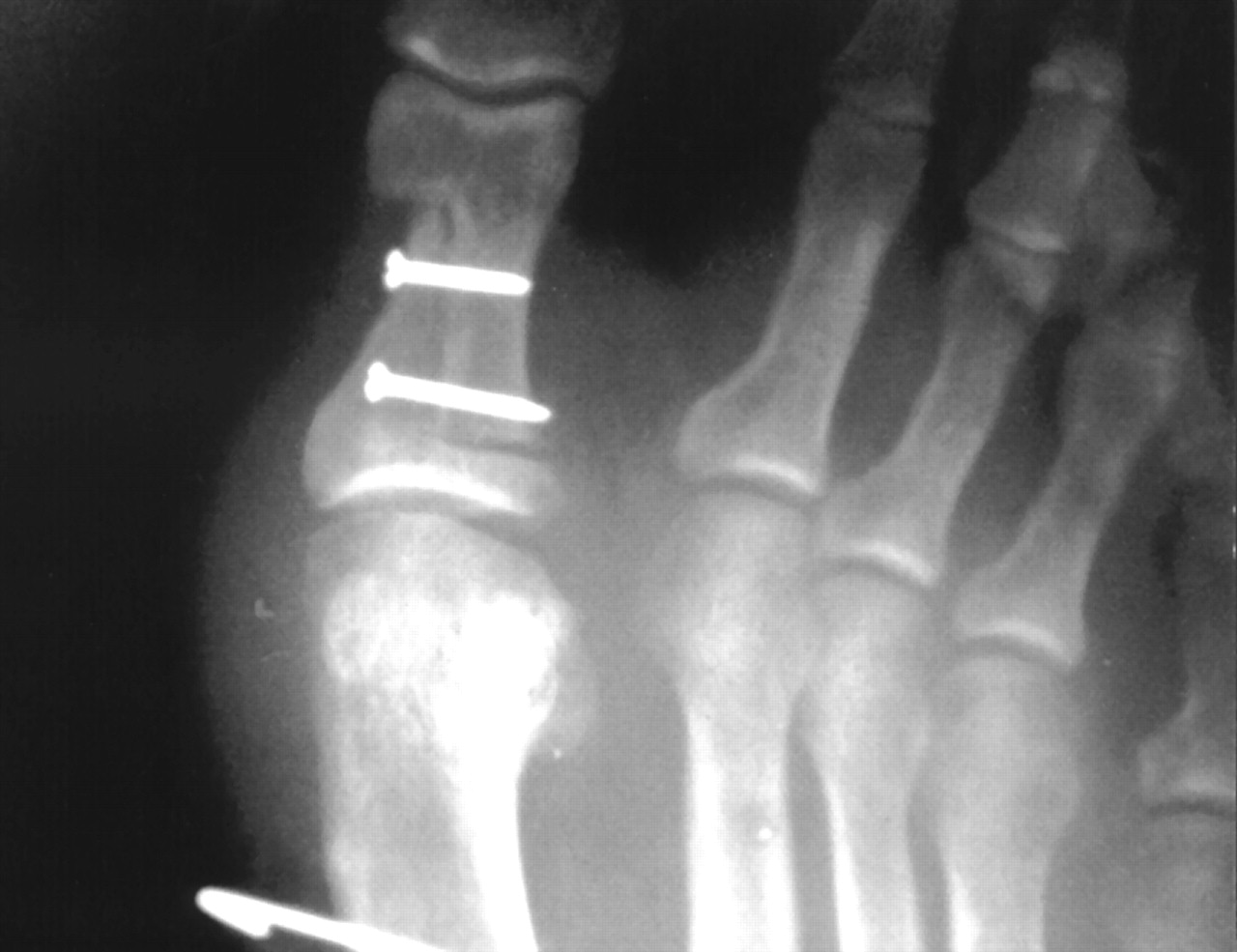
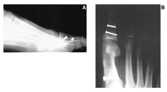
Figure 10.
Six-month postoperative lateral (A) and anteroposterior (B) radiographic views.
References
- Akin OF: The treatment of hallux valgus: a new operative procedure and its results. Med Sentinel33:678, 1925.
- Colloff B, Weitz EN: Proximal phalangeal osteotomy and hallux valgus. Clin Orthop54:105, 1967.
- Gerbert J, Melillo T: A modified Akin procedure for the correction of hallux valgus. JAPA61:132, 1971.
- Silberman FS: Proximal phalangeal osteotomy for the correction of hallux abductus. Clin Orthop85:98, 1972.
- Gerbert J, Mercado OA, Sokoloff TH: “The Surgical Treatment of Hallux Abducto-Valgus and Allied Deformities,” in Podiatric Medicine and Surgery: A Monograph Series, Vol 1, ed by MD Fielding, p 118, Futura Publishing, Mt Kisco, NY, 1973.
- Seelenfreund M, Fried A, Tikva P: Correction of hallux valgus deformity by basal phalanx osteotomy of the big toe. J Bone Joint Surg Am55:1411, 1973.
- Gerbert J, Spector E, Clark J: Osteotomy procedures on the proximal phalanx for correction of a hallux deformity. JAPA64:617, 1974.
- Purvis CG, Brown JH, Kaplan EG, et al: Combination Bonney-Kessel and modified Akin procedure for hallux limitus associated with hallux abductus. JAPA67:236, 1977.
- Clark J: “Akin-Type Procedures,” in Textbook of Bunion Surgery, ed by J Gerbert, p 143, Futura Publishing, Mt Kisco, NY, 1991.
- Yale J: Non-joint destructive procedure for long great toe with abductus deformity (a retrospective study). J Foot Surg18:7, 1979.
- Langford JH, Fenton CF: Hallux interphalangeal arthrodesis. JAPA72:155, 1982.
- McKeever DC: Arthrodesis of the first metatarsophalangeal joint for hallux valgus, hallux rigidus, and metatarsus primus varus. J Bone Joint Surg Am34:129, 1952.
- Brahms MA: Hallux valgus: the Akin procedure. Clin Orthop157:47, 1981.
- Levitsky DR: Rigid compression screw fixation of first proximal osteotomy for hallux abducto valgus. J Foot Surg21:65, 1982.
- McGlamry ED, Kitting RM, Butlin WE: Hallux valgus repair with correction of coexisting long hallux. JAPA60:86, 1970.
- Schenk R, Willenegger H: Zur histology der primaren knochen neilung. Langenbecks Arch Klin Chir Ver Dtsch Z Chir308:440, 1964.
- Perren SM, Matter P, Ruedi R, et al: Biomechanics of fracture healing after internal fixation. Surg Annu7:361, 1975.
- Hodor L, Hess T: Shortening Z-osteotomy for the proximal phalanx of the hallux using axial guides. JAPMA85:249, 1995.
© 2004 American Podiatric Medical Association