


Abstract
This article presents a series of case reports to describe the technique of ankle joint manipulation and its effects on common problems of the foot and ankle. The relationship between motion and pain is described, as are the effects of muscular inhibition on the presence of joint re- striction and their association with pain in various joints remote to the ankle joint.
As a method of managing musculoskeletal symptoms, manual manipulation of various joints of the body has been practiced worldwide for thousands of years. The relationship between joint motion and pain, particularly as associated with aging, was recognized by the ancient cultures of the world long ago. By using motion-enhancing exercises such as tai chi and yoga, some control of these symptoms could be achieved. In the modern Western world, stretching routines are commonly used to improve motion and to assist in the management of various muscle and joint symptoms. As a practical matter, all of the previous points suggest that some correlation exists between joint motion and pain perception.
Pain is transmitted from the periphery to the central nervous system by the thinly my-elinated or non- myelinated C-fiber nociceptors.[1] These C-fiber nerves conduct nocicep-tive impulses relatively slowly compared with other nerve transmission within the body. Nociception signals the body about the presence of noxious stimuli from excessive heat, pressure, and loss of tissue integrity. In addition, nociception is known to be modulable, with states ranging from hyperalgesia to the absence of sensation.[2] Therefore, the com-mon belief in patients having a specific high or low pain “threshold” may represent only their re- action to pain stimuli in certain situations. Changes in muscle tension, for in-stance, can create changes in this perceived threshold. Muscular hypertonicity has been shown to exacerbate pain sensation. Thus stress can heighten the perception of pain.
Approximately 85% of nociceptors synapse in the dorsal horn of the spinal cord at the wide dynamic range cell.[3] Interestingly, this is also where peripheral proprioceptors connect to the central nervous system. Proprioceptors are and myelinated fibers that rapidly transmit motion-related impulses. When proprioceptors are actively stimulated, the ability to sense pain is diminished. Although there are multiple neuroscience studies that describe these phenomena, many examples exist in everyday life. Infants in pain with colic are often relieved by being carried about or rocked. Injuring one’s finger is met with the al- most immediate response of shaking the hand rapidly or rubbing the site of pain. Although conscious under- standing of this interrelationship may not exist, it is natural to react in the manner described. The sensation of motion and the perception of pain are in-extricably linked in the human central nervous system.
Biomechanics of Ankle Dorsiflexion
The ankle joint is formed by the articulations among the tibia, fibula, and talus. The tibia and fibula form a mortise in which the talar dome dorsiflexes and plantarflexes. The articular surface of the talus is fashioned as a truncated pyramid, wider in the anterior surface and narrower in the posterior surface.[4] As ankle joint dorsiflexion occurs, the fibula translates, gliding superiorly and laterally. This results in a variety of functional changes relating to the attachments of the superior iliotibial band and biceps femoris,[5] but these are beyond the scope of this article. For the ankle, the main function is the widening of the mortise to receive the dorsiflexing talus. When normal translation of the fibula occurs, customary dorsiflex- ion is permitted. When fibular translation fails to occur, then equinus may be present.[6]
During walking, the entire body passes directly over the weightbearing foot with each step. For this to occur, the foot has been described as a series of pivotal rockers, permitting sagittal plane advancement during the single-support phase.[7] The initial rocker is represented by the round underside of the calcaneus. The final rocker is the dorsiflexion motion of the metatarsophalangeal joint. The ankle serves as the second rocker in this three-part system. Its for- ward flexion (dorsiflexion) permits the body to continue advancing over the foot despite the heel and forefoot being in contact with the ground. Failure of dorsiflexion in the ankle can cause a variety of pathomechanical situations.[8] For example, it can cause the forefoot to receive excessive loads far too early in the gait cycle, and it can create a phenomenon in which the foot must pronate excessively to accommodate the loss of sagittal plane ankle motion. Pivotal dorsiflexion of the ankle is an essential component of normal foot function.
Ankle Equinus and Peroneal Inhibition
Muscles receive two forms of stimulation from the central nervous system: facilitation (excitation) and inhibition (suppression).1 When muscles receive a facilitation signal, they act in a manner that can best be described as “strong.” Conversely, when muscles receive an inhibition signal, they function as though they were “weak.” This well-known response acts to balance motions within the body.[9] For instance, when typing and striking a key, the flexors to the fingers are facilitated and the extensors are inhibited. Over time and with repetition, the coordination of these events becomes very efficient.
When fibular translation is restricted, not only does a state of ankle equinus exist, but a secondary inhibition of the peroneal muscles can also occur. The peroneus longus muscle has been described as a pronator of the foot during open-kinetic-chain function. However, during closed-kinetic-chain function, it also serves to stabilize the first metatarsal against ground reaction force during walking. When peroneal inhibition exists, stabilization of the first metatarsal is compromised.[10] In addition, its ability to act as the antagonist to the tibialis posterior muscle is negatively affected. This can create an imbalance in the agonist-antagonist relationship between the tibialis posterior and peroneal groups. Imbalance such as this can create a chronic inverted position of the foot and ankle during walking. Patients who are affected by this disproportion often sense an “impending doom” of an ankle ready to suffer an inversion sprain. There can be instability and pain associated with walking on an imbalanced limb, yet the subtle nature of this problem is often missed in the clinical examination. Manipulation of the ankle has previously been shown to be an effective method of restoring adequate range of motion provided that the restriction is not caused by an osseous block directly in the ankle joint.[6] Manipulation not only promotes motion of the ankle but also appears to restore peroneal facilitation, thus relieving symptoms associated with the inhibited state.
Manipulation Technique
The processes of joint restriction, pain sensation, and muscular inhibition are described in detail to patients. With patients who experience disability for extended periods of time, reviewing the nature of the process is extremely important. They are apt to have feelings of extreme anger when they recognize that the treatment is relatively simple and that they seemingly suffered so long for no obvious reason. Proper preparation is essential for adequate patient management.
With the patient seated, the affected limb is positioned for manipulation. The knee is flexed and the foot is placed on the support surface of the chair. The fibular head is identified, and the peroneal notch is palpated. Care is taken to avoid the notch and the associated common peroneal nerve during the manipulation process.
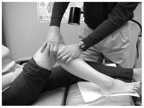
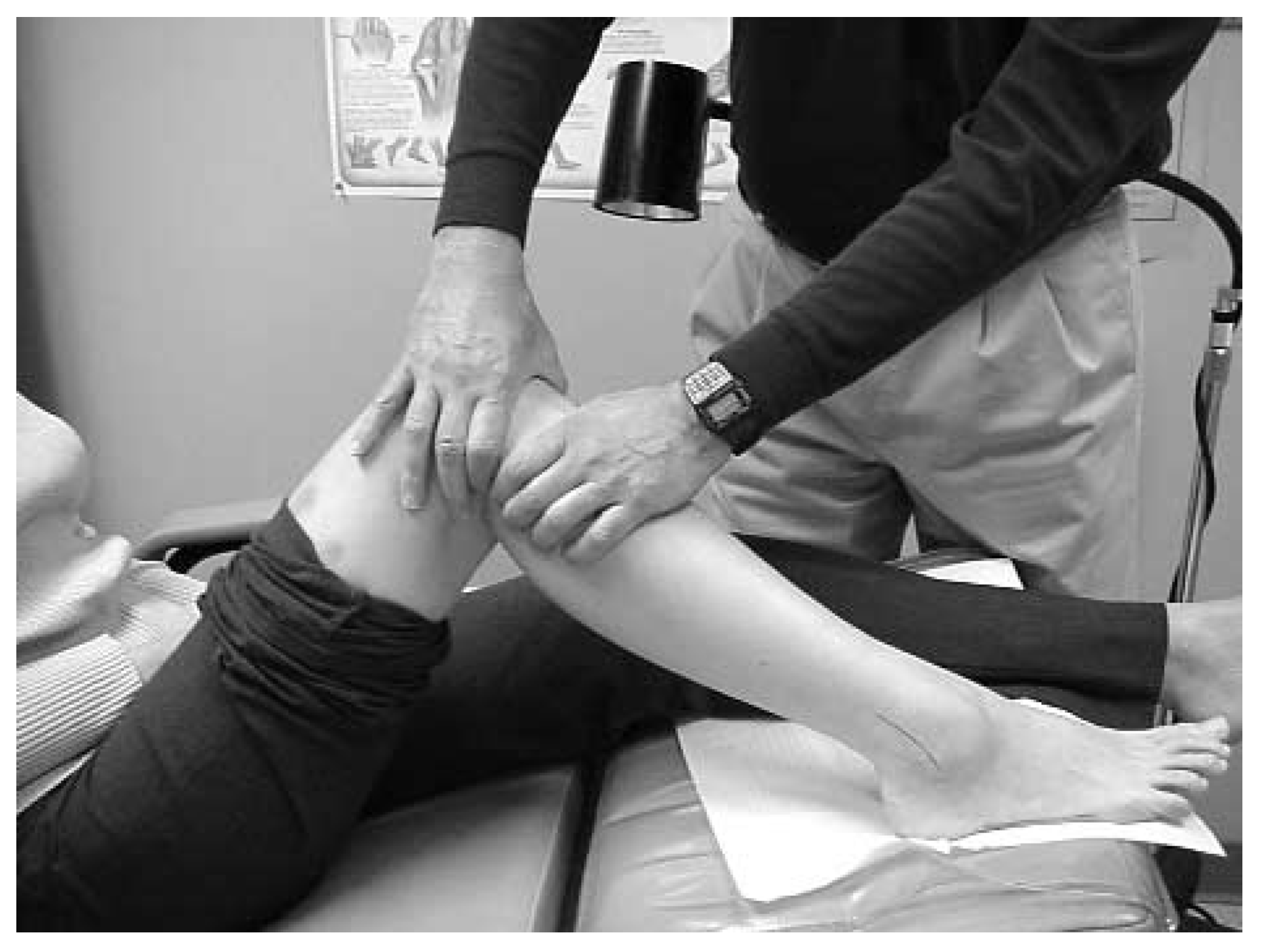
The manipulation will initially move the fibular head anteriorly from its posterior fixed position. The fibular head is grasped with either the index or the middle fingers of both hands, with one just above and the other just below the peroneal notch. Care is taken to reach behind the head itself but not into the muscular structures just posterior to the head. The thumbs are then positioned across the anteromedial aspect of the leg. Any “joint play” that may exist is then taken up by compressing the fibular head from posterior to anterior. Once the “end feel” of the fibular head is achieved, rapid adjustment of the head is performed in a posterior-to-anterior direction. The patient may experience a sudden pain sensation, but this often lasts only 1 to 2 sec. There is no residual sensation from the maneuver (Figure 1).
Figure 1.
To perform the ankle manipulation technique, the fibular head is grasped with either the index or the middle fingers, above and below the peroneal notch. The thumbs are anchored on the anterior aspect of the tibia. The adjustment is made by pulling the fibular head anteriorly, avoiding the common peroneal nerve in the process.
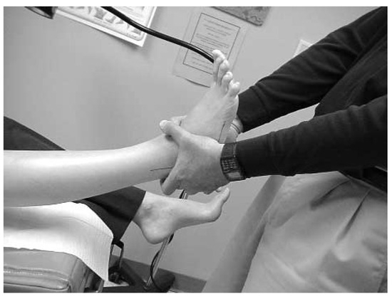
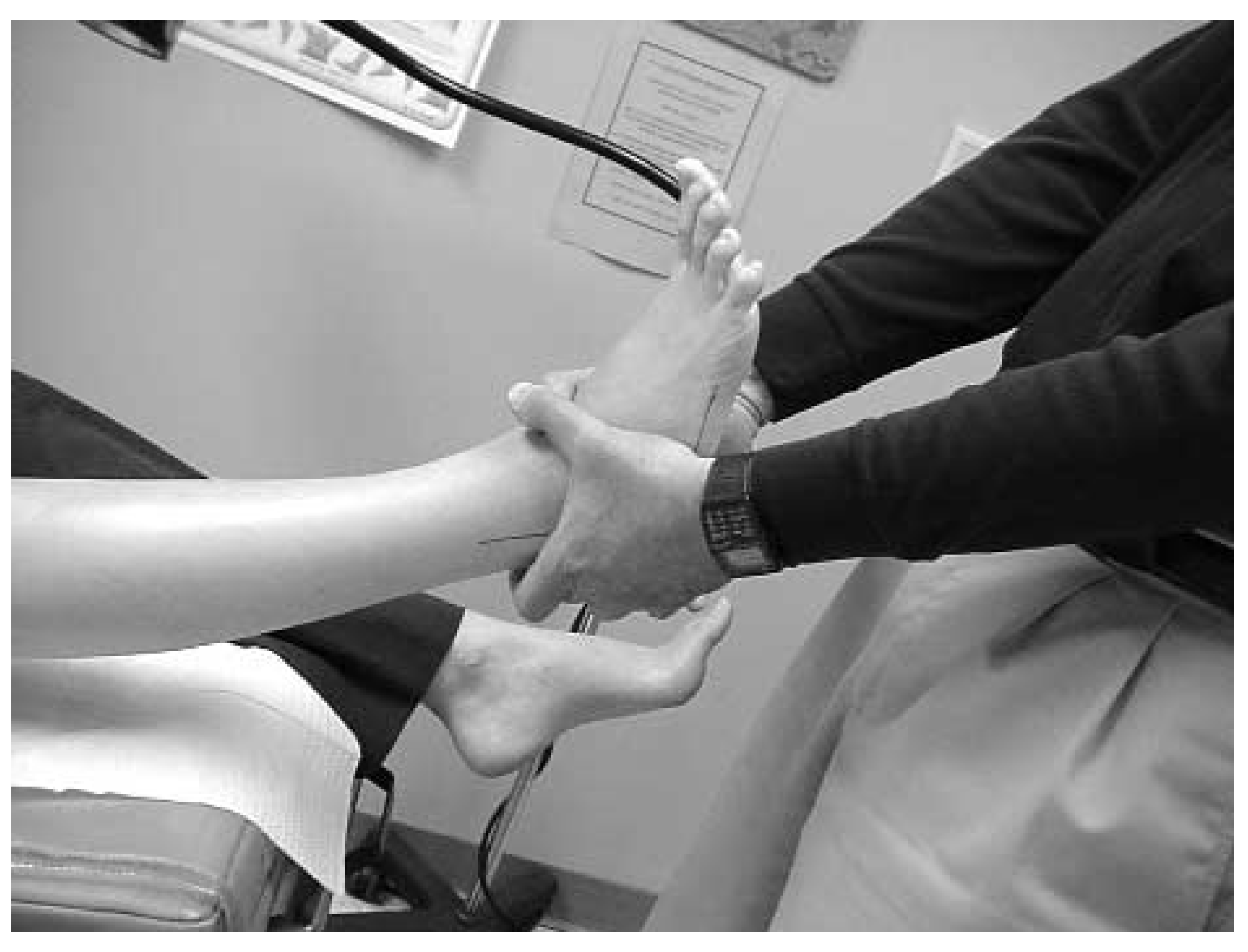
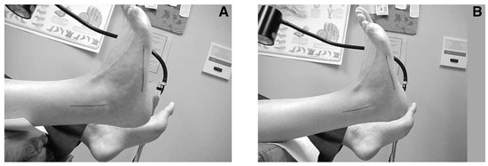
Once the proximal fibular head is mobilized, attention is then directed to the ankle joint. The foot is grasped posteriorly about the calcaneus with the long fingers. Anteriorly, the talar head and neck are identified and gripped with the thumbs, one crossed over the other. Longitudinal traction is then applied for 20 to 30 sec. The patient is encouraged to relax, as the clinician should support the entire limb weight during the traction process. Once the traction process is complete, the ankle is dorsiflexed gently to again achieve the end feel position and to take up any slack in dorsiflexion. The adjustment is performed in an almost rotary manner, with the talus being mobilized in an anterior-to-posterior (dorsiflexory) direction. Manipulation of the talus can result in an audible “click” or “thunk” that, although possibly startling, is not painful (Figure 2). The ankle is then im- mediately dorsiflexed (while holding the knee fully extended), with encouragement given to the patient to actively dorsiflex and plantarflex, with some clinician assistance. Changes in range of motion are often marked following this process (Figure 3).
Figure 2.
Longitudinal traction is applied to the ankle for 20 to 30 sec before the second part of the manipulation process is performed. The ankle is then dorsi- flexed to “end feel” and is adjusted in an anterior-to- posterior direction. There may be an audible click caused by the maneuver.
Figure 3.
A patient’s ankle in active dorsiflexion before (A) and after (B) ankle manipulation. Range of motion is clearly greater in the postmanipulation view.
Patients are immediately asked to walk once adequate range of motion has been restored. They are often amazed by the lack of pain and the return of mobility, and they may require reassurance as to the long-term effects of this procedure. Patients are shown proper triceps surae stretching techniques, with care taken to avoid overstretching and reinjury.
Case 1
A 42-year-old man presented for a second opinion regarding ongoing pain and instability in the ankle of 6 months’ duration. He was employed as a meter reader for the local gas company. He stepped backward and accidentally landed in a hole. He fell backward and to the right and experienced a severe inversion ankle in- jury. He was initially cared for in the emergency department, and findings from radiographs taken at the time of the injury were normal. The patient subsequently was seen by his primary-care physician and was then referred to an orthopedic surgeon for evaluation. He completed the standard RICE (rest, ice, compression, and elevation) format of care but still had difficulty walking after the injury. He had only limited success in two 1-month programs of physical therapy. The patient had severe pain with walking and the continual sense that he could sprain his ankle again. In addition, he described pain in the first metatarsophalangeal joint with any walking. He had under- gone magnetic resonance imaging and computed to-mography of the ankle, but no soft-tissue or osseous pathology could be found.
Clinical Examination Findings
The patient was an otherwise healthy man who was distraught about not being able to return to work.
There was very mild edema of the lateral aspect of the ankle and the dorsolateral aspect of the right foot. The drawer sign was negative. There was no evidence of anterior talofibular ligament injury or instability. Pain was evident over the sinus tarsi, but it was relatively mild. He had considerable pain on palpation of the peroneal tendons just posterior and inferior to the lateral malleolus. Peroneal strength was at one-quarter capacity on the right side, but comparison testing showed full capacity on the left side. There was only –3° of ankle joint dorsiflexion avail- able on the right, yet more than 10° was evident on the left. Palpation of the fibular head on the lateral side of the superior aspect of the lower leg just below the knee joint revealed a complete absence of translation during attempts at ankle joint dorsiflexion with the knee extended. Review of previous radiographs and scans failed to identify any other significant pathology. The first metatarsophalangeal joint also showed no outward signs of edema or erythema. Pain was evident on palpation of the dorsolateral aspect of the joint. Range of motion of the metatarsophalangeal joint was within normal limits, but pain was evident at the end of the range.
Treatment
The patient was treated with ankle manipulation in the manner described previously. There was immediate return of range of motion in the affected ankle. Peroneal strength spontaneously returned to full capacity. He was able to promptly stand and walk with- out pain. The sense of impending doom of a repeated injury swiftly resolved. The pain in the first metatarsophalangeal joint also spontaneously resolved. In addition to the standard calf-stretching exercises, the patient was shown simple peroneal strengthening exercises to maintain the required force for balanced ambulation. He was then referred to a physical therapy program and seen again 3 weeks later. At that time, he was walking without difficulty and was per- mitted to return to work at full capacity.
Case 2
A 9-year-old girl using crutches presented for a second opinion regarding a diagnosis of Kohler’s disease of the right navicular bone. She had been diagnosed as having this disease 3 months earlier by an orthopedic surgeon. She had been placed in a hard cast for 4 weeks and then in a removable walking cast for 6 additional weeks. She had been out of the walking cast for 2 weeks but could not bear full weight on the affected foot. She had mild edema and wore loose-fit- ting shoes.
On radiographs taken at the time of her initial presentation, the navicular bone appeared as a normally sized and formed bone for a 9-year-old girl. Comparison views were taken of the unaffected side, and there were no obvious differences between the navicular bones of the left and right feet.
Clinical examination revealed pain on palpation of the superior aspect of the right navicular bone. Ankle joint dorsiflexion with the knee extended was then evaluated and found to be approximately –3° on the right but a normal 10° on the left. With attempts at ankle joint dorsiflexion, her symptoms were reproduced.
The patient was treated with ankle manipulation using the method described previously. Range of motion instantly returned to normal. She was immediately able to stand and walk without pain. The patient was then shown proper stretching techniques, and she left the office without using her crutches. At a follow-up visit 3 weeks after treatment, she did not have any symptoms.
Case 3
A 46-year-old woman presented seeking a second opinion as to the management of an acutely inflamed first metatarsophalangeal joint of the right foot. She was a very active athlete who had previously been treated for obsessive-compulsive disorder that manifested as excessive exercise. Although this disorder had been well controlled, she continued a very “full” workout schedule and was distraught by the pain in her toe joint that prevented her from performing her exercise regimen. She had previously been told that she had osteochondritis dissecans and that she re- quired surgery to resolve her symptoms.
On examination, the first metatarsophalangeal joint was erythematous and edematous. Even small movements of the joint were painful, but when it was relaxed, the restriction of motion was only 10° (55° on the right side versus 65° on the left side). The ankle joint demonstrated 0° of dorsiflexion on the right side but 10° on the unaffected left side. Perone- al strength showed barely one-quarter capacity, with pain on attempts to evaluate. She stood and walked with the foot in the inverted position.
Radiographs demonstrated small osteophytes on the dorsum of the joint and some erosion of the head of the first metatarsal consistent with osteochondritis dissecans.
She was treated initially with manipulation of the ankle as described and was also given an adjustment to the first metatarsophalangeal joint that involved freeing the base of the first metatarsocuneiform joint and the metatarsophalangeal joint. She was immediately able to stand and walk without the marked in- version. Although her toe joint was sore, the intensity of her pain immediately decreased. The patient was then taped with low-Dye support. Custom foot orthoses were fabricated for her at a subsequent visit. Within several weeks she had complete resolution of pain and returned to her high level of exercise. She has been followed for several years, with only minor bouts of pain developing in the joint, which respond- ed immediately to manipulation.
Conclusion
Manual manipulation of the ankle has been described for use in the clinical setting for a variety of foot and ankle symptoms. Previous studies have shown that the manipulation provides for an immediate increase in available range of motion without side effects. The significance of the method of care, however, is that the interrelationships of the various joint and muscular components of the lower extremity are coordinated in a complex and inseparable fashion. Recognition that symptoms of the first metatarsophalangeal joint, for instance, may be related to chronic peroneal sup- pression due to fibular-related ankle equinus can be a useful implement in the podiatric physician’s diagnostic toolbox. Adding the fibular translation examination to the ankle joint range-of-motion assessment represents a simple method that can save many patients from chronic disability.
References
- BURT AM: Textbook of Neuroanatomy; WB Saunders: Philadelphia, 1993.
- ZIMMERMANN M: Pain mechanisms and mediators in osteoarthritis. Semin Arthritis Rheum 1989, 18, 22. [CrossRef] [Scilit] [PubMed]
- WILLARD, F.H. “Neuroendocrine-Immune Network, Nociceptive Stress and General Adaptive Response,” in Phys- iotherapy in Mental ffealth: A Practical Approach,; Everett, T., Dennis, M., Ricketts, E., Eds.; Butterworth Heinemann: Oxford, 1995; p. p 102. [Google Scholar]
- GRAY, H.; GOSS, C.M. “Osteology,” in Gray’s Anatomy, 28th Ed, p 264; Lea & Febiger: Philadelphia, 1970. [Google Scholar]
- DANANBERG, H.J. Lower Extremity Mechanics and Their Effect on Lumbosacral Function. In in Spine: State of the Art Reviews; Dorman, T., Ed.; Han- ley & Belfus: Philadelphia, 1995; Vol 9, No 2, p. 389. [Google Scholar]
- DANANBERG, H.J.; SHEARSTONE, J.; GUILIANO, M. Manipulation method for the treatment of ankle equinus. JAPMA 2000, 90, 385. [Google Scholar] [CrossRef] [Scilit] [PubMed]
- Slack. PERRY J: Gait Analysis: Normal and Pathologic Function; Thorofare, NJ, 1992. [Google Scholar]
- ROOT, M.L.; ORIEN, W.P. WEED JH: Normal and Abnormal Function of the Foot; Clinical Biomechanics Corp: Los Angeles, 1977. [Google Scholar]
- DORMAN T: Whiplash injuries: treatment with prolother- apy and a new hypothesis. J Orthop Med 1999, 21, 13. [CrossRef] [Scilit]
- DANANBERG, H.J.; PHILIPS, A.J.; BLAAKMAN, H. A Rational Approach to the Nonsurgical Treatment of Hallux Limitus. In Advances in Podiatric Medicine and Surgery; Kominsky, S.J., Ed.; CV Mosby: St Louis, 1996; Vol 2, p. 67. [Google Scholar]
© 2004 American Podiatric Medical Association