


Abstract
In a retrospective review of 102 patients treated for chondromalacia patellae and patellofemoral pain syndrome/retropatellar dysplasia (PFPS/RPD), the effectiveness of semiflexible foot orthoses was investigated. The combined disorders were diagnosed in 89.3% of the patients. Subjects were 46 women and 54 men, aged 12 to 87 years (mean, 37.9 years; SD, 15.9), who exhibited excessive forefoot varus or rearfoot varus. The initial screening and clinical diagnosis were based on an examination by an orthopedist. Particular attention was directed to patellar crepitation, patellofemoral malalignment, Q-angle measurements, limitation of range of motion, and knee effusion. Patients were evaluated for the onset and duration of patellofemoral pain and degree of knee joint disease. Semiflexible orthoses for each subject were fabricated, based on a clinical lower extremity biomechanical examination. At their follow-up visit, 76.5% were improved and 2% were asymptomatic, showing a significant decrease in the level of pain with orthoses intervention (chi-square P < .001). Although multiple treatment modalities are used for these patients, the results suggest that the use of semiflexible orthoses is significant in reducing symptoms of PFPS/RPD.
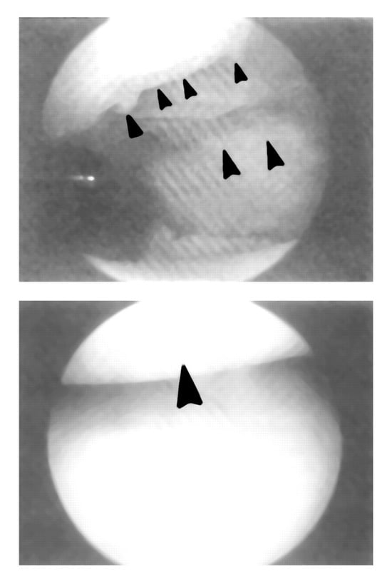
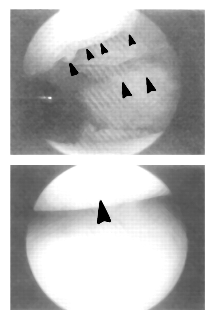
Chondromalacia patellae is a clinical condition of complex symptoms and physical signs, as well as an abnormality of patellofemoral cartilage. The clinical condition can be characterized by retropatellar and peripatellar pain and crepitation, pain or grating with tenderness on palpating the patellae, and subpatellar pain with athletic activity. Sitting for prolonged periods and walking up or down stairs usually aggravate the symptoms [1,2]. The term chondromalacia describes pathological changes that occur in the articular cartilage of the patella (Figure 1).
Figure 1.
Normal and abnormal (top view) knee articular cartilage (chondromalacia patellae) viewed arthroscopically. (Photo courtesy of Don Bunce, MD.).
The classic constellation of symptoms described previously are referred to as patellofemoral pain syndrome (PFPS); other names are retropatellar dysplasia (RPD), patellofemoral malalignment syndrome, and anterior knee pain. PFPS, which is commonly present in many active young patients, may show no evidence of chondromalacia on clinical examination of arthroscopy [2,3]. It is important to emphasize that chondromalacia patellae can exist as an incidental finding, even though the condition is often referred to as PFPS. Conversely, the symptom complex of chondromalacia patellae in some patients is not associated with patellar cartilaginous changes [2].
The associated lesions of the patellar cartilage involve swelling, fibrillation, fragmentation, and fissuring [2,4]. Outerbridge and Dunlop [5] described and graded the severity of the patellar articular cartilage lesion:
Grade 1: softening, swelling, or fibrillation of the articular cartilage
Grade 2: fragmentation and fissuring of an area <1.5 cm in diameter
Grade 3: fragmentation of an area >1.5 cm in diameter
Grade 4: cartilaginous erosion to the level of subchondral bone
Vuorinen et al. [6] found degenerative patellar changes in 29 of 31 knees at anatomopathological surgical examination.
Investigators propose many different etiologic factors for chondromalacia patella of knees. Direct trauma to the knee had precipitated symptoms of chondromalacia in nearly 50% of knees [7]. Goodfellow et al. [4] described a basal degeneration lesion of the patellar articular cartilage that is characterized by softening and swelling. They believed this type of lesion is a causative factor in chondromalacia. Patellofemoral malalignment and disturbances of the extensor muscle system of the knee have been implicated in the etiology [7,8]. Moreover, patellofemoral malalignment has been associated with an increase in varus alignment of the lower extremity and excessive subtalar joint (STJ) pronation during the stance phase of gait [9].
Abnormal coupling between axial rotation of the leg and inversion/eversion movement of the foot is another contributing factor to patellofemoral malalignment [10]. Studies have shown that this abnormal patellofemoral relationship results from the frontal and transverse motion of the tibia with respect to the femur and that the rotation of the tibia is related to the inversion/eversion rotation of the foot [10,11]. Tiberio hypothesized how a delay in the external tibial rotation could precipitate patellofemoral malalignment [12]. He attributed this to be the result of an excessive STJ pronation, along with an increase in internal tibial rotation, which creates a “biomechanical dilemma” for the tibiofemoral joint.
Patellofemoral malalignment is also associated with muscle strength imbalances and muscle–tendon structures. For example, atrophy or slow recruitment of the vastus medialis oblique (VMO) muscle may cause abnormal loading of the patellar articular cartilage, resulting in decreased strength and dysfunction of the extensor mechanism [13,14]. This causes the patella to drift laterally because the ability of the VMO to oppose pull of the vastus lateralis and lateral retinaculum is insufficient. Tightness of the lateral retinaculum may cause chondromalacia patellae as a result of an increase or decrease in the medial or lateral facet pressure [4,12]. A tight iliotibial band may also be a causative factor in patellofemoral malalignment [12].
Controversy in the literature is based on whether increasing or decreasing the Q angle can precipitate patellofemoral malalignment. What constitutes normal and abnormal measurements is also controversial in this angle. Normal Q angle has been reported to be between 10 and 18 degrees [15,16]. Woodall cautioned against the diagnostic significance of the Q angle because its measurement is assessed statically at knee extension and can therefore be altered as the quadriceps muscle group ensures activity [17]. On the other spectrum, some studies have linked PFPS with Q angles measuring as high as 20 degrees [16,18]. These are the consequences of excessive lateral forces on the patella from increased femoral anteversion and increased external tibial torsion that is exerted by a large Q angle. The use of foot orthoses in patients with PFPS has decreased Q-angle measurements significantly [19].
To correct for excessive STJ pronation and patellofemoral malalignment, an orthotic foot device can be inserted between the foot and shoe to help maintain the STJ in its neutral position throughout the stance phase of the gait cycle [20]. Root et al. [21] defined STJ neutral position as when the STJ was neither pronated nor supinated. Novick and Kelley studied the effectiveness of orthotics during the loading phase of gait. They reported how changes in force moments controlled movement of the rearfoot and reduced calcaneal eversion accelerations and velocities provided relief of clinical symptoms in the lower limb [22]. In addition to reducing maximum pronation or calcaneal eversion, orthotics have also decreased maximum internal tibial rotation acceleration and velocity and increased the transverse tibial rotation acceleration [1,23].
The literature on the kinematic analysis of the lower extremity and its role in reducing pain in overuse injuries is extensive. In a prospective study of 20 patients with musculoskeletal lower extremity pain incurred from recreational running, Nawoczenski et al. [10] were first to examine the effectiveness of orthotics on three-dimensional kinematics of the lower limb. They reported that the effect of orthotics occurred significantly between tibial internal/external abduction/adduction, and calcaneal inversion/eversion, as well as from heel contact to maximum internal tibial rotation. The changes in these kinematic and kinetic variables postulate why foot orthoses provide relief of the lower limb clinical symptoms. In a similar study examining the effectiveness of orthotics on kinematic behavior of the lower leg and rearfoot, McPoil and Cornwall [24] proved a significant correlation between the transverse tibial rotation and motion of the rearfoot during the stance phase of gait.
As many athletes with musculoskeletal knee injuries are successfully treated with foot orthotics, the question arises as to how these devices affect mechanics of the knee joint and relieve clinical symptoms. Most investigations on orthotic effectiveness in reducing pain of the lower extremity suggest a strong biomechanical relationship between the foot and knee [1,4,9,10,12,20,24,25]. Clement et al. [9] retrospectively surveyed 1,650 patients with overuse running injuries, PFPS being the most common injury. Although they reported beneficial success in controlling patellofemoral pain from foot orthotic treatment, the authors found that certain injuries were more frequent in one sex or the other and suggested that future studies should differentiate injuries by sex. In a clinical study of 20 adolescent girls in whom patellofemoral pain syndrome was diagnosed, Eng and Pierrynowski [23] found that the use of soft foot orthoses, for a period of 8 weeks, coupled with isometric quadriceps muscle stretching, reduced the pain far more than just an exercise program alone. The authors reported that the reduction in pain with orthotic treatment was due to reduced frontal and transverse plane motions at the knee, talocrural, and STJ during walking and running. DeHaven et al. reported an 82% success rate from soft and flexible foot orthotic treatment on 100 athletes diagnosed with chondromalacia patellae [13]. However, other treatments, such as quadriceps and hamstrings strengthening exercises, running, and knee braces, were included as part of the conservative management program. Only 8% required surgical intervention [13]. Eggold [20] examined 146 athlete runners; of the 56 treated for knee pain, there was 100% reduction in symptoms following orthotic treatment.
The treatment of chondromalacia patellae consists of conservative care to correct patellofemoral malalignment and manage symptoms, and surgical methods to treat lesions of the patellae and allow for realignment. The conservative treatment includes patient education, rest, quadriceps muscle strengthening and stretching, bracing and orthoses, McConnel taping, and antiinflammatory medications [13,14].
A variety of surgical procedures is used to treat lesions of the patellar articular surface in chondromalacia. McCarroll et al. [26] selected a particular procedure, depending on the severity of the disease. They found trephine and drilling sufficient methods for grade 2 disease; trephine and drilling or facetectomy for grade 3 disease; and facetectomy for grade 4 disease. They concluded that young, active patients who place more stress on the patellofemoral joint receive less satisfactory results than older, sedentary patients. Lateral retinaculum release proved beneficial in 79% of 14 knees with patellofemoral pain [25]. Patients who complained of pain from retropatellar articular surface in grade III chondromalacia and severe arthrosis benefit significantly by anterior advancement of the tibial tuberosity [16]. This procedure decreases the compressive forces that the patellofemoral joint endures and increases the strength of the quadriceps muscle group through the patellar ligament. Partial chondrectomy and patellectomy have been used to treat chondromalacia patellae [19,27,28,29].
The goal of this study is to determine the efficacy of foot orthoses in treating patellofemoral knee pain. Although multiple treatment modalities are used for these patients, it is hoped the study verifies that orthoses are significant in reducing symptoms. Furthermore, it is thought that the materials and corrective measures used in foot orthoses may prove useful to other practitioners fabricating orthoses for knee pain. Foot orthoses are often prescribed and used for the conditions discussed here, but objective information on the effectiveness of such orthoses cannot be inferred from a literature review.
Materials and Methods
Between 1990 and 1995, orthopedists diagnosed PFPS in 102 patients who were chosen to serve as subjects in this retrospective study. These individuals sought professional consultation from local orthopedic specialists, podiatrists, and physical therapists for activity-related musculoskeletal lower limb pain of at least 2 months duration. Criteria for participation in this study included no history of congenital deformity of the lower extremity nor traumatic orthopedic injuries to either lower extremity at least 6 months before participation in the study. At the time of the study, neither the subjects nor the investigators knew they would be involved in this retrospective review.
The initial screening and clinical diagnosis of PFPS/RPD were first based on an examination by an orthopedist and later by a podiatrist for prescription of thermoplastic functional orthoses. Both knees were clinically examined by the orthopedist. All knees exhibited clinical signs and symptoms of chondromalacia patellae. Particular attention was directed to the patellar crepitation, compression, and pain, tracking, and malalignment of the knees, Q-angle measurement, knee effusion, lateral retinaculum tightness, and range of motion limitation. Radiographic studies were ordered and reviewed by the orthopedist when indicated. Patients were evaluated for the onset and duration of their patellofemoral pain, previous treatment, degree of degenerative knee joint disease, foot type, and type of orthoses previously tried. Forefoot and rearfoot deformities (varus and valgus) along with structural tibial, knee (genu varum/valgum), rotational (internal/external), and limb length inequality were also noted.
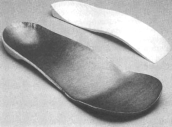
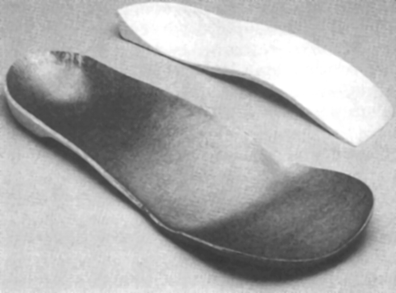
The subjects were considered appropriate candidates for orthotic intervention based on history of musculoskeletal symptoms and a preliminary clinical lower extremity screening examination. Orthoses for each subject were fabricated from nonweightbearing cast impressions and assessed for conformity to prescription and correct fit. Orthoses were casted in the neutral position, prone, and prescribed by the senior investigator. The functional thermoplastic polypropylene devices of various flexibilities and corrections (forefoot and rearfoot posting) were fabricated by Plus Labs (Rancho SantaFe Springs, CA) (Figure 2) by an Ortho CAD/CAM System (American Digital, Inc). Subjects were given instructions to progressively increase wear time of the orthoses during the subsequent 4 weeks. They were advised to discontinue wearing the new orthoses if they experienced any discomfort or new musculoskeletal pain. Patients’ charts were retrospectively reviewed to assess improvement and reduction of symptoms (Table 1). The “postdispensal” visit was 2 to 4 weeks after receiving the orthoses. Note was taken if patients needed adjustment to the orthoses and whether patients indicated they were “asymptomatic,” “improved,” “no different,” or “worse” at their follow-up appointment. However, patients were not prompted to categorize themselves. Appropriate statistical tests were performed after the data were accumulated.
Figure 2.
Typical foot orthosis used in study.
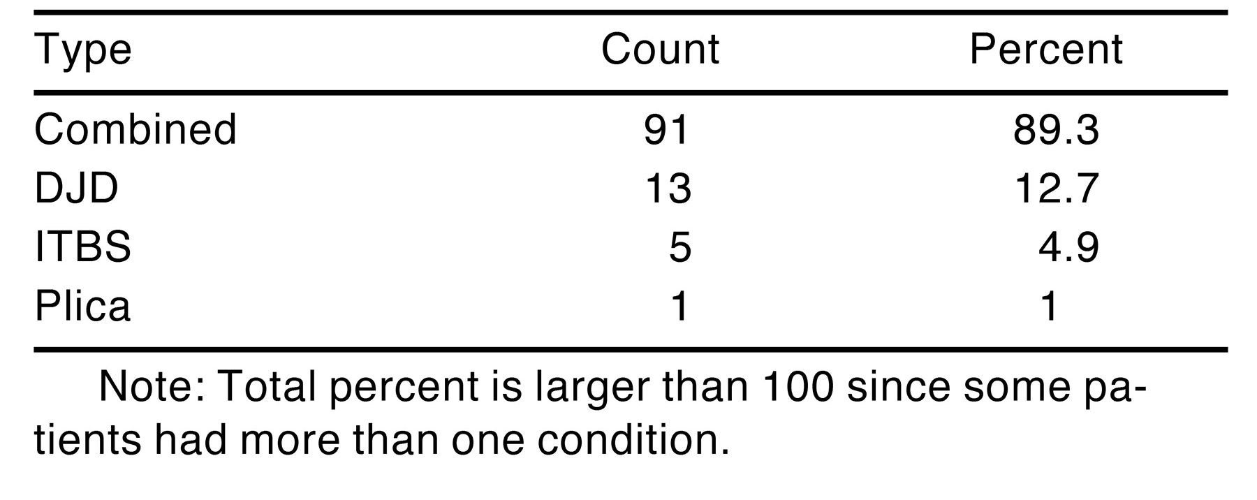
Table 1.
Types of Diagnosed Conditions.
Results
Data were analyzed for 102 patients: 46 women and 52 men; for 4 patients, the gender was not noted. The average age of patients was 37 years (if the age of the patient was not noted on the initial visit, it was computed between birth date and final visit date).
A majority of the patients had the combined diagnoses of chondromalacia patellae/PFPS/RPD. These patients comprised 89.3% of the cases, followed by degenerative joint disease (12.7%), illiotibial band syndrome (4.9%), and plica (1%); the total is greater than l00% because some patients had more than one condition (Table 1). The average duration of symptoms before treatment was 20.9 months, with the longest being 240 months (20 years).
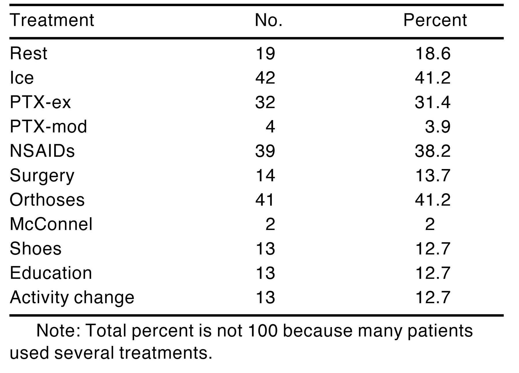
The types of treatment tried before functional orthotic therapy and their distribution are depicted in Table 2.
Table 2.
Types of Treatments Previously Tried.
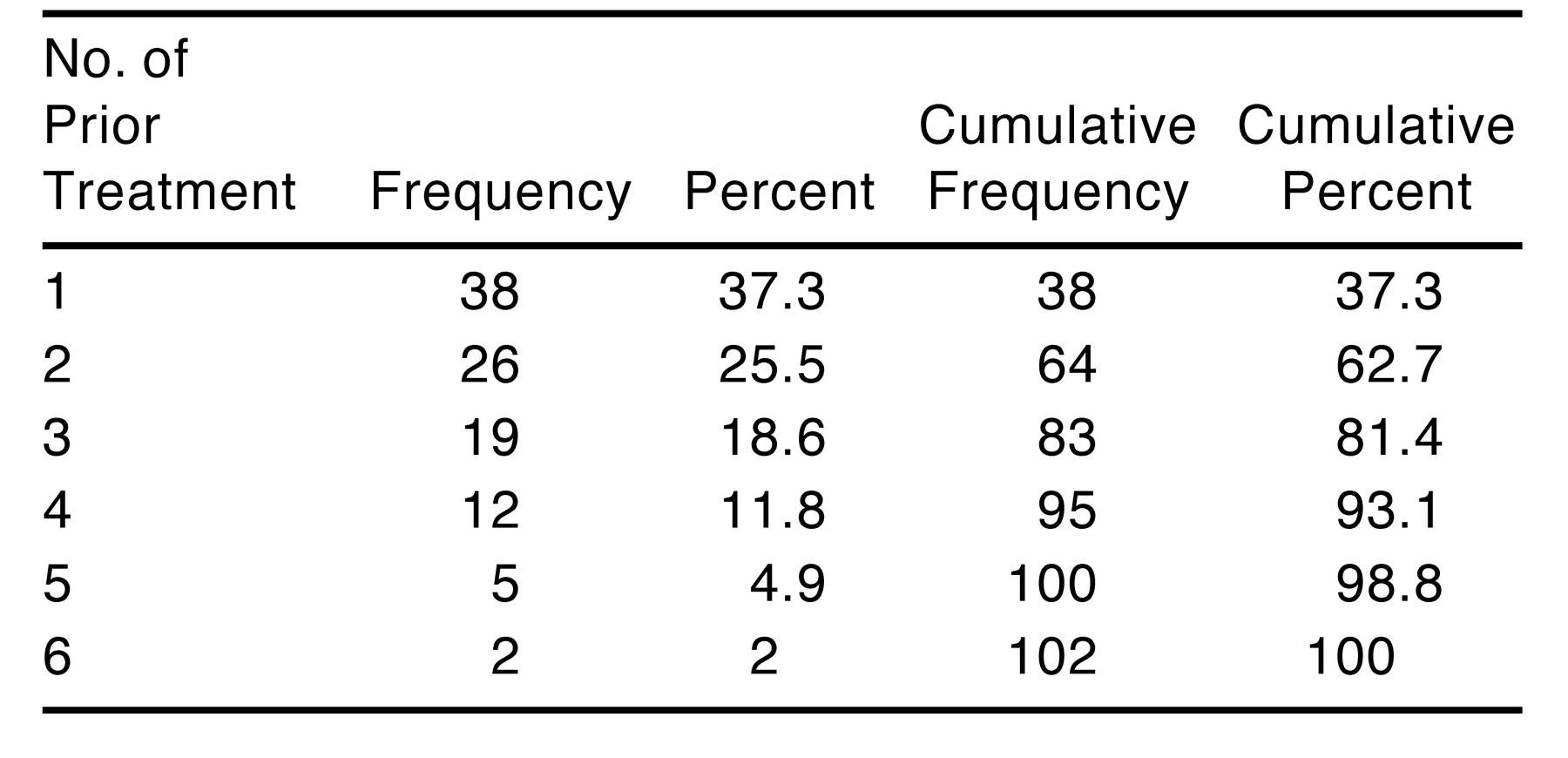
Ice and over-the-counter devices (or other orthoses made elsewhere) were the most common forms of treatment (41.2%). Nonsteroidal antiinflammatory drugs (NSAIDs) and physical therapy exercises were the next most common (38.2% and 31.4%, respectively). Surgery had been performed on 13.7% of the patients. A number of patients in the study had tried more than one treatment; only 38 had tried only one form of therapy (Table 3).
Table 3.
Number of Types of Previous Treatment.
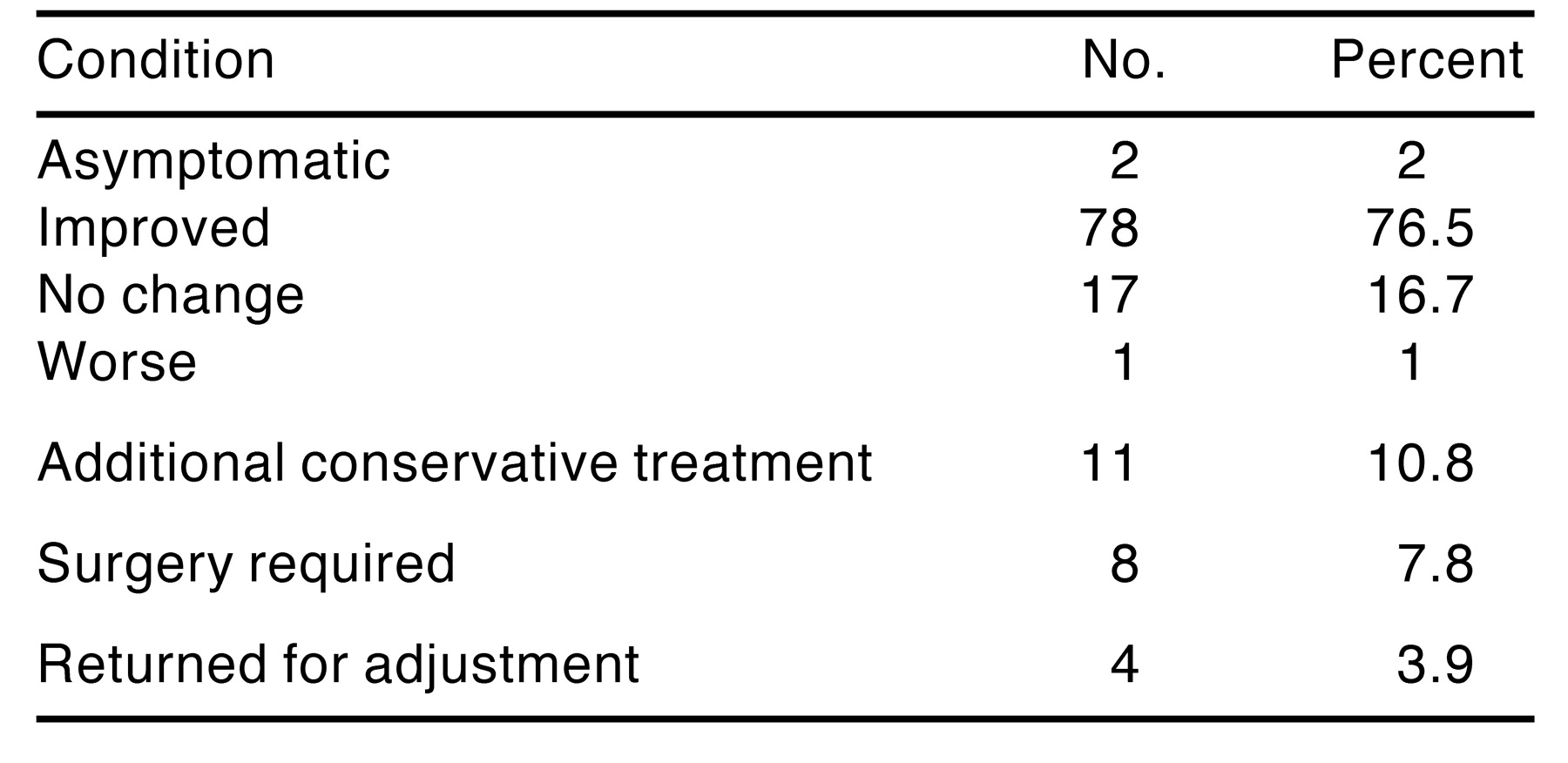
A statistically significant number of patients were either asymptomatic or improved by their follow-up visit, which ranged from 2 to 4 weeks after orthoses dispensal. Chi-squared analysis yielded P < .001. Because of the small sample size, Fisher’s Exact Test was performed, with a result of P = 12%. In this group, 2% of the patients said they were asymptomatic, and 76.5% said they were improved; 17 patients (16.7%) said they experienced no change, and 1 patient complained of being worse. Only 4% of the patients necessitated an orthotic adjustment (Table 4).
Table 4.
Results After Treatment With Orthoses.
There was insufficient information available in charts to indicate which patients felt they were able to return to their regular desired activities. Additional conservation treatment was used by 10.8% of the patients, and 7.8% subsequently underwent surgery. It is not known how these patients fared.
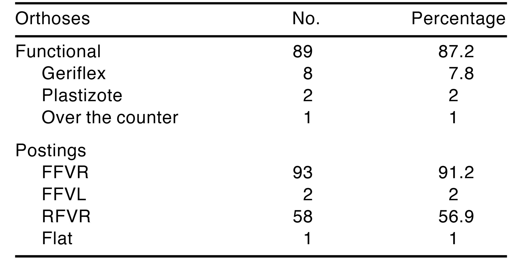
The most common orthosis was thermoplastic polypropylene (thickness range, 1–2 mm; semiflexible). This type of device was used by 87.2% of the patients. Geriflex or Plastizote devices (which had increased flexibility) were used in 10 patients. (All devices were made by the same laboratory and casted by the senior investigator.) The most common type of posting was intrinsic forefoot varus (91.2%). Rearfoot varus posting was noted to be used on 56.9%; only 2% had forefoot valgus posting (Table 5).
Table 5.
Orthoses Type and Posting.
Discussion
The results of this study are promising. Although the patients were not initially treated with a study in mind, the fact that approximately 76.5% improved with orthosis intervention set an initial standard for future studies. This seems similar to other previous reports. Because neither the patients nor the practitioners were prompted to subjectively categorize themselves at their follow-up visit, the results from this study may be viewed more objectively.
Clearly, this study has several limitations. The results merely indicated one practitioner’s success rate with one basic type of orthosis and casting method. However, others criticize that studies on the effectiveness of foot orthoses are limited by variables between practitioners, materials, and casting methods. Only 4% of the devices needing adjustment would be construed as relatively low. Anecdotally, one may expect a higher number of adjustments, as many patients reportedly complained of inability to use orthoses because of leg soreness and poor fit in shoes.
The results indicate that only 56.9% of the patients had rearfoot varus posting; review of the data for some of the patients showed this result to be misleading. Some of the patients were listed as having an orthosis with rearfoot varus only if the posting was greater than 2 degrees. Therefore, the data from this particular point are skewed. The senior investigator tends to post the rearfoot in 2 degrees of varus unless there is a tibial varum, external rotation, or genu varum/valgum deformity. He has found there is less adjustment soreness, which appears substantiated by the results.
At 37 years, the average patient’s age in this study is relatively young. One may find differences in treating adolescent girls versus older adults with foot orthoses. Furthermore, one may see better (or worse) results over time. One does not know from this study whether the patients continued to wear their orthoses.
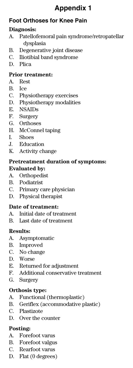
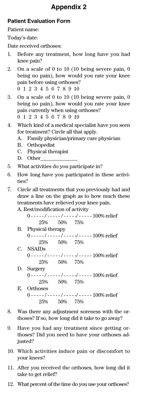
From the data collected, one may postulate that there may be a correlation between a forefoot varus deformity (since 91.2% of the patients have correction for this) and PFPS. This should be evaluated more closely. Future studies are planned, including having patients evaluate themselves with a visual analogue scale. Particular attention will be given to pre- and posttreatment symptoms and activity level. Also, the time frame it takes to obtain relief may be of value to athletic patients with these conditions. A larger sample size would help strengthen statistical conclusions. A sample of the patient survey is included in Appendix 2.
Conclusion
This study examined the efficacy of orthotic intervention in treating PFPS. In this study, 102 subjects were identified and examined, based on a history of musculoskeletal symptoms; 2% of the patients said they were asymptomatic, and 76.5% said they were improved; 17 patients (16.7%) said they experienced no change, and 1 patient said he or she was worse.
These findings suggest that foot orthoses are an effective means of relieving clinical symptoms of PFPS/RPD. Furthermore, the study reveals the eventual improvement of PFPS/RPD in many young people. The results point to the importance of future studies evaluating a larger sample size of patients with a visual analogue scale.
We are aware of the variability in findings of orthotic effectiveness in other studies. This may be due to the differences in orthotic material, the orthotic posting, casting fabrication methods, and the types of footwear. Furthermore, other studies have involved a variety of subjects with an assortment of foot structures that were studied with various shoes and orthotic inserts. Nonetheless, it is hoped that the results here will motivate others to pursue more study of the efficacy of orthotic intervention for knee pain.
Appendix 1
Appendix 2
References
- Bates, B.; Osterning, L.; Mason, B.; James, L. Foot orthotic devices to modify selected aspects of the lower extremity mechanics. Am J Sports Med 1979, 7, 338–342. [Google Scholar] [CrossRef] [Scilit] [PubMed]
- Stougard J: Chondromalacia of the patellae. Acta Orthop Scand 1975, 46, 685–694.
- Metcalf, R.W. An orthoscopic method for lateral release of the subluxating patella. Clin Orthop 1982, 167, 9. [Google Scholar] [CrossRef] [Scilit]
- Goodfellow, J.; Hungerford, D.S.; Woods, C. Patellofemoral joint mechanics and pathology. 2: Chondromalacia patellae. J Bone Joint Surg [Br] 1976, 58, 291. [Google Scholar] [CrossRef] [Scilit]
- Outerbridge, R.E.; Dunlop. JAY: The problem of chondromalacia patellae. Clin Orthop 1975, 110, 117. [Google Scholar] [CrossRef] [Scilit]
- Vuorinen, O.P.; Tunturi, T.; Harkkonen, M. Chondromalacia patellae: Results of operative treatment. Arch Orthop Traum Surg 1985, 104, 175–181. [Google Scholar] [CrossRef] [Scilit]
- Insall, J.N.; Aglietti, P.; Tria, A.J. Patellar pain and incongruence. 2: Clinical application. Clin Orthop 1983, 176, 225–232. [Google Scholar] [CrossRef] [Scilit]
- Hvid, I.; Anderson, L.I.; Schmidt, H. Chondromalacia patellae: The relationship to abnormal patellofemoral joint mechanics. Acta Orthop Scand 1981, 52, 661–666. [Google Scholar] [CrossRef] [Scilit]
- Clement, D.B.; Taunton, J.E.; Smart, G.W.; McNicol, K.L. A survey of overuse running injuries. Physician Sports Med 1981, 9, 47–58. [Google Scholar] [CrossRef] [Scilit]
- Nawoczenski, D.A.; Cook, T.M.; Saltzman, C.L. The effect of foot orthotics on three-dimensional kinematics of the leg and rearfoot during running. J Orthop Sports Phys Ther 1995, 21, 317–327. [Google Scholar] [CrossRef] [Scilit]
- McCulloch, M.; Brunt, D.; Vander Linden, D. The effect of foot orthotics and gait velocity on lower limb kinematics and temporal events of stance. J Sports Phys Ther 1993, 17, 2–10. [Google Scholar] [CrossRef] [Scilit] [PubMed]
- Tiberio D: The effect of excessive subtalar joint pronation on patellofemoral mechanics: A theoretical model. J Orthop Sports Phys Ther 1987, 9, 160–165. [CrossRef] [Scilit]
- DeHaven, K.E.; Dolan, W.A.; Mayer, P.J. Chondromalacia patellae in athletes. Clinical presentation and conservative management. Am J Sports Med 1979, 7, 5. [Google Scholar] [CrossRef] [Scilit]
- Fischer, R.L. Conservative treatment of patellofemoral pain. Orthop Clin North Am 1986, 17, 269–272. [Google Scholar] [CrossRef] [Scilit]
- Norkin, C.C.; Levangie, P.K. Joint structure and function: A comprehensive analysis, 2nd ed.; FA Davis: Philadelphia, 1992. [Google Scholar]
- Woodall W, Welsh J: A biomechanical basis for rehabilitation programs involving the patellofemoral joint. J Orthop Sports Phys Ther 1990, 11, 535–541. [CrossRef] [Scilit]
- Heartley, F.W.; Allen, P.R.; Patrick, J.H. Tibial tubercle advancement for anterior knee pain: A temporary or permanent solution. Clin Orthop 1986, 208, 215. [Google Scholar] [CrossRef] [Scilit]
- Tria, A.J.; Palumbo, R.C.; Alicera, J.A. Conservative care for patellofemoral pain. Orthop Clin North Am 1992, 23, 545–554. [Google Scholar] [CrossRef] [Scilit] [PubMed]
- D’Amico, J.C.; Rubin, M. The influence of foot orthoses on quadriceps angle. J Am Podiatr Med Assoc 1986, 76, 337–340. [Google Scholar] [CrossRef] [Scilit]
- Eggold JF: Orthotics in the prevention of runners’ overuse injuries. Physician Sports Med 1981, 9, 125–131.
- Root, M.L.; Orien, W.P.; Weed, J.H.; Hughes, R.J. Biomechanical examination of the foot Vol 1; Clinical Biomechanics: Los Angeles, CA, 1971. [Google Scholar]
- Novick, A.; Kelley, D. Position and movement changes of the foot with orthotic intervention during the loading response of gait. J Orthop Sports Phys Ther 1990, 11, 301–312. [Google Scholar] [CrossRef] [Scilit]
- Eng, J.J.; Pierrynowski, M.R. Evaluation of soft foot orthotics in the treatment of patellofemoral pain syndrome. Phys Ther 1993, 73, 67–70. [Google Scholar] [CrossRef] [Scilit]
- McPoil, T.G.; Cornwall, M.W. Footwear and foot orthotic effectiveness research: A new approach. J Orthop Sports Phys Ther 1995, 21, 337–344. [Google Scholar]
- Fulkerson, J.P. The etiology of patellofemoral pain in young active patients: A prospective study. Clin Orthop Relat Res 1983, 179, 129–133. [Google Scholar] [CrossRef] [Scilit]
- McCarroll, J.R.; Odonohue, D.H.; Crana, W.A. The surgical treatment of chondromalacia of the patellae. Clin Orthop Relat Res 1983, 175, 130–134. [Google Scholar] [CrossRef] [Scilit]
- Bently, G. Chondromalacia patellae. J Bone Joint Surg [Am] 1970, 52, 221. [Google Scholar] [CrossRef] [Scilit]
- Jensen, D.B.; Albrektsen, S.B. The natural history of chondromalacia patellae: A 12-year follow-up. Acta Orthop Belg 1990, 56, 503–506. [Google Scholar]
- Kramer, P.J. Patellar malalignment syndrome: Rationale to reduce excessive lateral pressure. J Orthop Sports Phys Ther 1986, 8, 301–309. [Google Scholar] [CrossRef] [Scilit]

© 2003 American Podiatric Medical Association