


Abstract
The purpose of this study was to determine the effectiveness of two types of foot orthoses in controlling the magnitude and rate of internal tibial rotation, measured by the tibial pointer device, during walking. Ten subjects between the ages of 23 and 43 years volunteered to participate in the study. Prior to data collection, each subject was issued two types of foot orthoses: a pair of rigid, plastic orthoses with posting in either the forefoot or the rearfoot, and a pair of soft, accommodative, premolded orthoses with no posting. All subjects wore standardized footwear. Following a controlled break-in period for both footwear and orthoses, each subject was asked to walk at a self-selected speed over a 12-m walkway while the movement of internal tibial rotation was recorded with a video camera during five trials. The results indicated that both the rigid plastic and the accommodative foot orthoses significantly reduced the magnitude and the rate of internal tibial rotation. No significant difference was noted between the soft and rigid foot orthoses conditions.
Foot orthoses have become an accepted adjunct in the treatment of foot and ankle injuries associated with abnormal foot function [1]. Overuse injuries that affect the foot, ankle, and lower extremity are thought to result from inflammation of the soft tissues, osseous structures, and contractile tissues caused by increased tissue stress [1]. Abnormal foot function is thought to be one of the primary factors leading to increased tissue stress. The purpose of foot orthoses as part of the total management scheme is to control the degree of stress applied to these inflamed tissues by controlling excessive foot motion [2]. Because the most common factor cited in the literature as the cause of increased tissue stress is excessive foot pronation, [3,4] foot orthoses are often designed to control both the magnitude and the rate of pronation [5]. The magnitude or amount of foot motion has been linked to increased stress on soft and bony tissues, whereas the rate or acceleration of foot pronation has been implicated as a factor in the development of contractile tissue stress.
While foot orthoses appear to be an important component of the management program for overuse injuries caused by excessive foot pronation, the validation of these devices is a primary concern for any health professional who prescribes or fabricates them. The current trend in health-care reimbursement programs mandates that the clinician demonstrate the efficacy and cost-effectiveness of prescribed treatments, including foot orthoses. Unfortunately, much of the literature advocating the use of foot orthoses has relied on anecdotal evidence [6,7,8,9,10,11,12,13,14,15]. Although these reports provide the clinician with information on current or new trends in the prescription and use of foot orthoses, they do not provide any quantitative evidence regarding the effectiveness of foot orthoses. Several studies have also been reported that retrospectively and prospectively surveyed patients who were dispensed foot orthoses as part of their treatment programs [3,16,17]. These survey studies have effectively illustrated the level of comfort obtained through use of orthoses as well as the patient’s perceived level of effectiveness of the orthoses, but they also fail to provide any quantitative evidence regarding the effectiveness of foot orthoses in providing motion control.
The majority of studies attempting to quantify the effectiveness of foot orthoses in controlling excessive foot pronation during walking have involved the kinematic analysis of rearfoot motion. The most common method used for rearfoot motion analysis involves measuring inversion and eversion of the rearfoot (calcaneus) in relation to the lower leg. In these studies, subjects are usually filmed from behind as they walk either on a treadmill or over ground. To measure rearfoot motion, markers are placed along a line that bisects the posterior lower leg and the rearfoot. The angle between these two lines is calculated and used as a measure of complex rearfoot motion. This method works well for barefoot walking, but difficulties arise when the subject is required to wear shoes, such as in studies evaluating the effectiveness of orthoses. The extensive use of this method of studying rearfoot motion is most likely due to the method’s relative ease of use and its use of essentially the same angle measured by clinicians during static weightbearing evaluations.
Several recent studies have used this method of motion analysis to assess the effectiveness of foot orthoses during walking [18,19,20,21]. While these studies attempted to quantify the changes in rearfoot motion effected by the use of foot orthoses, a major limitation of the studies was the difficulty in measuring the movement of the foot within the shoe. Because the method used in each of these studies involved placing markers on the counter of the shoe, rather than directly on the rearfoot, the movement recorded was probably more representative of shoe movement than of changes in foot motion caused by the foot orthoses. In addition, some of these studies failed to standardize the footwear worn by the subjects or to control the technique used to fabricate the foot orthoses being investigated. Both of these factors could significantly decrease the validity of these studies.
Johanson et al [22] modified the shoe used by each subject in their study to address the problems associated with placing markers on the shoe when assessing the effectiveness of foot orthoses. In each shoe, a 5-cm portion of the shoe padding and counter was excised and the opening was reinforced with translucent urethane to retain the integrity of the shoe counter. The effects of different orthotic posting methods on 22 subjects who had a forefoot varus deformity of at least 8° were then tested using kinematic analysis of the rearfoot. Each subject walked on a treadmill for the following four conditions using the modified footwear: 1) orthotic shell with no posting, 2) orthotic shell with forefoot posting, 3) orthotic shell with rearfoot posting, and 4) orthotic shell with both forefoot and rearfoot posting. The researchers’ findings indicated that the orthoses with posting in both the forefoot and the rearfoot or just the rearfoot were more effective in controlling rearfoot pronation than orthoses with posting only in the forefoot. However, the greatest change in rearfoot pronation occurred when subjects went from the barefoot condition to wearing the shoe with orthotic shell and no posting (1.3°). There were no statistically significant differences among the four conditions tested. The addition of a forefoot-only or a rearfoot-only post resulted in differences of only 0.2° and 0.55°, respectively. The change in rearfoot pronation as a result of using both a forefoot and a rearfoot post was 0.95°. The results of this study show that the most important component of the orthosis in controlling rearfoot pronation was the shell itself. The shell used in the study was semirigid and provided total contact with the plantar surface of the foot, including under the medial longitudinal arch. Thus the results suggest that total-contact foot orthoses would be more effective than actual posting or wedging in controlling rearfoot pronation caused by a forefoot varus deformity.
In a more recent study, Brown et al [23] used the same type of footwear and the same methods as Johanson et al [22] to assess the effectiveness of a custom-made, semirigid “biomechanical” orthosis in comparison with an over-the-counter arch support. The biomechanical foot orthoses were fabricated by the same commercial laboratory from a cast obtained while each subject was in the subtalar joint neutral position. Twenty-four subjects with a predetermined forefoot varus deformity of at least 8° walked on a treadmill while rearfoot motion was recorded for the following three conditions: 1) shoe-only, 2) shoe with custom-made orthosis, and 3) shoe with over-thecounter arch support. The results indicated no difference among the three conditions for either maximum pronation or maximum pronation velocity. The maximum pronation values for the shoe-only, arch support, and custom-made orthosis conditions were 11.2°, 11.0°, and 10.7°, respectively. The greatest decrease in maximum pronation velocity was obtained with the over-the-counter device. The authors concluded that further research was necessary to identify the biomechanical effects responsible for the clinical success of foot orthoses and to improve the design of foot orthoses for controlling rearfoot pronation.
The results of these two investigations seem to suggest that foot orthoses that provide total contact with the plantar region of the foot are as effective as biomechanical foot orthoses with posting. In addition, the results of the study by Brown et al [23] indicate that over-the-counter foot orthoses are as effective in controlling foot pronation as custom-made devices. While both of these studies have contributed greatly to the understanding of the effect of foot orthoses on foot motion within the shoe, there are several concerns regarding the methods used in these studies. One concern is that both of the studies used two-dimensional (2-D), rather than three-dimensional (3-D), video analysis. In addition, data in both of these studies were obtained by recording results of treadmill walking. The results of previously reported studies, however, justify the use of 2-D video analysis as long as the motion analysis is limited to the first 60% of the stance phase of walking [24,25]. In addition, Lemke et al [26] reported that kinematic analysis of the rearfoot is similar whether data are collected by having subjects walk on a treadmill or over ground. The most obvious issue in these studies is whether excising a portion of the shoe counter affects the performance of the foot orthosis in controlling excessive foot pronation. Because the shoe provides the platform for the foot orthosis, changes or modifications to the platform could substantially affect the performance of the orthosis. Even if the technique used in the previously mentioned studies to excise a portion of the counter did not affect the integrity of the shoe platform, this technique would not be a practical solution for clinicians interested in assessing the effect of the orthoses they have prescribed. The development of a clinical method for assessing the effectiveness of foot orthoses for any patient is critical to justify reimbursement for these devices.
In an attempt to develop a clinical method to indirectly assess foot motion within the shoe, Cornwall and McPoil [27] described the use of a tibial pointer device. The purpose of the tibial pointer device is to measure transverse tibial rotation during dynamic activity. The premise of this device is that a relationship exists between transverse tibial rotation and motion of the rearfoot. By establishing this relationship for each individual patient or subject as he or she walks barefoot, the tibial pointer system can be used to indirectly measure rearfoot motion when the patient or subject is wearing shoes, with or without foot orthoses. Thus it is not necessary to modify the shoe in order to assess the effectiveness of foot orthoses clinically. The tibial pointer device described by these authors consisted of a 25 × 25 × 25-mm aluminum block with two 110-mm aluminum rods positioned at a 90° angle to each other. There were 15-mm-diameter spherical reflective markers attached to the end of each aluminum rod. The aluminum block was placed at the level of the tibial tubercle and secured with Velcro straps (Velcro USA Inc, Manchester, New Hampshire). In their study, 8 subjects (16 feet) had reflective markers placed on the lower leg and calcaneus; the tibial pointer device was positioned over the anterior aspect of the tibial tubercle. Each subject walked barefoot over a 12-m walkway and was filmed as he or she walked by video cameras positioned from the front and from behind. The tibial pointer and rearfoot motion data were analyzed using 2-D analysis. The results indicated a high degree of association between transverse tibial rotation and rearfoot motion patterns for all 16 feet (r = 0.95). The authors noted, however, that the relationship between transverse tibial rotation and rearfoot motion varied among the subjects. In the same study, two case studies were presented in which the tibial pointer device was used to assess the effectiveness of foot orthoses.
Although the tibial pointer device described by Cornwall and McPoil [27] appears to be a viable and clinically relevant method of assessing the effectiveness of foot orthoses, a major concern is the reliability and validity of the tibial pointer device with the use of 2-D analysis. Three-dimensional analysis of motion has been called the superior method for representing the actual movement of the lower extremity during walking [28]. Sawert et al [29] conducted a study involving 20 subjects to determine whether the reliability and validity of 2-D analysis of transverse tibial rotation during walking was comparable with those of tibial rotation assessed using 3-D analysis. They reported a high degree of association (r = 0.85) between the mean pattern of 2-D tibial rotation, as measured using the tibial pointer device, and the mean pattern of 3-D tibial rotation, determined using a standardized method of analysis [28]. They concluded that the use of the tibial pointer device with 2-D motion analysis techniques provided a reliable and valid method of assessing transverse tibial rotation.
The results of the studies conducted by both Johanson et al [22] and Brown et al [23] to determine the effectiveness of orthotic devices suggest that several issues regarding the design of foot orthoses remain unclear. Are foot orthoses that provide total contact with the plantar region of the foot as effective in controlling foot pronation as foot orthoses that use posting or wedging for motion control? Are soft, accommodative foot orthoses as effective in controlling foot pronation as devices constructed from rigid plastic materials? These questions indicate the need for further research that attempts to delineate those elements critical for the design of foot orthoses. The use of the tibial pointer device provides a reliable, valid, and clinically feasible method of assessing the degree of motion control provided by foot orthoses.
Thus the purpose of the present study was to determine the effectiveness, using the tibial pointer device, of two types of foot orthoses in controlling the magnitude and rate of internal tibial rotation during walking. The two types of foot orthoses were evaluated in ten subjects using standardized footwear. The foot orthoses selected for this investigation had marked differences in both material density and those features incorporated into their design for motion control. The assumption was made that the primary factor in the effectiveness of foot orthoses was the degree of control of foot pronation. Thus only variables associated with internal tibial rotation were evaluated because of the link between internal tibial rotation and foot pronation.
Materials and Methods
Subjects
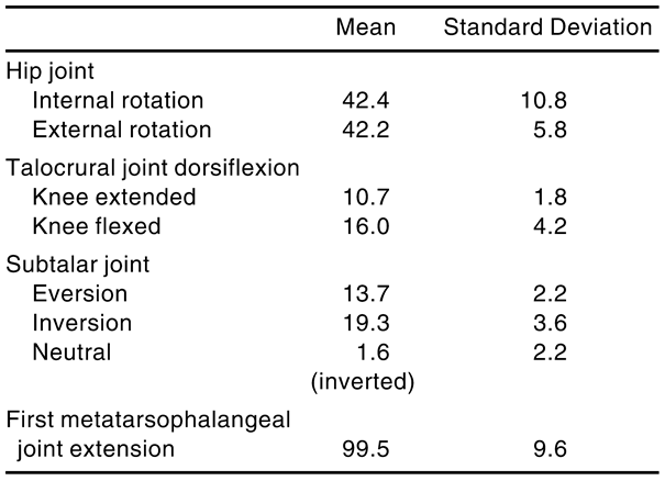
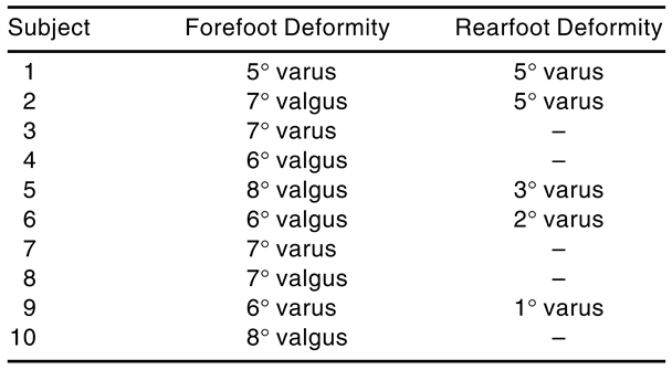
Ten subjects were chosen from a group of 110 volunteers who underwent a bilateral lower-extremity musculoskeletal screening prior to selection. The ten subjects selected, five women and five men, met all of the following screening criteria: 1) no history of congenital deformity; 2) no history of trauma or pain of either lower extremity or the lumbosacral region within the 12 months preceding the start of the investigation; 3) no apparent hallux valgus based on visual assessment; 4) symmetrical internal and external rotation of the hip; 5) no malleolar torsion; 6) 10° of talocrural joint dorsiflexion, with the knee extended; 7) 65° of first metatarsophalangeal joint extension; 8) an observed forefoot or rearfoot deformity; and 9) wear either a size 71/2 (US) women’s or a size 9 (US) men’s shoe. The musculoskeletal screening examination was performed by the same investigator (T.G.M.); the procedures used in the examination have been previously described by McPoil and Brocato [30]. The mean age of the ten subjects was 30.3 years, with a range of 23 to 43 years. Table 1 lists the mean values for the musculoskeletal screening examination, and Table 2 lists the forefoot and rearfoot deformities determined for all ten subjects. The study procedure was approved by the institutional review board of Northern Arizona University in Flagstaff, and all subjects signed an informed consent form prior to joining the study.
Table 1.
Mean Values (°) for the Musculoskeletal Screening Examination (Right Foot Only).
Table 2.
Forefoot and Rearfoot Deformities for All Subjects (Right Foot Only).
Instrumentation
Tibial Pointer Device. The same method reported by Cornwall and McPoil [27] was used in this study, except for the following modification. To decrease the momentum applied to the tibial pointer device, especially during the loading response period of walking, the aluminum block was embedded into 3-mm-thick hand-splinting material, which was shaped to fit the anterior aspect of the upper leg. To improve comfort, the side of the splinting material that was placed next to the subject’s skin was covered with a 3-mm sheet of microcellular rubber. The entire system was placed on the subject’s leg so that the aluminum block was positioned at the level of the tibial tubercle; it was secured using elasticized Velcro straps.
Motion-Recording Equipment. A video camera (Panasonic Corp, Cypress, California, model B4-400) was placed directly in front of the subject as he or she walked down a 12-m walkway. The video camera was connected to a videocassette recorder (Panasonic Corp, model AG-1960). Video images from the camera were recorded at 60 fields per second with a shutter speed of 1/500 sec. The camera was calibrated using a known linear distance in the field of view to correct for any lens aberrations that might affect the kinematic measurements. The accuracy of the filming and digitizing procedures used in this study was assessed by filming a series of known angular measurements. The results of this test showed a difference of less than 0.5° between all of the calibrated and actual measurements obtained.
Footwear and Foot Orthoses
Two different pairs of foot orthoses were fabricated for each subject. The rigid orthoses condition consisted of a pair of UCO-BKS medium-width foot orthoses (UCO International, Prospect Heights, Illinois, model 23/4383) modified with a heat gun so that each subject was comfortable walking in them. The UCOBKS foot orthoses are premolded and constructed from a 2.5-mm rigid, high-molecular-weight polyethylene material. Once the subject was comfortable with the fit of the orthoses, appropriate forefoot and/or rearfoot posts were added to each orthosis based on the findings of the initial musculoskeletal screening. Prefabricated thermocork wedges (UCO model 20/1324), with a durometer of 55 to 60 (Shore A gauge), were glued to the anterior and/or posterior aspect of the foot orthoses and ground to the appropriate angle by the same investigator. The top of the UCO-BKS orthosis was then covered with a 2-mm layer of UCOLITE (UCO model 32/0050-202; durometer 35, Shore A gauge).
The soft orthoses condition consisted of a pair of UCO-BF foot orthoses (model 20/0682). The UCO-BF orthoses are premolded, total-contact devices that are fabricated with material having a durometer of 30 to 35 (Shore A gauge). Forefoot and/or rearfoot posting was not added to the soft orthoses. Although each subject was permitted to request modifications to the BF orthoses at the initial fitting, no modifications were made for any of the ten subjects.
The footwear used for the study was the Reebok Boston Road running shoe (Reebok International Ltd, Stoughton, Massachusetts, model 1-26033 for women, model 1-26035 for men). This shoe was selected because the men’s and women’s models have the same last shape. In addition, the shoe is slip-lasted with a moderate amount of rearfoot stabilization, is constructed with a dual-density ethylene vinyl acetate (EVA) midsole, and has a removable, compressionmolded EVA sock liner.
Procedure
Before the start of data collection, each subject was fitted with a pair of Boston Road running shoes and was asked to wear the shoes for a 40-hour break-in period. Subjects were instructed to count only those hours during which they were involved in activities such as walking, aerobics, and other types of fitness workouts. Once the subjects had accumulated 40 hours of wear time, they were scheduled for the first data-collection session.
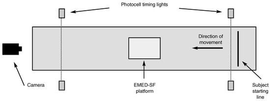
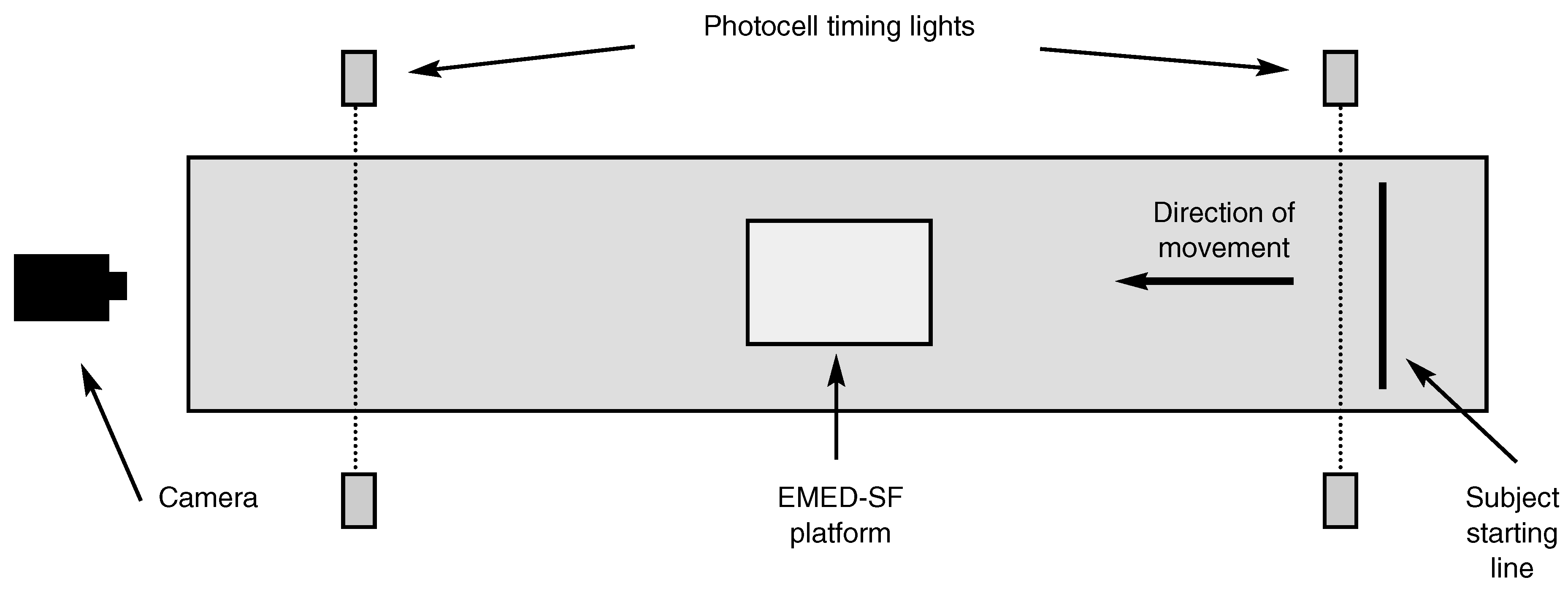
Upon arriving for the first data-collection session, each subject had his or her height and weight measured and recorded. Subjects were then instructed to practice walking barefoot at a self-selected speed along the 12-m walkway for several minutes. Figure 1 illustrates the arrangement of the equipment used for data collection. Walking speed was monitored using two infrared photocells positioned 6.1 m apart and connected to a digital timer. When between-trial walking speed was consistent (variation of less than 5% between trials), the tibial pointer device previously described was attached over the tibial tubercle of each subject’s right leg. The subject was then asked to walk barefoot over the walkway while movement of the tibial pointer device was recorded for five consecutive trials using a video camera positioned in front of the subject and operating at a speed of 60 Hz. Once the barefoot walking trials were completed, the subject then donned the running shoes, with the tibial pointer device still positioned on the right leg, and again practiced walking over the walkway. When the between-trial walking speed was consistent (variation of less than 5% between trials) compared with that recorded for the barefoot trials, each subject was asked to walk over the walkway for the shoeonly condition. The movement of the tibial pointer device was again recorded with the video camera during five consecutive trials. To determine the consistency of each subject’s gait velocity during video data collection, the duration of the stance phase was recorded using the EMED-SF capacitance transducer platform (Novel USA, Inc, Minneapolis, Minnesota), which was located at the midpoint of the walkway.
Figure 1.
Illustration of the arrangement of equipment used for data collection.
Upon completion of data collection for the shoeonly condition, each subject was given a pair of rigid orthoses, properly posted for his or her foot alignment, as well as a pair of soft orthoses. Each subject was instructed to follow the same 40-hour break-in period for each pair of orthoses as previously described for the footwear. Subjects were permitted to wear the foot orthoses only in the study running shoes during the orthosis break-in period. The final testing session was scheduled when each subject had recorded a total wearing time of 120 hours for the study running shoes, including 40 hours for each of the two pairs of foot orthoses.
Before final data collection, subjects were asked to practice walking with their study footwear over the 12-m walkway while the gait velocity was monitored. After a consistent gait velocity was attained, the tibial pointer device was attached to the subject’s right leg and the subject was again instructed to practice walking over the walkway. When the between-trial walking speed was consistent (variation of less than 5% between trials) compared with the speed previously recorded for the shoe-only condition, each subject walked over the walkway wearing each pair of foot orthoses. The order of testing for the foot orthoses conditions was randomly determined for each subject. Video data were recorded for the right foot for five consecutive trials for both the soft orthoses and rigid orthoses conditions. To determine the consistency of each subject’s gait velocity during video data collection, the duration of the stance phase was again recorded using the EMED-SF capacitance transducer platform that was located at the midpoint of the walkway.
Data Analysis
The stance-phase durations for each trial were calculated using the NovelWin EMED-SF software program (Novel USA, Inc). The video data were digitized using the Peak Performance automated analysis software (Peak Performance Technologies, Englewood, Colorado) to obtain 2-D coordinates of the two markers of the tibial pointer device. The 2-D coordinates were used to determine the perceived distance between the two pointer arms. Transverse tibial rotation values were then calculated using the algorithm previously described by Cornwall and McPoil [27]. Actual values for 2-D transverse tibial rotation were obtained by subtracting the static position of the tibial pointer device markers while the subject was filmed standing from the dynamic tibial pointer device angles determined for each frame of the five trials for each condition. The dependent variables calculated from the resulting dynamic transverse tibial rotation data for each of the four conditions were 1) maximum internal tibial rotation and 2) maximum internal tibial rotation acceleration.
Statistical Analysis
Reliability of Stance-Phase Duration. Type (3,1) intraclass correlation coefficients [31] and the coefficient of variation were calculated to determine the consistency of the stance-phase durations for the 20 trials collected using the EMED-SF platform. The coefficient of variation is the standard deviation expressed as a percentage of the mean; according to Rothstein [32], it is an appropriate statistical method of analyzing the reliability of multiple measurements. The level of reliability for the intraclass correlation coefficients was classified using the characterizations reported by Landis and Koch [33]. These characterizations were as follows: “slight,” if the coefficient ranged from 0 to 0.20; “fair,” if the coefficient ranged from 0.21 to 0.40; “moderate,” if the coefficient ranged from 0.41 to 0.60; “substantial,” if the coefficient ranged from 0.61 to 0.80; and “almost perfect,” if the coefficient ranged from 0.81 to 1.00.
Evaluation of the Effectiveness of Foot Orthoses. A one-way repeated measures analysis of variance (ANOVA) was used to evaluate the effectiveness of the foot orthoses in controlling both the magnitude and rate of transverse tibial rotation. A Tukey’s post hoc comparison was used to determine differences among the conditions tested. An alpha level of .05 was used for all tests of statistical significance.
Results
Reliability of Stance-Phase Duration
The mean stance-phase duration for the barefoot trials of all ten subjects was 0.552 sec, with a standard deviation of 0.031 sec. The mean stance-phase duration for the shoed trials of all ten subjects was 0.545 sec, with a standard deviation of 0.036 sec. The intraclass correlation coefficient calculated for the barefoot condition was 0.88, and that calculated for the shoed conditions was 0.89. The coefficient of variation for the barefoot condition was 5.9%, and that for the shoed conditions was 6.5%.
Evaluation of the Effectiveness of Foot Orthoses
The means and standard deviations for the dependent variables maximum internal tibial rotation and maximum internal tibial rotation acceleration are listed in Table 3. The results of the ANOVA conducted on the dependent variable maximum internal tibial rotation were significant for differences among the four experimental conditions (F = 5.0; df = 3; P < .007). The results of the Tukey’s post hoc analysis indicated the following: 1) the barefoot and shoe-only conditions were significantly different (P < .05) from the two foot orthoses conditions; and 2) no significant difference was found between the barefoot and shoeonly or the soft orthoses and rigid orthoses conditions. The results of the ANOVA conducted on the dependent variable maximum internal tibial rotation acceleration were also significant for differences among the four experimental conditions (F = 7.9; df = 3; P < .001). The results of the Tukey’s post hoc analysis indicated the following: 1) the barefoot condition was significantly different (P < .05) from the shoe-only, soft orthoses, and rigid orthoses conditions; 2) the shoe-only condition was significantly different (P < .05) from the soft orthoses and rigid orthoses conditions; and 3) no significant difference was found between the soft orthoses and rigid orthoses conditions.
Table 3.
Mean Values for Maximum Internal Tibial Rotation and Maximum Internal Tibial Rotation Acceleration.
Discussion
The authors’ first concern in interpreting the results of this study was whether each subject’s stance-phase durations were consistent for the four conditions evaluated, especially considering that data collection occurred on two separate days. The consistency of stance-phase durations was a critical issue because increased variability between the two days of data collection could indicate that the subject’s walking speed was different on the two test days. The characterizations of the stance-phase duration intraclass correlation coefficients for the barefoot and shoed conditions, which were 0.88 and 0.89, respectively, would be “almost perfect” according to the criteria established by Landis and Koch [33]. The coefficient of variation values for the barefoot and shoed conditions were only 5.9% and 6.5%, respectively. On the basis of the intraclass correlation coefficient and coefficient of variation values obtained, the authors concluded that the stance-phase durations were extremely consistent among the four conditions for all subjects and that further analyses of the results could be made.
The purpose of this study was to determine the effectiveness, using the tibial pointer device, of two types of foot orthoses in controlling the amount and acceleration of internal tibial rotation during walking. With regard to controlling the amount of internal tibial rotation, the results of this study indicate that premolded, total-contact foot orthoses are as effective as devices constructed from rigid materials. In addition, the use of posting or wedging did not improve the effectiveness of the foot orthoses in controlling foot pronation. These results are in agreement with the findings previously reported by Brown et al, [23] who reported no difference in the degree of motion control provided by a custom-made, semirigid orthosis with posting compared with a soft, over-the-counter device. In a more recent study, Nigg et al [34] reported similar findings in a group of runners who used shoe inserts constructed from foam of different densities. They found that total foot pronation was not significantly affected by differences in shoeinsert density, which ranged from soft to hard.
Unlike the results previously reported by Johanson et al, [22] the shoe-only condition in the current study actually increased maximum internal tibial rotation by 1° compared with the barefoot condition. The authors attribute this finding to the fact that the 120-hour break-in period for the athletic footwear used in the study may not have been long enough to cause adequate breakdown of the lateral aspect of the midsole. Nigg and Morlock [35] have previously reported that increased stiffness of the lateral aspect of the midsole can increase the moment for foot pronation at heel contact.
While both foot orthoses conditions decreased the acceleration of foot pronation in comparison with the barefoot and shoe-only conditions, the greatest degree of deceleration was achieved using the soft, total-contact device. This finding is also in agreement with the findings of the study by Brown et al, [23] which reported that the greatest reduction in foot pronation velocity was achieved using an orthosis constructed from soft materials.
The results of the investigations conducted by Johanson et al [22] and Brown et al [23] suggested that two important issues regarding the design of foot orthoses needed clarification. The first issue was whether foot orthoses that provide total contact with the plantar region of the foot were as effective in controlling foot pronation as foot orthoses fabricated with posting or wedging. The second issue concerned the density of the material used to fabricate foot orthoses. It is widely believed by consumers and clinicians alike that a foot orthosis fabricated from a harder material provides a greater degree of foot motion control than devices constructed from softer materials. The findings of the current study, as well as those of previous investigations, indicate that the material density used to fabricate the foot orthosis does not affect the amount of motion control afforded the patient. In addition, posting or wedging specific foot deformities as part of the foot orthosis design does not appear to be any more effective in providing motion control than providing total contact with the plantar surface of the foot. The work of Lundberg et al [36] supports the concept of the foot orthosis providing total foot contact, particularly in the area of the medial longitudinal arch. They used roentgen stereoradiography to assess the movement of all of the bones on the medial side of the foot during the motions of foot pronation and supination. They concluded that the greatest amounts of motion occurred in the talonavicular joint, followed by the subtalar joint. They also noted that the joints proximal and distal to the medial cuneiform substantially contributed to the total motion recorded.
It is important to note that the total-contact, soft, accommodative foot orthoses used in this study were not a throwback to the old “arch support” system used in the early part of this century as well as today. The old “arch support” was fabricated from unyielding rigid materials and provided little more than static support to the plantar surface of the foot, including the medial longitudinal arch. The major limitation of the “arch support” was that it failed to permit dynamic foot movement during activity [37]. If a foot orthosis is to provide total contact with the plantar surface of the foot, including the medial longitudinal arch, the materials used in fabrication must be of the appropriate density to permit dynamic movement of the foot and, at the same time, not allow excessive motion of the medial longitudinal arch, which could indicate excessive foot pronation [38].
The dilemma facing clinicians who desire to use foot orthoses fabricated from soft materials is the issue of durability. Simply put, foot orthoses fabricated from rigid materials last longer, and this is an important factor when dealing with consumers. If clinicians believe that soft orthoses would be the best option for the management of a patient’s problem, the findings of this study indicate that they may prescribe these orthoses with the knowledge that the degree of motion control will not be diminished. The results of the current investigation should also serve as a stimulus to the manufacturers of foot orthoses to develop products of soft materials that offer greater durability.
A major assumption in the methods used in this investigation was that transverse tibial rotation could be used as an indirect measure of rearfoot pronation. Although the previously published article by Cornwall and McPoil [27] reported a high degree of correlation between transverse tibial rotation and rearfoot motion, other researchers [39,40] have reported that the relationship between transverse tibial rotation and rearfoot motion is quite variable. To take into account the possible individual variation in this relationship, the authors employed a within-subject or repeated measures design that allows each subject to serve as his or her own control. It is strongly suggested that future studies using the tibial pointer device described in this article as part of the research methods include the same type of statistical design to take into account the possible between-subject variability in the relationship between transverse tibial rotation and rearfoot motion.
Conclusion
This study has demonstrated that the tibial pointer device, when used to indirectly measure foot movement, can be an effective tool for the assessment of the effectiveness of foot orthoses when each subject serves as his or her own control. The results of this study further confirm that use of soft, accommodative, premolded foot orthoses, in addition to shoes, can reduce the magnitude and acceleration of transverse internal tibial rotation. Furthermore, the use of rigid materials in the construction of foot orthoses and the use of posting or wedging are no more effective than soft, accommodative, premolded devices in controlling the magnitude and acceleration of transverse internal tibial rotation during walking.
References
- MCPOIL, TG. HUNT GC: Evaluation and management of foot and ankle disorders: present problems and future directions. J Orthop Sports Phys Ther 1995, 21, 381. [Google Scholar] [CrossRef] [Scilit]
- JONES, LJ. “Prescription Writing for Functional and Accommodative Foot Orthoses,” in Clinical Biomechanics of the Lower Extremities,; Valmassy, RL, Ed.; CV Mosby: St Louis, 1996; p. 295. [Google Scholar]
- DONATELLI, R; HURLBERT, C; CONAWAY, D. ET AL: Biomechanical foot orthoses: a retrospective study. J Orthop Sports Phys Ther 1988, 10, 205. [Google Scholar] [CrossRef] [Scilit] [PubMed]
- PRESTON ET: Flat foot deformity. Am Fam Physician 1974, 9, 143.
- WU, KK. “General Considerations of Foot Orthoses,” in Foot Orthoses: Principles and Clinical Applications,; Wu, KK, Ed.; Williams & Wilkins: Baltimore, 1990; p. 97. [Google Scholar]
- DONOVAN, JC; SCARDINA, RJ; FRYKBERG, RG. ET AL: New England Deaconess Hospital podiatry service: sport orthotic device. JAPA 1979, 69, 571. [Google Scholar]
- FERGUSON, H; BLAKE, RL. Update and rationale for the inverted functional foot orthosis. Clin Podiatr Med Surg 1994, 11, 311. [Google Scholar] [CrossRef] [Scilit] [PubMed]
- WALLACE L: Foot orthoses: are they effective? Clin Manage 1983, 3, 25.
- PRATT DJ: Functional foot orthoses. Foot 1995, 5, 101. [CrossRef] [Scilit]
- KIRBY KA: The medial heel skive technique: improving pronation control in foot orthoses. JAPMA 1992, 82, 177. [CrossRef] [Scilit]
- BAYCROFT, CM. Foot orthotics and musculo-skeletal function. J Manual Med 1990, 5, 22. [Google Scholar]
- 12. HANNAFORD DR: Soft orthoses for athletes. JAPMA 1986, 76, 566. [CrossRef] [Scilit]
- DENNIS, KJ; COOKE, RA; VALMASSY, RL. ET AL: Biplane Posting Elevator: a new method of rearfoot post application. JAPMA 1985, 75, 272. [Google Scholar] [CrossRef] [Scilit]
- BLAKE, RL. Inverted functional orthoses. JAPMA 1985, 75, 275. [Google Scholar]
- DARRIGAN, RD; GANLEY, JV. Functional orthoses with intrinsic rearfoot post. JAPMA 1985, 75, 619. [Google Scholar] [CrossRef] [Scilit] [PubMed]
- RICHIE, DH, JR.; OLSON, WR. Orthoses for athletic overuse injuries: comparison of two component materials. JAPMA 1993, 83, 492. [Google Scholar] [CrossRef] [Scilit]
- BLAKE, RL; DENTON, JA. Functional foot orthoses for athletic injuries: a retrospective study. JAPMA 1985, 75, 359. [Google Scholar] [CrossRef] [Scilit] [PubMed]
- BLAKE, RL. FERGUSON HJ: Effect of extrinsic rearfoot posts on rearfoot position. JAPMA 1993, 83, 447. [Google Scholar] [CrossRef] [Scilit] [PubMed]
- MCCULLOCH, MU; BRUNT, D; LINDEN, DV. The effect of foot orthotics and gait velocity on lower limb kinematics and temporal events of stance. J Orthop Sports Phys Ther 1993, 17, 2. [Google Scholar] [CrossRef] [Scilit] [PubMed]
- ENG, JJ. PIERRYNOWSKI MR: The effect of soft foot orthotics on three-dimensional lower-limb kinematics during walking and running. Phys Ther 1994, 74, 836. [Google Scholar] [CrossRef] [Scilit]
- NOVICK, A; KELLEY, D. Position and movement changes of the foot with orthotic intervention during loading response of gait. J Orthop Sports Phys Ther 1990, 11, 301. [Google Scholar] [CrossRef] [Scilit]
- JOHANSON, MA; DONATELLI, R; WOODEN, MJ; et al. Effects of three different posting methods on controlling ab-normal subtalar pronation. Phys Ther 1994, 74, 149. [Google Scholar] [CrossRef] [Scilit]
- BROWN, GP; DONATELLI, R; CATLIN, PA; et al. The effect of two types of foot orthoses on rearfoot mechanics. J Orthop Sports Phys Ther 1995, 21, 258. [Google Scholar] [CrossRef] [Scilit]
- CORNWALL, MW. MCPOIL TG: Reducing two-dimensional rearfoot motion variability during walking. JAPMA 1993, 83, 394. [Google Scholar] [CrossRef] [Scilit] [PubMed]
- CORNWALL, MW. MCPOIL TG: Comparison of 2-dimensional and 3-dimensional rearfoot motion during walking. Clin Biomech 1995, 10, 36. [Google Scholar] [CrossRef] [Scilit]
- LEMKE, K; CORNWALL, MW; MCPOIL, TG. ET AL: Comparison of rearfoot motion in overground versus treadmill walking. JAPMA 1995, 85, 243. [Google Scholar] [CrossRef] [Scilit]
- CORNWALL, MW. MCPOIL TG: Footwear and foot orthotic effectiveness research: a new approach. J Orthop Sports Phys Ther 1995, 21, 337. [Google Scholar] [CrossRef] [Scilit] [PubMed]
- VAUGHAN, CL; DAVIS, BL. O’CONNOR JC: Dynamics of Human Gait. Human Kinetics: Champaign, IL, 1992. [Google Scholar]
- SAWERT, MK; CORNWALL, MC; MCPOIL, TG. Validation of two-dimensional measurement of transverse tibial rotation during walking using three-dimensional movement analysis. Lower Extremity 1995, 2, 285. [Google Scholar]
- MCPOIL, TG; BROCATO, R. The Foot and Ankle: Biomechanical Evaluation and Treatment. In Orthopaedic and Sports Physical Therapy; Gould, J, Ed.; CV Mosby: St Louis, 1990; p. 313. [Google Scholar]
- SHROUT, PE; FLEISS, JL. Intraclass correlations: uses in assessing rater reliability. Psychol Bull 1979, 86, 420. [Google Scholar] [CrossRef] [PubMed]
- ROTHSTEIN, JM. Measurement and Clinical Practice: Theory and Application. In Measurement in Physical Therapy; Rothstein, JM, Ed.; Churchill Living-stone, New York, 1985; p. 1. [Google Scholar]
- LANDIS, JR. KOCH GG: The measurement of observer agreement for categorical data. Biometrics 1977, 33, 159. [Google Scholar] [CrossRef] [Scilit] [PubMed]
- NIGG, BM; KHAN, A; FISHER, V. ET AL: Effect of shoe insert construction on foot and leg movement. Med Sci Sports Exerc 1998, 30, 550. [Google Scholar] [CrossRef] [Scilit] [PubMed]
- NIGG, BM; MORLOCK, M. The influence of lateral heel flare in running shoes on pronation and impact forces. Med Sci Sports Exerc 1985, 17, 427. [Google Scholar] [CrossRef] [Scilit]
- LUNDBERG, A; SVENSSON, OK; BYLUND, C. ET AL: Kinematics of the ankle/foot complex: part 2. pronation and supination. Foot Ankle Int 1989, 9, 248. [Google Scholar] [CrossRef] [Scilit] [PubMed]
- SIMS, DS; CAVANAGH, PR. “Selected Foot Mechanics Related to the Prescription of Foot Orthoses,” in Disorders of the Foot and Ankle,, 2nd Ed; Jahss, MH, Ed.; WB Saunders: Philadelphia, 1991; p. 469. [Google Scholar]
- MCPOIL TG: “The Foot and Ankle,” in Orthopaedic and Sports Physical Therapy,, 3rd Ed; Malone, TR, McPoil, TG, Nitz, AJ, Eds.; CV Mosby: St Louis, 1997; p. 259. [Google Scholar]
- VAN LANGELAAN EJ: A kinematic analysis of the tarsal joints: an x-ray photogrammetric study. Acta Orthop Scand Suppl 1983, 204, 1.
- LUNDBERG, A; SVENSSON, OK; BYLUND, C. ET AL: Kinematics of the ankle/foot complex: part 3. influence of leg rotation. Foot Ankle Int 1989, 9, 304. [Google Scholar] [CrossRef] [Scilit] [PubMed]


© 2000 American Podiatric Medical Association