


Abstract
Although emerging evidence suggests a causal relationship between arch structure and musculoskeletal injury, few investigations have adequately assessed arch function during gait. In this study, digitized videofluoroscopy was used to evaluate the sagittal plane motion of the calcaneus during gait. Nine female subjects requiring diagnostic foot radiographs underwent videofluoroscopy. The calcaneal inclination angle, arch height ratio, and tarsal index were digitally analyzed for all radiographic images. Calcaneal pitch was correlated to both the arch height ratio and the tarsal index. Repeated measures analysis of variance helped to identify a significant reduction in the mean calcaneal pitch during the midstance and early propulsive periods of gait. These findings suggest that although calcaneal pitch may be used as an indicator of rearfoot position, biomechanical classification of foot types based on radiographs may result in erroneous conclusions concerning foot function.
In recent times, the medial longitudinal arch has been the focus of considerable clinical attention. While emerging evidence appears to suggest a causal relationship between medial longitudinal arch structure and musculoskeletal pathology [1,2,3,4], the exact mechanism of injury remains controversial. Traditionally, research has focused primarily on assessing arch structure, with minimal regard for function. Indeed, the two appear to be used synonymously throughout the literature, with foot types frequently being classified biomechanically according to their static appearance [5]. Recent evidence, however, suggests that a foot’s static structure bears little relation to its dynamic function [6,7]. Thus static measures of arch structure may not necessarily reflect the arch’s dynamic function.
The advent of digitized fluoroscopy to evaluate the biomechanics of the foot is a response to the growing belief that, in the relationship of mechanical behavior to injuries from overuse, it is not the quantity of motion that is important, but the nature of the motion, that is, how the extremes of motion are reached [8]. Digitized videofluoroscopy provides a method of assessing kinematic parameters in vivo, thus reducing much of the error associated with conventional motion-recording techniques [9]. The error inherent in videofluoroscopic systems has been reported to be as low as 1% and is typically on the order of 0.87° and 0.44 mm for angular and linear measurements, respectively [10,11].
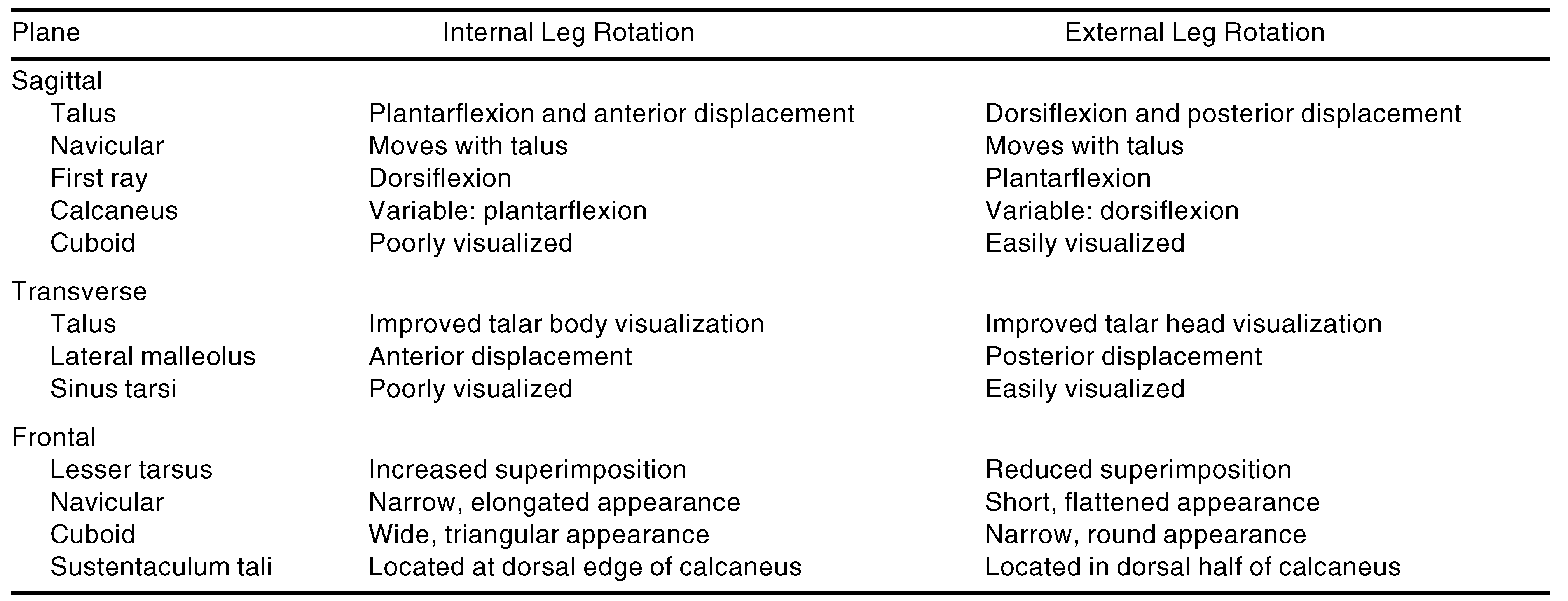
Although digitized fluoroscopy has frequently been used in empirical investigations of the lumbar and cervical spine [10,12,13,14], few investigations have applied the technique to the arches of the human foot. Green et al [15] employed fluoroscopy in assessing the tarsal motion of four subjects with various clinically determined biomechanical pathologies. Although the study was restricted to investigating the effect of internal and external rotation of the leg, the authors qualitatively described the resultant tarsal motion in each body plane (Table 1). The study, although methodologically limited, demonstrated the efficacy of fluoroscopy in identifying motion patterns within the foot and reinforced kinesiologic principles related to pronation and supination of the foot.
Table 1.
Summary of Tarsal Motion in Each Body Plane.
More recently, Perlman et al [16] used digitized videofluoroscopy in evaluating sagittal plane motion of the calcaneus during gait. Calcaneal pitch was assessed for 27 gait cycles of 15 subjects. Measurements were taken from the relevant images at three points in each gait cycle: immediately prior to, during, and following the middle of midstance. Although a significant decrease in the mean calcaneal pitch was noted at each point, several limitations prohibit the generalization of these findings to the entire stance phase of gait. Chief among these is that three arbitrary points in the gait cycle were selected. This provided limited information about calcaneal movement throughout the entire stance phase of gait.
Another limitation involved the underlying assumption that calcaneal pitch can be used as an indicator of rearfoot motion. Although several radiographic texts have suggested that a low calcaneal pitch reflects pronation of the foot and that a high pitch reflects supination [17,18], in vivo studies have not definitively confirmed this. DiGiovanni and Smith [19], in assessing normal biomechanical angles of 51 adult feet, noted that pronation and supination of the static weightbearing foot resulted in discrepant motion of the calcaneus, a finding consistent with those of Green et al. [15]. Consequently, changes in calcaneal inclination may not necessarily reflect changes in the rearfoot during gait.
The aim of the current investigation is twofold: first, to evaluate the relationship between rearfoot motion and calcaneal pitch, and second, to investigate the sagittal plane motion of the calcaneus throughout the entire midstance period of gait.
Methods
Subjects
Nine female volunteers aged 29 to 48 years who required diagnostic foot x-rays were recruited. In accordance with the rules of the Queensland University of Technology ethics committee, written consent was obtained from each subject following verbal and written explanation of the project.
Procedures
The study used the methodology outlined by Perlman et al. [16]. Initially, a weightbearing lateral radio-graph was taken of the right foot. Next, following a suitable period for the subjects to become acclimated, two series of fluoroscopic images of the right foot were obtained while each subject walked at a self-selected speed along an elevated walkway. Although no attempt was made to standardize cadence or discourage participants from looking at the target area, subjects were encouraged to continue walking past the 9-inch fluoroscopic head. Both x-ray and fluoroscopic equipment employed the same exposure parameters: 150 cm, 58 kV, and 3.0 mA.
Radiographs were subsequently recorded in videotape format, and each of the 25 fluoroscopic images per step was numbered. Trials were reviewed for image quality and were included if images provided clear visualization of the cortical edges of the calcaneus, talus, navicular, and cuboid. The majority of nonusable images resulted either from an inability to see the cortical edges of bone or from a radiographic flare phenomenon. The radiographic flare phenomenon is akin to a photographic flash and is the result of saturation (or overexposure) of the image-intensifying screen [20]. In total, 17 images were retained from each subject during a single gait cycle.
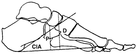
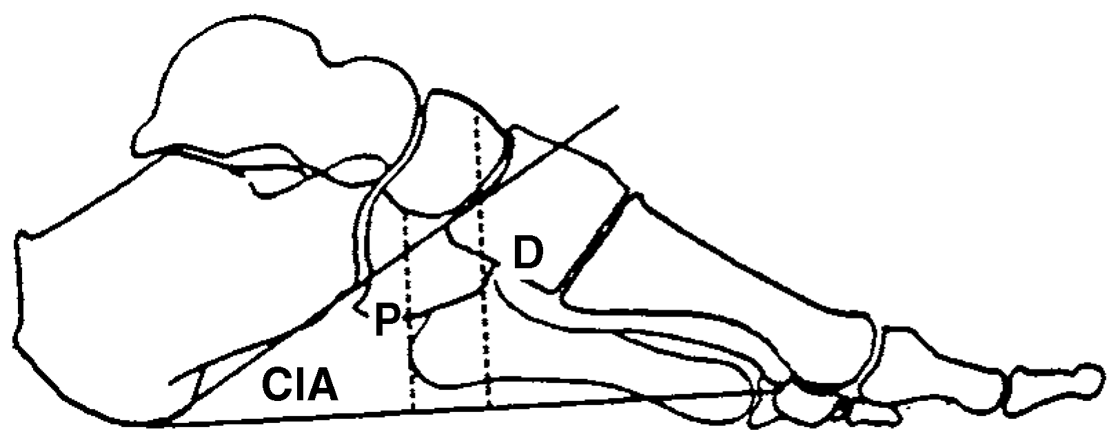
Images were subsequently digitized and digitally enhanced with Corel Draw software (Corel Draw, Ottawa, Ontario, Canada) in conjunction with a Miro DC 10 video-capture board (Pinnacle Systems, Inc, Mountain View, California). The calcaneal inclination angle was measured for all images (Fig. 1) [21]. Images within a trial were measured on two occasions, with both the trial order and the image sequence randomized to reduce the possible effects of fatigue and practice. Radiographic images were measured in the same manner.
Figure 1.
Calcaneal inclination angle (CIA) and arch height ratio. The arch height ratio is represented by P/D, where P represents the distance to the midpoint of the plantar aspect of the navicular (measured in millimeters) and D represents the distance to the midpoint of the dorsal aspect of the navicular (measured in millimeters).
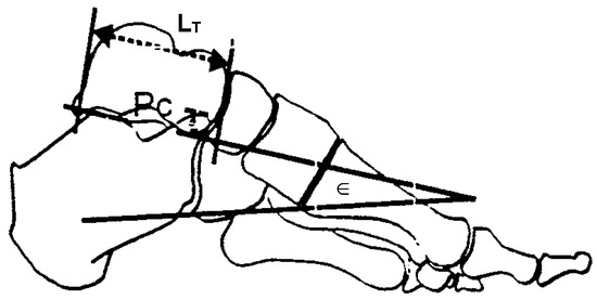
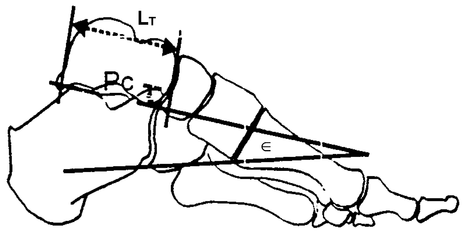
Additional measures of the arch height ratio and the tarsal index were also recorded. The tarsal index, as outlined by Benink [22], was derived as follows: the angle of inclination of the talocalcaneal joint (∈) was determined by measuring the angle of the tangent to the posterior articular surface of the calcaneus and the anterior articular surface of the plantar aspect of the talar head. The extent of superimposition of the talar head and anterior calcaneus (Pc) was also measured. The tarsal index (TI) was calculated with the formula TI = 100 × Pc/LT × tangent ∈ (Fig. 2). The arch height ratio represents a composite of two measures of arch height and may be defined as the ratio of 1) the distance from the plantar aspect of the navicular tuberosity to a line connecting the crista of the first metatarsal head and the medial calcaneal tubercle to 2) the distance from a point on the dorsal navicular, equidistant from the proximal and distal margins, to a line connecting the crista of the first metatarsal head and the medial calcaneal tubercle (Fig. 1) [23]. Angles and ratios between lengths were employed to eliminate error arising from magnification effects [21].
Figure 2.
The tarsal index as outlined by Benink [22]. LT, Talar length in millimeters; Pc, perpendicular distance of the overprojection of the talar head and anterior aspect of the calcaneus relative to a line joining the posterior facet of the calcaneus and the plantar aspect of the talar head (measured in millimeters); ∈, talocalcaneal inclination angle (measured in degrees).
Analysis
Intraclass correlation coefficients were used to assess the intrarater reliability. Pearson product moment correlations were used to evaluate the relationship among calcaneal pitch, arch height ratio, and tarsal index for radiographic images. The mean and the standard deviation for calcaneal pitch were calculated for each fluoroscopic image. Repeated measures analysis of variance was employed to identify possible changes in the mean calcaneal inclination during gait.
Results
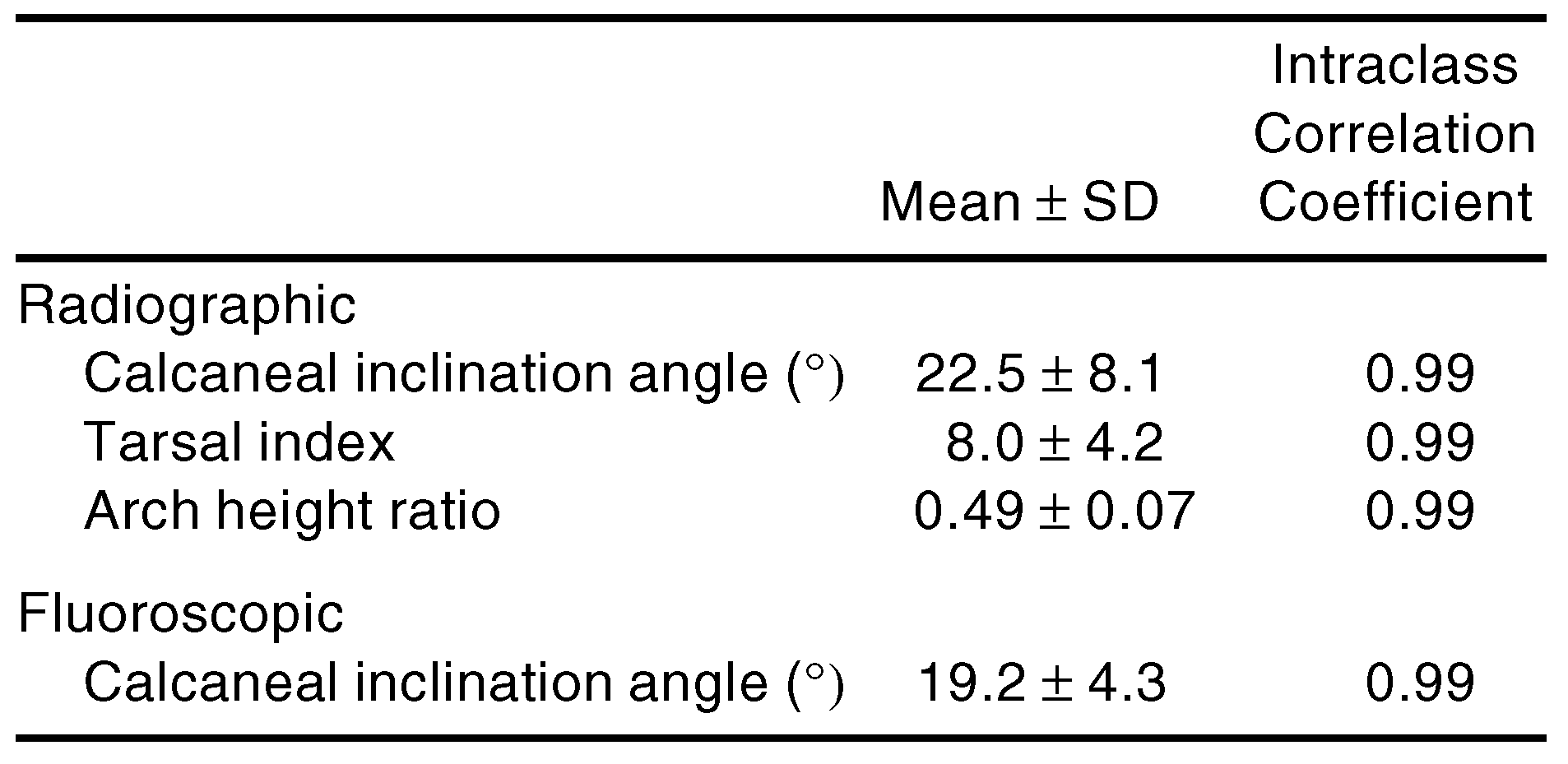
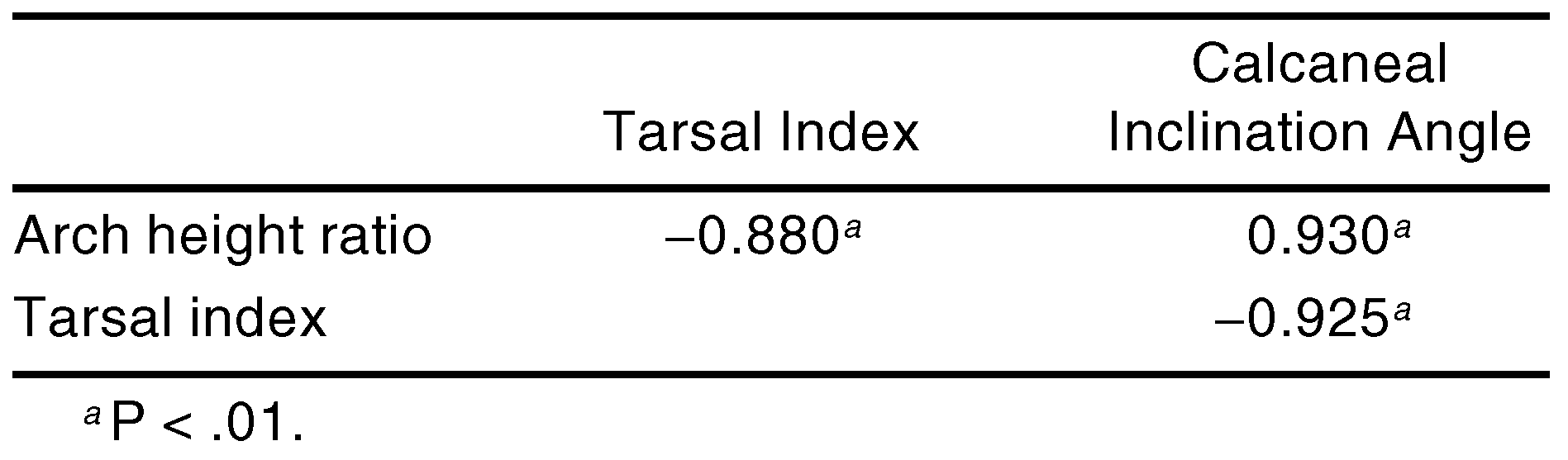
The intraclass correlation coefficients for the radiographic and fluoroscopic variables are listed in Table 2. All intraclass correlation coefficients are in excess of 0.99 and suggest high intrarater reliability [24]. Means and standard deviations for radiographic measurements are also presented in Table 2. Pearson product moment correlations between radiographic variables were uniformly high and are summarized in Table 3.
Table 2.
Mean, Standard Deviation, and Intraclass Correlation Coefficient for Radiographic and Fluoroscopic Angles.
Table 3.
Correlation Matrix for Radiographically Derived Measures.
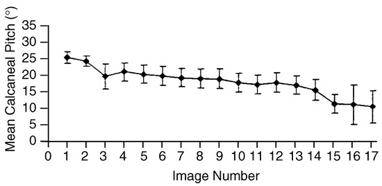
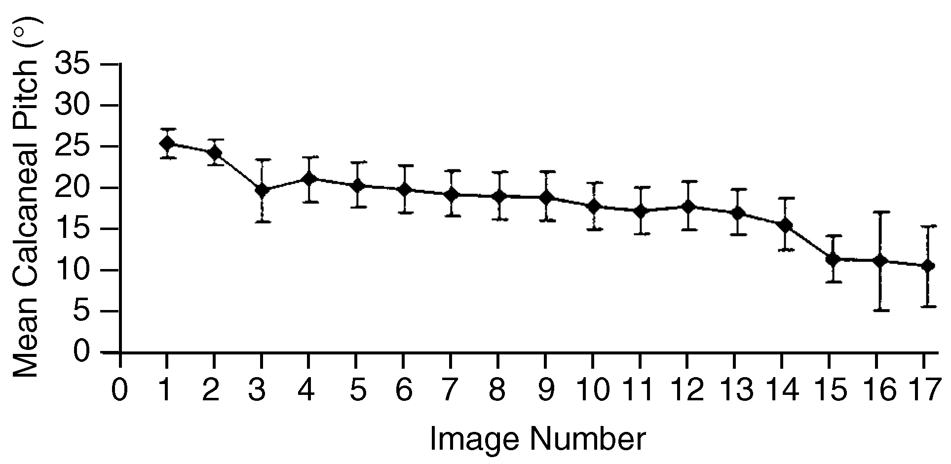
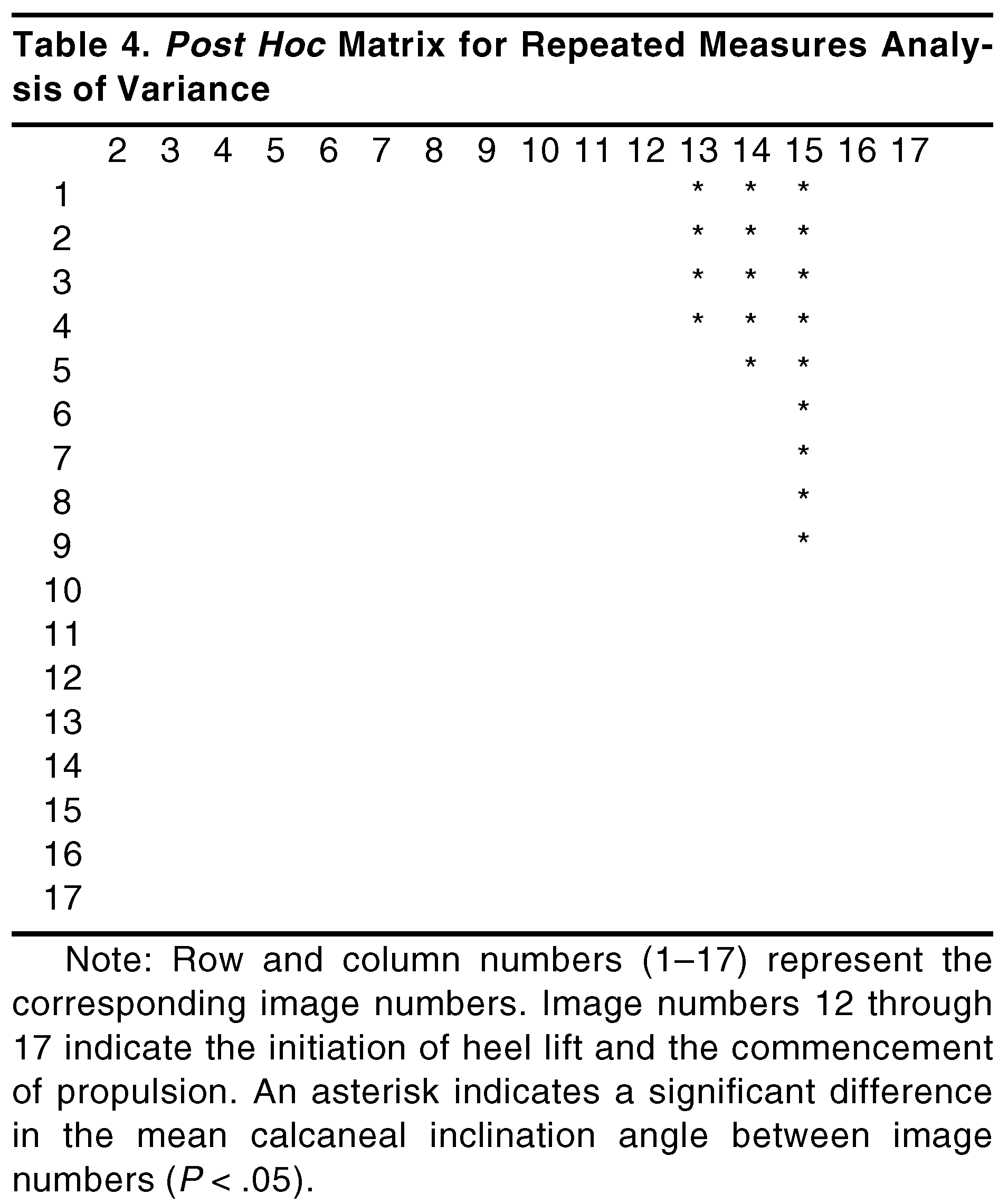
The mean calcaneal pitch for each fluoroscopic image is presented in Figure 3. Repeated measures analysis of variance identified a significant main effect for time (F16,9 = 5.18, P < .05). Student-Newman- Keuls post hoc analysis revealed calcaneal pitch to be significantly lower during the early propulsive period of gait (Table 4).
Figure 3.
Mean calcaneal pitch and standard error for each of the 17 images obtained. Image numbers 12 through 17 indicate the initiation of heel lift and the commencement of propulsion.
Table 4.
Post Hoc Matrix for Repeated Measures Analysis of Variance.
Discussion
Although an almost linear relationship between the mechanical behavior of the tarsus and the tarsal index has been previously reported [22,25], few investigations have employed the tarsal index in assessing rearfoot structure. Benink [22], while investigating the tarsal mechanics of cadavers, observed that a reduction in the tarsal index was associated with external rotation of the lower limb. More recently, van den Bogert et al [25] noted a high correlation between the tarsal index and the inclination of the subtalar joint axis. Clinically, low tarsal indices have been found to be associated with ankle instability and cavovarus deformities of the rearfoot [25,26]. The current investigation demonstrated a high correlation between the tarsal index and arch height; this supports the intimation that a high tarsal index is associated with a low medial column.
Table 2 shows the relationship between the radiographically derived measures of calcaneal pitch, arch height ratio, and tarsal index. The calcaneal inclination angle was highly correlated to both the tarsal index and the height of the medial longitudinal arch. Given that both measures are reported to reflect tarsal pronation and supination [22,27,28,29], it appears that calcaneal pitch may be used as an indicator of rearfoot position, with a reduction in an individual’s calcaneal inclination likely to reflect tarsal pronation.
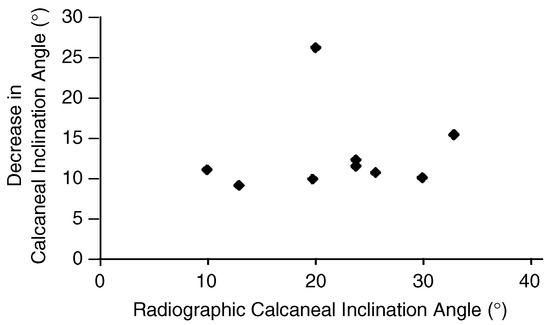
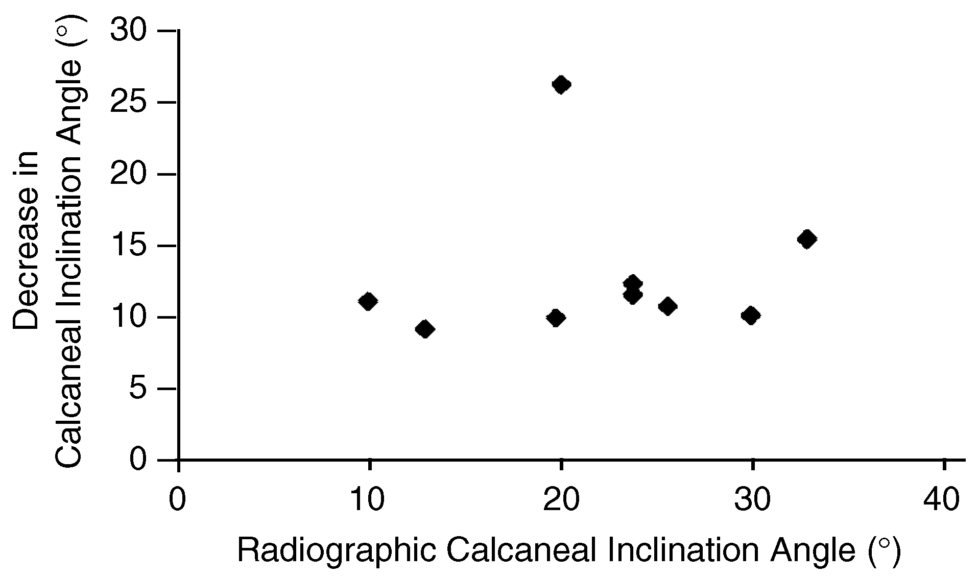
There was, however, no evidence to support the biomechanical classification of pronated or supinated foot types based on radiographic measures of calcaneal pitch [17]. All subjects displayed a reduction in calcaneal pitch throughout midstance, irrespective of their radiographically derived angle (Fig. 4). Consequently, it may be misleading to assume functional differences between feet that have been classified on the basis of static radiographic appearance. This is consistent with the work of Cavanagh et al [6], in which radiographically derived determinants of arch structure accounted for only 38% of the variance associated with pressures plantar to the first metatarsal head.
Figure 4.
Decrease in calcaneal pitch with gait as a function of each subject’s radiographic angle.
Few studies have adequately assessed arch function during the dynamics of gait. The current investigation noted a continued reduction in calcaneal pitch throughout midstance and early propulsion. While this may represent the pathologic gait of the “late pronator” as proposed by Perlman et al,[16] it may equally reflect normal sagittal plane motion of the calcaneus during dynamic conditions. Kitaoka et al [23], in a three-dimensional study evaluating the effect of load on arch kinematics of 15 cadavers, noted progressive lowering of the medial column with increasing load. Similarly, Kayano [30], in a goniometric study of foot function, noted that medial arch elongation coincided with the peaks in ground reaction during gait. Thus the rapid reduction in calcaneal pitch observed during the early midstance and propulsive periods (Fig. 3) may reflect a normal response to the increased ground-reactive force reported to occur at each of these times. Further research is required to elucidate whether the observed phenomenon is representative of normal or pathologic motion patterns.
Radiographic and anatomic studies have demonstrated changes in calcaneal inclination on the order of 2° to 6° with maximal pronation and supination of the foot [28,31,32]. The present study, however, noted a mean reduction of 12.5° in calcaneal pitch between the early midstance and propulsive periods of gait (Fig. 2). Although the increase in calcaneal motion observed during gait might reflect pathologic function, it may also reflect inherent differences between static and dynamic measurement conditions. Thus the observed increase in calcaneal motion during gait may reflect a dynamic force-attenuation mechanism within the human tarsus [33].
Conclusion
Although the importance of calcaneal pitch to arch structure has long been recognized, sagittal plane motion of the calcaneus has been viewed as relatively inconsequential to foot mechanics and has received limited empirical attention. The current investigation noted a significant reduction in the calcaneal inclination angle throughout the midstance and early propulsive periods of gait. While this may represent pathologic function within the foot, it may equally reflect normal rearfoot motion. Future investigations should be directed toward assessing function in heterogeneous, asymptomatic populations to determine normal sagittal plane motion of the calcaneus. The application of faster shutter speeds and a larger fluoroscopic field would allow visualization of the entire foot throughout propulsion, thus providing additional insight into the mechanics of the foot.
References
- COWAN DN, JONES BH, ROBINSON JR: Foot morphological characteristics and risk of exercise-related injury. Arch Fam Med 2: 773, 1993.
- GILADI M, MILGROM C, STEIN M, ET AL: The low arch, a protective factor in stress fractures. Orthop Rev 14: 709, 1985.
- HOPPER D, BRYANT A, ELLIOT B: Foot types and lower limb injuries in elite netball players. JAPMA 84: 355, 1994.
- SIMKIN A, LEICHTER I, GILADI M, ET AL: Combined effect of foot arch structure and an orthotic device on stress fractures. Foot Ankle 10: 25, 1989.
- SONG J, HILLSTROM HJ, SECORD D, ET AL: Foot type biomechanics: comparison of planus and rectus foot types. JAPMA 86: 16, 1996.
- CAVANAGH PR, MORAG E, BOULTON AJM, ET AL: The relationship of static foot structure to dynamic foot function. J Biomech 30: 243, 1997.
- KNUTZEN KM, PRICE A: Lower extremity static and dynamic relationships with rearfoot motion in gait. JAPMA 84: 171, 1994.
- SMITH LS, CLARKE TE, HAMILL CL, ET AL: The effects of soft and semi-rigid orthoses upon rearfoot movement in running. JAPMA 76: 227, 1986.
- MASLEN BA, ACKLAND TR: Radiographic study of skin displacement errors in the foot and ankle during standing. Clin Biomech 9: 291, 1994.
- KANAYAMA M, ABUMI K, KANEDA K, ET AL: Phase lag of the intersegmental motion in flexion-extension of the lumbar and lumbosacral spine: an in vivo study. Spine 21: 1416, 1996.
- CHOLEWICKI J, MCGILL SM, WELLS RP, ET AL: Method for measuring vertebral kinematics from videofluoroscopy. Clin Biomech 6: 73, 1991.
- CHOLEWICKI J, MCGILL SM: Lumbar posterior ligament involvement during extremely heavy lifts estimated from fluoroscopic measurements. J Biomech 25: 17, 1992.
- CROFT AC, KRAGE JS, PATE D, ET AL: Videofluoroscopy in cervical spine trauma: an interpreter reliability study. J Manipulative Physiol Ther 14: 390, 1994.
- ROOZMON P, GRACOVETSKY SA, GUOW GJ, ET AL: Examining motion in the cervical spine: I. imaging systems and measurement techniques. J Biomed Eng 15: 5, 1993.
- GREEN DR, SGARLATO TE, WITTENBERG M: Clinical biomechanical evaluation of the foot: a preliminary radiocinematographic study. JAPA 65: 732, 1975.
- PERLMAN PR, SISKIND V, JORGENSEN A, ET AL: Changes in the calcaneal pitch during stance phase of gait: a fluoroscopic analysis. JAPMA 86: 322, 1996.
- VITO G, KALISH S: “Biomechanical Radiographic Evaluation,” in The Biomechanics of the Foot and Ankle, ed by R Donatelli, p 66, FA Davis, Philadelphia, 1990.
- WEISMAN SD: “Biomechanically Acquired Foot Types,” in Radiology of the Foot, 2nd Ed, ed by SD Weisman, p 98, Williams & Wilkins, Baltimore, 1989.
- DIGIOVANNI JE, SMITH SD: Normal biomechanics of the adult rearfoot: a radiographic analysis. JAPA 66: 812, 1976.
- OKKALIDES D: Assessment of target recovery after TV camera saturation in x-ray fluoroscopy using a personal computer with frame-grabber. Br J Radiol 69: 415, 1996.
- SALTZMAN CL, NAWOCZENSKI DA, TALBOT KD: Measurement of the medial longitudinal arch. Arch Phys Med Rehabil 76: 45, 1995.
- BENINK RJ: The constraint mechanism of the human tarsus: a roentgenological experimental study. Acta Orthop Scand Suppl 56: 1, 1985.
- KITAOKA HB, LUNDBERG A, LUO ZP, ET AL: Kinematics of the normal arch of the foot and ankle under physiologic loading. Foot Ankle Int 16: 492, 1995.
- LANDIS JR, KOCH GG: The measurement of observer agreement for categorical data. Biometrics 33: 159, 1977.
- VAN DEN BOGERT AJ, SMITH GD, NIGG BM: In vivo determination of the anatomical axes of the ankle joint complex: an optimization approach. J Biomech 27: 1477, 1994.
- LARSEN E, ANGERMANN P: Association of ankle instability and foot deformity. Acta Orthop Scand 61: 136, 1990.
- COBEY JC, SELLA E: Standardizing methods of measurement of foot shape by including the effects of subtalar rotation. Foot Ankle 2: 30, 1981.
- HLAVAC HF: Differences in x-ray findings with varied positions of the foot. JAPA 57: 465, 1967.
- NESTOR CJ: Rearfoot complex: a review of its interdependent components, axis orientation and functional model. Foot 7: 86, 1997.
- KAYANO J: Dynamic function of medial foot arch. J Jpn Orthop Assoc 60: 1147, 1986.
- ALTMAN MI: Sagittal plane angles of the talus and calcaneus in the developing foot. JAPA 58: 463, 1968.
- OLDENBROOK LL, SMITH CE: Metatarsal head motion secondary to rearfoot pronation and supination: an anatomical study. JAPA 69: 24, 1979.
- SIMKIN A, LEICHTER I: Role of the calcaneal inclination in the energy storing capacity of the human foot: a biomechanical model. Med Biol Eng Comput 28: 149, 1990.

© 1999 American Podiatric Medical Association