


To the Editor:
Leiomyomas are benign neoplasms arising from smooth (nonstriated) muscle [1]. They are rather uncommon outside the uterus and gastrointestinal tract, but when they occur outside these areas, they are most frequently found in the subcutaneous tissue and skin [2,3]. Within soft tissue, leiomyomas are usually confined to the superficial layers and occur only rarely in the deeper structures [4]. Most of these lesions arise from structures in these areas that normally contain smooth muscle, such as the arrectores pilorum, the tunica dartos, the erector muscle of the nipple, and the media of large and small vessels [4]
These lesions may be classified as either superficial or vascular. Superficial leiomyomas are usually derived from arrectores pilorum, dartoid, vulvular, and mamillary smooth muscle. These neoplasms are sometimes also referred to as multiple cutaneous leiomyomas [2,3,5]. They can be found in any location but are most common in the extremities, the trunk, and the head and neck regions [4].
Leiomyomas of the vascular or solitary type are usually larger than the nodules of the multiple type and tend to show no familial tendency, unlike the superficial type. These angioleiomyomas occur in the extremities in 90% of cases, with the majority in the lower extremity [4].
Clinically, leiomyomas are smooth, freely movable, encapsulated lesions that vary in size from a few millimeters to approximately 3 cm [3,5,6,7]. The nodules usually develop slowly into firm masses and vary in color from pink to brown [3,5]. These neoplasms may have a translucent or waxy appearance [5].
Leiomyomas are typically associated with pain that occurs spontaneously and may be paroxysmal in nature, with sharp, stabbing, or pinching qualities [3,5]. The pain is usually attributed to the tumor’s directly compressing a cutaneous nerve or to an external force that creates pressure on the lesion [5,8]. However, it has also been suggested that the pain is related not to neural elements but rather to periodic transient contraction of the vessels, with resultant tissue anoxia [9].
The etiology of vascular leiomyomas is uncertain. However, they are commonly attributed to the following causes: trauma, infection, hormones, familial and hereditary factors, arteriovenous malformations, and possibly induction of mesenchymal cells of loose connective tissue to form smooth muscle [3,10]. The incidence of leiomyomas appears to be unrelated to race or ethnic group; they seem to occur most frequently in women between the ages of 30 and 50 years [3,11].
Case Report
A 38-year-old man presented with a chief complaint of a painful soft-tissue mass on the back of his right heel that had been present for 2 months. This lesion caused the patient a great deal of pain, which he described as a deep, local, aching type of discomfort. It was aggravated by pressure and certain types of footwear. The patient believed that the lesion was secondary to his driving 100 miles per day. He stated that the lesion had increased in size. He also reported that a similar lesion in the same region had been excised by his family physician a few months previously.
On physical examination, a protuberant soft-tissue mass was noted at the retrocalcaneal aspect of the right heel. The mass measured 1 cm in diameter and was approximately 5 mm high. The mass had an erythematous base with a rim of hyperkeratotic tissue. Palpation elicited mild-to-moderate discomfort. There was no evidence of drainage, odor, or sinus tract formation. The patient’s general physical condition and health were good. The radiographic examination revealed no osseous involvement.
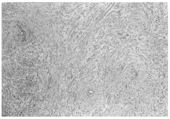
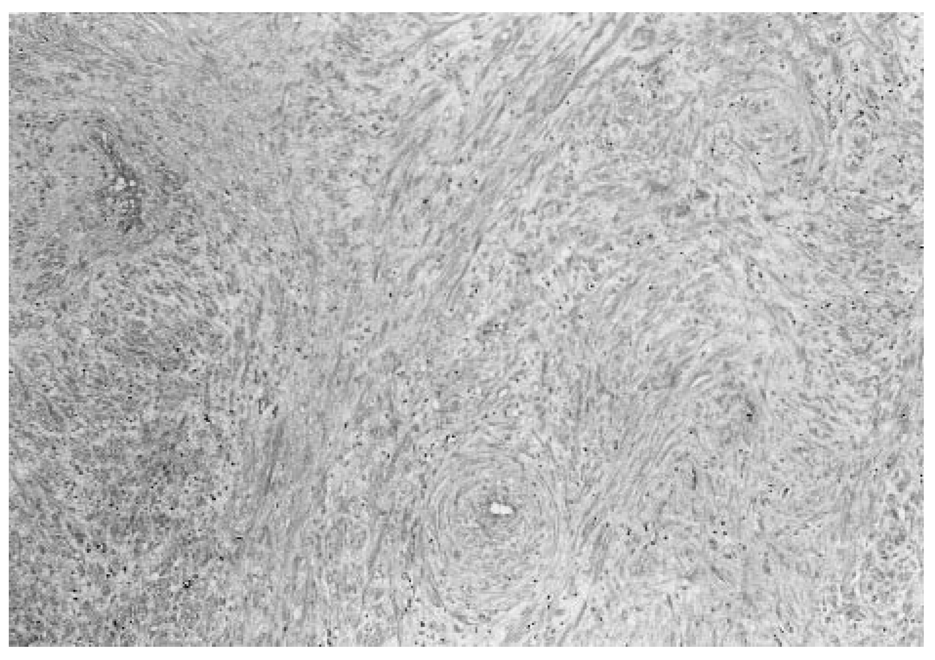
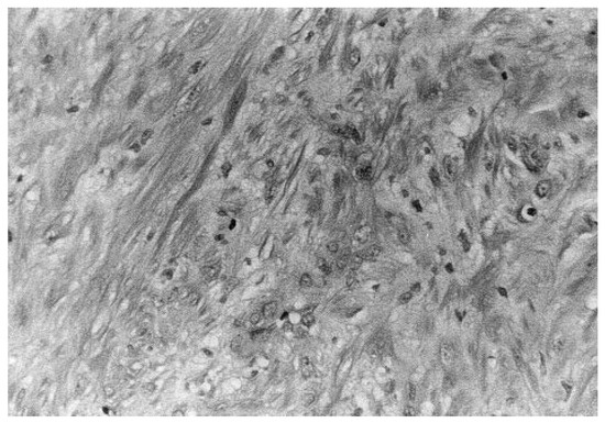
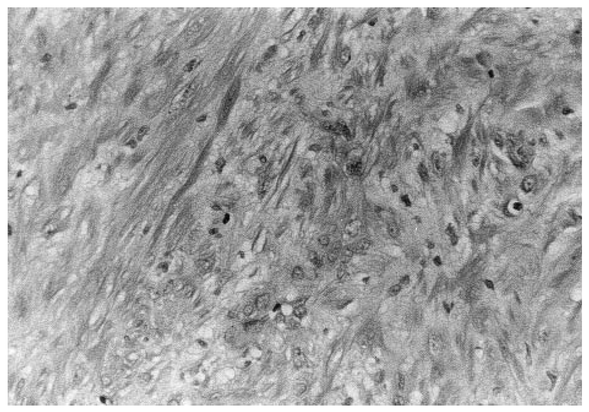
The patient was scheduled for surgical excision of the mass. The preoperative examination and laboratory findings were within normal limits. The lesion was removed under local anesthesia. The lesion was found by the pathology department to be consistent with angioleiomyoma (Figure 1 and Figure 2).
Figure 1.
Low-power view of the leiomyoma showing nodular proliferation in the reticular dermis and the presence of vascular channels (H&E, × 100).
Figure 2.
High-power view of the leiomyoma showing spindle cells with regular-to-oval nuclei and cytoplasmic vacuolation (H&E, × 400).
Discussion
Complete excision is the treatment of choice, with the rate of recurrence after this procedure low. However, in his study of ten patients with leiomyomas, Bulmer12 noted four cases of recurrence despite apparently complete excision. In the case presented here, a similar lesion had been excised previously.
However, the procedure was performed by another physician, and it is unknown whether the excision was complete.
It is generally accepted that leiomyomas rarely become malignant [3,13,14]. Leiomyosarcomas account for only 2% to 12% of all soft-tissue sarcomas [4]. However, it has been reported that the lesion may undergo malignant change with metastasis to lymph nodes, bone, and viscera [13]. Thus proper follow-up and pathologic identification are essential.
Also noteworthy is that in the present case the initial diagnosis was a ganglionic cyst. Because leiomyomas often simulate ganglionic cysts, lipomas, fibromas, or glomus tumors, they are frequently misdiagnosed [3]. MacDonald and Sanderson [6] have stated that the incidence of leiomyoma is almost certainly greater than that of glomus tumors, which are more readily diagnosed in the differentiation of small, painful subcutaneous nodules. In fact, glomera are uncommon in the lower extremity except subungually, or on the dorsal aspect of a digit. Painless swelling in the region of a joint is likely to indicate ganglia [12]. Transillumination can aid in the differentiation of cysts and solid lesions. A thorough description of the lesions that may be confused with leiomyomas has been provided by Visalli and McCarthy [15].
Summary
Leiomyomas are smooth-muscle tumors that may be encountered in the practice of podiatric medicine when they affect the arrector pili muscles or the smooth muscle of a vessel wall. In the present case, the lesion was located on the heel, an area with no hair growth; this further supports the diagnosis, as the most likely origin of the lesion was the smooth muscle of a blood vessel [16]. Vascular leiomyomas show a greater predilection for the lower extremities than their superficial counterparts. Surgical excision is the treatment of choice for these benign lesions, whose prognosis is excellent. When a patient presents with a painful mass in the lower extremity, the diagnosis of leiomyoma should be considered.
Acknowledgment
Thomas Ziegler, MD, of the Department of Pathology at Millcreek Community Hospital, Erie, Pennsylvania, for his help with the pathologic interpretation, and Mark Seidlinger for preparation of the photomicrographs.
References
- Stedman’s Medical Dictionary, 25th Ed ed; Williams & Wilkins: Baltimore, 1990.
- LATTES, R. Myomatoses. In Tumors of the Soft Tissues, ed by W Hartman; Armed Forces Institute of Pathology: Washington, DC, 1982; p. 60. [Google Scholar]
- TSOUTSOURIS, GV. Vascular leiomyoma. J Foot Surg 1982, 21, 37. [Google Scholar] [PubMed]
- ANDERSON, RE. Tumors and Tumorlike Conditions of the Soft Tissues. In Pathology; Kissan, J, Ed.; CV Mosby: St Louis, 1990; Vol 2, p. 2480. [Google Scholar]
- DOCKERY, GL; WENDELL, RE. Benign vascular leiomyomaangioma of the digit: a case report. JAPA 1979, 69, 438. [Google Scholar]
- MACDONALD, DM; SANDERSON, KV. Angioleiomyoma of the skin. Br J Dermatol 1974, 91, 161. [Google Scholar] [CrossRef] [Scilit] [PubMed]
- MCCAIN, LR; GALINSKI, AW. Pedal leiomyoma: a case report. JAPA 1969, 59, 399. [Google Scholar]
- GIRELLI, LP. Angiomyoma (vascular leiomyoma) excision from the human foot: a case report. J Foot Surg 1975, 14, 67. [Google Scholar]
- BRENNER, MA; RABINOWITZ, AD. Interdigital angioleiomyoma. Cutis 1986, 37, 350. [Google Scholar] [PubMed]
- HABERSHAW, GM; HURCHIK, JM; NASSER, I. Pedal leiomyoma. J Foot Ankle Surg 1994, 33, 260. [Google Scholar] [PubMed]
- SWEENEY, J; KEATING, SE. Angioleiomyoma. J Foot Surg 1983, 22, 21. [Google Scholar]
- BULMER, JH. Smooth muscle tumours of the limbs. J Bone Joint Surg Br 1967, 49, 52. [Google Scholar]
- RAKOW, RB. Leiomyoma of the foot: a case report. JAPA 1966, 56, 24. [Google Scholar]
- DAVIS, JL; RUSSELL, RD; GIACOPELLI, JA. Calcific leiomyoma: a unique case report. JAPMA 1995, 85, 96. [Google Scholar] [CrossRef] [Scilit] [PubMed]
- VISALLI, AJ; MCCARTHY, DJ. Podiatric considerations of angioleiomyoma. J Foot Surg 1992, 31, 372. [Google Scholar] [PubMed]
- SPINOSA, FA. Leiomyoma of the foot. J Foot Surg 1985, 24, 68. [Google Scholar]
© 1999 American Podiatric Medical Association