


Abstract
Complex regional pain syndrome is a chronic pain syndrome that is often instigated by postoperative or post-traumatic events. The disease process can progress through three stages, the first of which tends to respond best to treatment. A review of the literature is presented, followed by a report of a patient who developed symptoms of complex regional pain syndrome following a water-skiing accident.(J Am Podiatr Med Assoc 89(10): 534-539,1999)
Recent taxonomic revisions in the International Association for the Study of Pain classification of chronic pain syndromes placed reflex sympathetic dystrophy under the term “complex regional pain syndrome.” By definition, complex regional pain syndrome requires the presence of regional pain and sensory changes following a noxious event. Complex regional pain syndrome type I (CRPS I) corresponds to reflex sympathetic dystrophy, occurring without a definable nerve lesion. Complex regional pain syndrome type II (CRPS II, formerly called causalgia) occurs in cases in which there is a definable nerve lesion [1]. Complex regional pain syndrome type I is most commonly attributed to accidental trauma. Additional reported causes have included surgical intervention, diabetic neuropathy, disc herniation, cerebrovascular accident, myocardial infarction, and multiple sclerosis [2].
Sympathetic Neuroanatomy
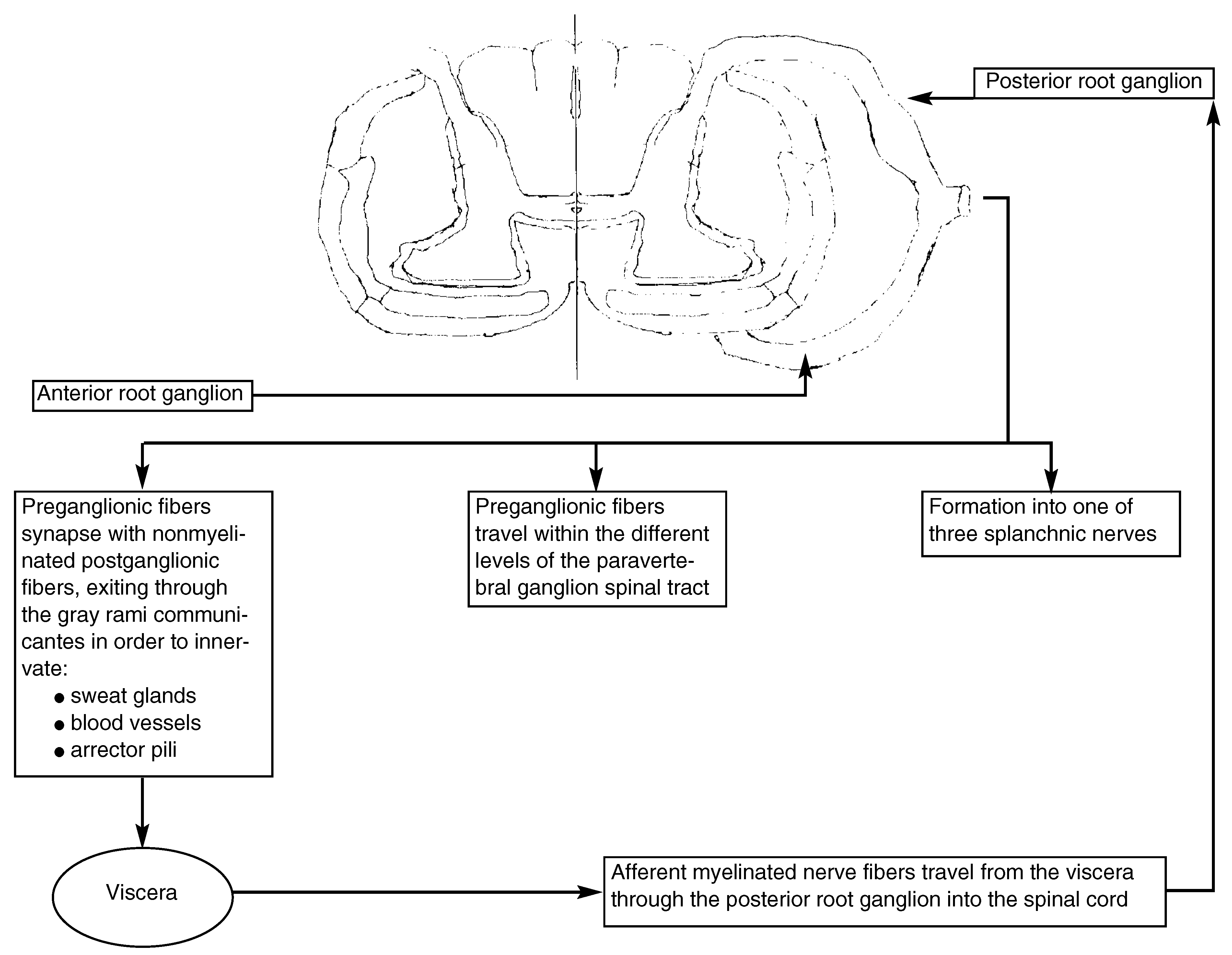
The sympathetic branch of the autonomic nervous system involves preganglionic efferent cell bodies located from T1 through L2 or L3 within the lateral gray column of the spinal cord [3]. Preganglionic fibers exit the spinal cord through the anterior nerve root before entering the paravertebral sympathetic ganglion. Within the ganglion, the sympathetic nervous system branches into three distinct distribution patterns (Fig. 1) [4]:
1. Preganglionic fibers synapse with nonmyelinated postganglionic fibers, which exit the paravertebral ganglion through gray rami communicantes and travel with somatic spinal nerves to innervate smooth muscle in blood vessels, sweat glands, and arrector pili apparatus within the skin.
2. Preganglionic nerves travel in either a cephalic or a caudal direction to synapse with postganglionic fibers at different levels of the paravertebral spinal tract.
3. Preganglionic fibers pass through the paravertebral ganglion, ultimately forming one of three splanchnic nerves (greater, lesser, and least). The greater splanchnic nerve is responsible for stimulating the secretion of norepinephrine and epinephrine from the adrenal medulla.
Sympathetic efferent nerve fibers ultimately synapse with one of three major adrenergic receptors (α-, β1-, and β2-adrenergic receptors) located at sympathetic effector organ sites. Systemic arterioles that go to muscles are constricted by α-adrenergic receptors and dilated by β2-adrenergic receptors. Systemic arterioles within the skin are generally constricted with sympathetic stimulation.
Afferent myelinated nerve fibers travel from the viscera, through the paravertebral ganglion and posterior root ganglion, and into the spinal cord. Here they either synapse with a local reflex arc or ascend to higher levels within the central nervous system.
Figure 1.
Basic anatomy of the sympathetic nervous system.
Pathophysiology
The etiology of CRPS I is not completely understood. Contemporary research indicates that pain associated with CRPS I is transmitted by the peripheral nervous system during the acute or early stage and by both the peripheral and central nervous systems during the middle and late stages. Much of this theory is attributed to physiologic changes following neuronal insult. Injured or irritated nerves tend to establish decreased nerve conduction thresholds secondary to chemical sensitization (bradykinins and prostaglandins) and up-regulation of α-adrenergic receptors [5,6,7,8,9]. Schwartzman [10] believes that this protective mechanism sustains peripheral nociceptive stimulation long enough to initiate the production of new (amino acid-based) neuropeptides, selectively targeting N-methyl-D-aspartate (NMDA) amino-acid nociceptive receptors. This, in turn, allows the central nervous system to generate CRPS I-related pain by activating amino-acid nociceptive receptor sites (ie, NMDA receptors). Conventional treatment modalities (ie, physical therapy, sympathetic blocks, and drugs affecting αand β-adrenergic receptor sites) are ineffective against central nervous system amino-acid nociceptive receptors, along with any neuropeptides responsible for stimulating them. This may explain the relative ineffectiveness of these treatment modalities when they are employed during the middle and late stages of CRPS I.
Clinical Presentation
A common denominator among patients with CRPS I is pain that is disproportionate to the physical findings. The pain is of a constant burning nature, and is aggravated by motion or even changes in emotional states. Patients experience symptoms when subjected to stimuli that are not normally capable of eliciting pain, a condition called allodynia. Noxious stimulation produces an exaggerated response, which persists after cessation of the stimulus (hyperpathia). Well-defined, localized edema (or ligature sign) that is associated with CRPS I is attributed to a reflex neurogenic inflammatory response whereby chemicals, including substance P and calcitonin G peptide, promote vasodilatation and capillary permeability.10 Motor dysfunction reduces the ability to initiate and sustain movement of the affected limb. Increased muscle tone, reflexes, and associated tremors are attributed to the Orbeli effect, whereby sympathetically driven norepinephrine release weakens aerobic (type I) muscles and increases the activity of anaerobic (type II) muscles [11].
Untreated CRPS I can progress through three stages [2,12]. Stage I (the acute stage) can last up to 6 months. In stage I, the patient experiences constant burning, localized edema, muscle spasms, and dry skin. Radiographs are usually normal; however, technetium-99m bone scans may indicate increased uptake within smaller joints. Stage II (the dystrophic stage) is characterized by progressive burning and aching, marked allodynia, and hyperpathia. The skin becomes cool and appears pale, gray, and cyanotic. Muscle wasting may ensue owing to limited activity. Radiographic changes may indicate “patchy” or “spotty” osteoporosis. Stage III (the atrophic stage) involves irreversible tissue changes. The skin remains cool, glossy, and cyanotic. Radiographic changes demonstrate diffuse osteoporosis with increased uptake on bone scan images.
Diagnosis and Treatment
Gibbons et al [13] reported the alleviation of CRPS I symptoms after the administration of a lumbar sympathetic block. Intravenous phentolamine (α-adrenergic antagonist) injections may be administered for diagnostic purposes in those patients who are unable to receive sympathetic lumbar blocks. Radiographic and bone scintigraphic studies can be used as an adjunct to confirm a diagnosis or monitor treatment. Much of the deossification that is noted on radiographic examination is secondary to widespread hyperemia. Examples include deossification of the cancellous portion of long bones secondary to hyperemia of the terminal branches of nutrient arteries, deossification of metaphyseal zones of the tibia secondary to metaphyseal vessel hyperemia, deossification beneath subchondral regions secondary to hyperemia of terminal vessels to joints, or any combination of these deossification mechanisms. Todorovic-Tirnanic et al [14] concluded that bone scans are preferred over plain film radiographs for determining the early diagnosis of postfracture stage I CRPS I. Stage II CRPS I demonstrated comparable diagnostic capabilities of both bone scintigraphy and radiography. Leitha et al [15] proposed a combination of three technetium-99m scintigraphic patterns yielding a high rate of diagnostic accuracy independent of sex, age, and precipitating factors: diffuse uptake in the carpal or tarsal region, diffuse uptake in small joints, and an increased activity ratio in the late blood pool phase.
The key to reversing symptoms related to CRPS I is early diagnosis and treatment. Casting, prolonged static bracing, and forceful passive range of motion should be avoided. Pharmaceutical treatments with steroids are thought to reduce vascular permeability to inflamed nerve endings [16]. Calcium channel blockers have been used to increase peripheral blood flow and antagonize the overall effect of norepinephrine on vascular smooth muscle [16,17]. Physical therapy modalities, including ultrasound, active range-of-motion exercises, hydrotherapy, contrast baths, and transcutaneous electrical nerve stimulation, are all adjuncts to treatment. Hassenbusch et al [18] reported good-to-fair alleviation of painful symptoms involving vasomotor tone and patient activity levels with the use of peripheral nerve stimulation in patients with symptoms from the distribution of one major peripheral nerve injury. Chemical sympathectomy has involved paravertebral blocks, intravenous regional anesthesia (Bier blocks), and peripheral nerve blocks. Recent experimental attempts to block the NMDA chronic pain receptors have been partially successful yet plagued by undesirable side effects [10]. Burnstock [19] reported moderate success with antagonistic agents developed to target adenosine triphosphate-activated purine receptors on nociceptive neurons.
Case Report
A 51-year-old man presented to the emergency department at the Veterans Affairs Medical Center in Brooklyn, New York, on June 16, 1997, with a painful, swollen left ankle. The patient related a history of forceful plantarflexion of his left foot and ankle following a water-skiing accident 2 days prior to presentation. His medical and surgical histories were unremarkable. His social history was significant for smoking (half a pack per day); there was no history of alcohol or intravenous drug abuse. The patient had no known allergies to medications.
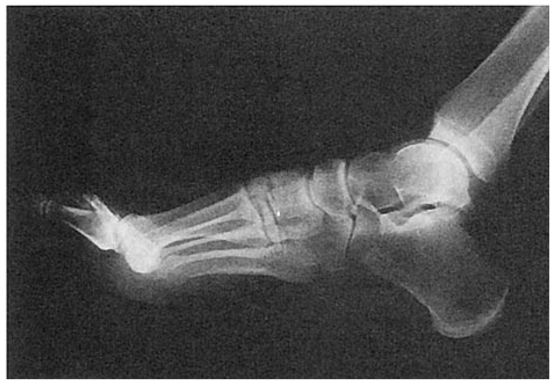
The physical examination revealed localized edema of the left foot and ankle. Tenderness was elicited on palpation of the lateral and dorsal aspects of the left foot. The dorsalis pedis pulse was 1/4. There was no palpable posterior tibial pulse. Capillary filling time was approximately 2.0 to 2.5 sec. Range of motion in the ankle and subtalar joint was guarded. Sensation and motor control of all five toes were intact, with no signs of any open lesions around the area of chief complaint. Radiographic findings were negative for any fractures or dislocations of the left foot and ankle (Fig. 2).
Left ankle sprain was diagnosed and the patient was placed in a posterior splint and asked to follow up with the referring service the following day. On June 17, 1997, the patient was seen in the clinic of the referring service. Further evaluation of radiographs prompted a change in the diagnosis to a nondisplaced left second metatarsal base fracture and a displaced cuboid fracture fragment. The patient was prescribed 40 tablets of ibuprofen, 600 mg, and placed in a below-the-knee fiberglass cast with a toeplate extension. The patient returned to the consulting clinic on June 26, 1997 (10 days after injury), complaining of tightness of the cast and associated burning pain across the tarsometatarsal joint. The patient also was unable to bear weight on the left foot because of the increased pain. After reviewing the films a third time, the referring service changed the diagnosis to a left ankle sprain with a symptomatic os cuboides secundarium. The patient was again placed in a below-the-knee fiberglass cast and instructed to remain nonweightbearing. On July 31, 1997 (45 days after injury), the consulting service removed the cast and placed the patient in a CAM Walker (Smith & Nephew, Inc, Vista, California), with instructions to walk as tolerated.
Figure 2.
Lateral view of the left foot 2 days following injury.
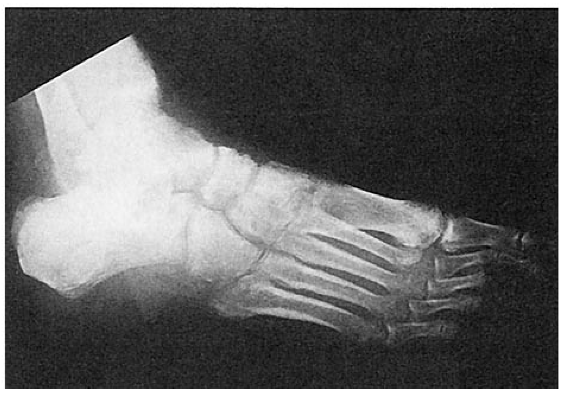
The patient presented to the podiatric surgical clinic at the Veterans Affairs Medical Center on August 20, 1997 (65 days after injury). Radiographs obtained that day were compared with previous radiographs; the new radiographs revealed moderate “spotty” osteoporosis of the metatarsal heads along with a “hazy” decalcification of the midtarsal joint, cortical margins of the cuneiforms, and dorsal aspects of both the navicular and the talus (Fig. 3). No fracture could be visualized at the base of the second metatarsal. The entire left foot was extremely tender on palpation. Dorsalis pedis and posterior tibial pulses were 2/4 with positive calor and localized edema; there were no dermatologic changes of the left foot. The diagnosis of stage I reflex sympathetic dystrophy was made and the patient was given a posterior tibial block consisting of 5 mL of 1% lidocaine without epinephrine. The patient was placed on a physical therapy regimen consisting of active range-of-motion exercises, contrast baths, and electrical muscle stimulation for 90 min, twice a day. The patient was fitted with an Unna boot around the left forefoot and instructed to walk with crutches. The patient was compliant with the treatment plan and reported improvement within 3 weeks of beginning treatment.
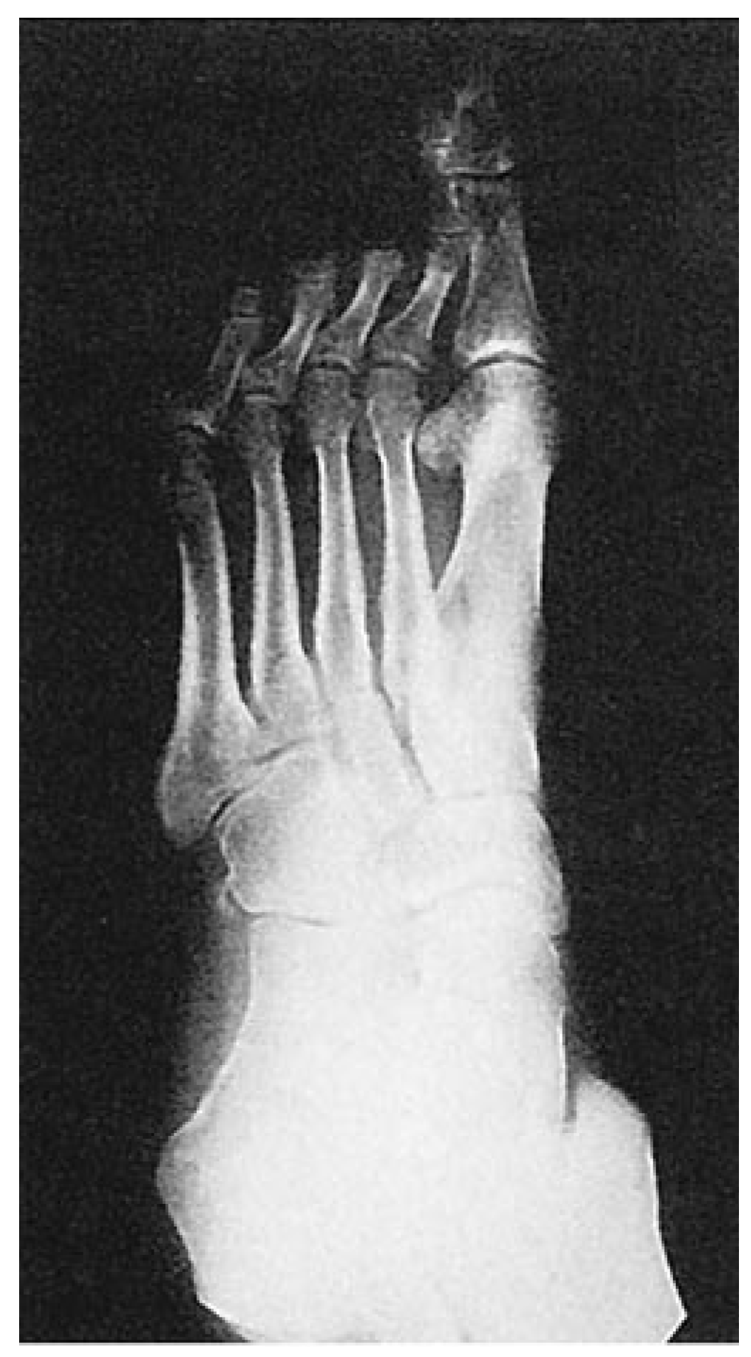
The patient received two additional sympathetic posterior tibial blocks, and gradually regained the ability to bear weight by September 10, 1997 (86 days following injury and 21 days following institution of the physical therapy regimen). The patient continued electrical muscle stimulation and range-of-motion exercises, but discontinued contrast baths after 4 weeks of physical therapy. Serial radiographs taken on September 10, 1997, revealed gradual remineralization of the metatarsal heads and midtarsus, with significant joint-space resolution (Fig. 4). On October 15, 1997 (121 days following injury and 5 weeks following institution of the physical therapy regimen), the patient regained full weightbearing capability and reduced the electrical muscle stimulation to once a day. At the seventh week following institution of the physical therapy regimen, calcium supplementation was added in an effort to facilitate remineralization of the left foot. On November 19, 1997 (156 days following injury and 10 weeks following institution of the physical therapy regimen), the patient received a fourth posterior tibial block injection, which dramatically improved dermatologic turgor and texture of the affected foot.
Figure 3.
Oblique view of the left foot 65 days following injury.
Discussion
It is important to note that trauma was the instigating factor in the case of the patient described in this report. The patient’s symptoms worsened during the period of time in which he was placed in a below-theknee cast. Although the patient did not receive a diagnostic lumbar sympathetic block, the diagnosis was made on the basis of history, clinical signs and symptoms, radiographic studies, and symptoms that were monitored following treatment. The patient’s condition improved significantly with injections and physical therapy. Calcium supplementation was implemented to help improve calcification of the left midtarsus. It is important to advocate stringent compliance with the treatment regimen. In this case, the patient was compliant with his physical therapy regimen and overall treatment plan. The patient’s physical signs and symptoms on presentation to the podiatric surgical clinic were consistent with the acute or early stage of CRPS I. Timing is a significant factor in the treatment of CRPS I, because early diagnosis and treatment are associated with a better prognosis than treatment during later stages.
Figure 4.
Oblique view of the left foot 21 days following institution of the physical therapy regimen.
References
- STANTON-HICKS, M; JANIG, W; HASSENBUSCH, S.; et al. Reflex sympathetic dystrophy: changing concepts and taxonomy. Pain 1995, 63, 127. [Google Scholar] [CrossRef] [Scilit] [PubMed]
- ROGER, JN; VALLEY, MA. Reflex sympathetic dystrophy. Clin Podiatr Med Surg 1994, 11, 73. [Google Scholar] [CrossRef] [Scilit]
- CERVERO, F. Sensory innervation of the viscera: peripheral basis of visceral pain. Physiol Rev 1994, 74, 95. [Google Scholar] [CrossRef] [Scilit] [PubMed]
- SNELL, RS. “The Autonomic Nervous System,” in Clinical Neuroanatomy, 3rd Ed, ed by RS Snell, p 485, Little, Brown, Boston, 1987. Snell, RS, Ed.;
- BAROLAT, G; SCHWARTZMAN, R; WOO, R. Epidural spinal cord stimulation in the management of reflex sympathetic dystrophy. Stereotact Funct Neurosurg 1989, 53, 29. [Google Scholar] [CrossRef] [Scilit] [PubMed]
- LIVINGSTON, WK. Pain Mechanisms: A Physiological Interpretation of Causalgia and Its Related States; Plenum Press: New York, 1976. [Google Scholar]
- MELZAK, R.; WALL, PD. Pain mechanism: a new theory. Science 1965, 150, 971. [Google Scholar] [CrossRef] [Scilit] [PubMed]
- DOUPE J, CUWLEN CR, CHANCE GQ. Post-traumatic pain and causalgic syndromes. J Neurol Neurosurg Psychiatry 1944, 7, 33. [Google Scholar] [CrossRef] [Scilit] [PubMed]
- DEVOR, M. Nerve pathophysiology and mechanisms of pain in causalgia. J Auton Nerv Syst 1983, 7, 371. [Google Scholar] [CrossRef] [Scilit] [PubMed]
- SCHWARTZMAN, R. Presentation at the Clinical Symposium on Reflex Sympathetic Dystrophy Syndrome, October 17, 1995, Baltimore.
- BEJ, MD; SCHWARTZMAN, RJ. Abnormalities of cutaneous blood flow regulation in patients with reflex sympathetic dystrophy as measured by laser Doppler fluxmetry. Arch Neurol 1991, 48, 912. [Google Scholar] [CrossRef] [Scilit] [PubMed]
- KURVERS, HA; JACOBS, MJ; BEUK, RJ.; et al. The spinal component to skin blood flow abnormalities in reflex sympathetic dystrophy. Arch Neurol 1996, 53, 58. [Google Scholar] [CrossRef] [Scilit] [PubMed]
- GIBBONS, JJ; WILSON, PR. RSD score: a criteria for the diagnosis of reflex sympathetic dystrophy and causalgia. Clin J Pain 1992, 8, 260. [Google Scholar] [CrossRef] [Scilit] [PubMed]
- TODOROVIC-TIRNANIC, M; OBRADOVIC, V; HAN, R.; et al. Diagnostic approach to reflex sympathetic dystrophy after fracture: radiography or bone scintigraphy? Eur J Nucl Med 1995, 22, 1187. [Google Scholar] [CrossRef] [Scilit] [PubMed]
- LEITHA, T; STAUDENHERZ, A; KORPAN, M; et al. Pattern recognition in five-phase bone scintigraphy: diagnostic patterns of reflex sympathetic dystrophy in adults. Eur J Nucl Med 1996, 23, 256. [Google Scholar] [CrossRef] [Scilit] [PubMed]
- PURDY, CA; MILLER, SJ. “Reflex Sympathetic Dystrophy Syndrome,” in Comprehensive Textbook of Foot Surgery, 2nd Ed, Vol 2, ed by ED McGlamry, AS Banks, MS Downey, p 1124, Williams & Wilkins, Baltimore, 1992.
- JOHNSON, KA; ALEXANDER, IJ. “Reflex Sympathetic Dystrophy,” in Disorders of the Foot and Ankle: Medical and Surgical Management, Vol 3, ed by MH Jahss, p 2187, WB Saunders, Philadelphia, 1991. Jahss, MH, Ed.;
- Hassenbusch, SJ; Stanton-Hicks, M; Schoppa, D; et al. Long-term results of peripheral nerve stimulation for reflex sympathetic dystrophy. J Neurosurg 1996, 84, 415. [Google Scholar] [CrossRef] [Scilit] [PubMed]
- BURNSTOCK, G. A unifying purinergic hypothesis for the initiation of pain. Lancet 1996, 347, 1604. [Google Scholar] [CrossRef] [Scilit] [PubMed]
© 1999 American Podiatric Medical Association