


Abstract
Tailor’s bunion or bunionette are terms that describe a pathologic enlargement occurring laterally on the fifth metatarsophalangeal joint. Regardless of the etiology that precipitates the deformity, the resulting abnormal protrusion of soft tissue or bone can result in pain for the patient. Symptoms can range from mild discomfort to severe, debilitating pain. The patient may present with pain dorsolaterally, laterally, or plantarly. The symptoms are mechanically induced, and are often associated with hyperkeratotic lesions and adventitious bursae. Patients complain most often that they cannot find comfortable shoes. The authors compare the effectiveness of fixated versus nonfixated distal osteotomies of the fifth metatarsal for the correction of tailor’s bunion. This study shows that fixation can help control postoperative dorsal displacement of the fifth metatarsal capital fragment (p < 0.0001) and produce less shortening of the metatarsal resulting in fewer complications.
Etiology
The etiology of tailor’s bunion is multifactorial [1,2,3,4,5,6]. Most proposed etiologies fall into one of two categories: structural (implying an anatomic variant or abnormality within the bone itself) or biomechanical. In 1949, Davies [7], Dickson and Diveley [8], Brown [9], Sponsel [10], and Wu [11] proposed various etiologies ranging from embryonic splaying of the fifth metatarsal to poorly fitting shoes. LeLievre [12] and DuVries [13] each proposed three etiologic mechanisms including the presence of a supernumerary bone and lateral bending of the fifth metatarsal. The latter mechanism was supported also by Yancy [14] and Sgarlatto [15]. Nestor et al [16] reported that lateral deviation exists, but showed no correlation between lateral deviation and the development of symptoms.
Other mechanisms include the presence of prominent lateral condyles with an increase in the fourth intermetatarsal angle, a lack of insertion or malinsertion of the transverse head of the adductor hallucis, abhorrent biomechanics, neoplasms, and neurologic disorders [1,2,3,6,17,18,19,20].
Clinical Evaluation
Patients with symptomatic tailor’s bunions complain of throbbing, cramping, or piercing pain in relation to the fifth metatarsal head. [1,2,5,21,22] Symptoms are more frequently seen in women and are associated with the narrower toebox common in women’s shoes. [1] Repetitive pressure and abnormal shearing between soft tissue and bone create a secondary hyperkeratotic lesion with or without an inflamed palpable adventitious bursa. [4] Erythema and edema of the affected area are often present.
When plantar irritation and associated lesions are present, weightbearing will be the aggravating factor, and a change in style of shoes may not alleviate the patient’s symptoms. In this case, an intractable plantar keratosis is most often present, along with a fifth metatarsal head that is palpable on the plantar aspect. [2]
Often associated with tailor’s bunions is a varus or adducto varus fifth digit with an heloma durum at the dorsolateral aspect of the interphalangeal joint. [5] Thul and Hoffman [23] suggest a correlation between tailor’s bunions and traumatically induced neuromas of the proper digital branch of the superficial division of the lateral plantar nerve. This is not supported by subsequent findings in the literature.
Radiographic Evaluation
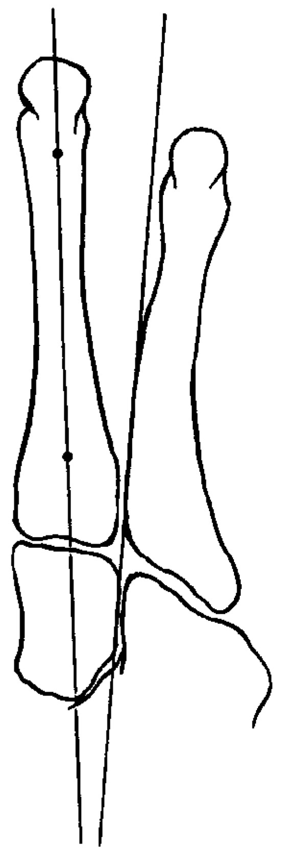
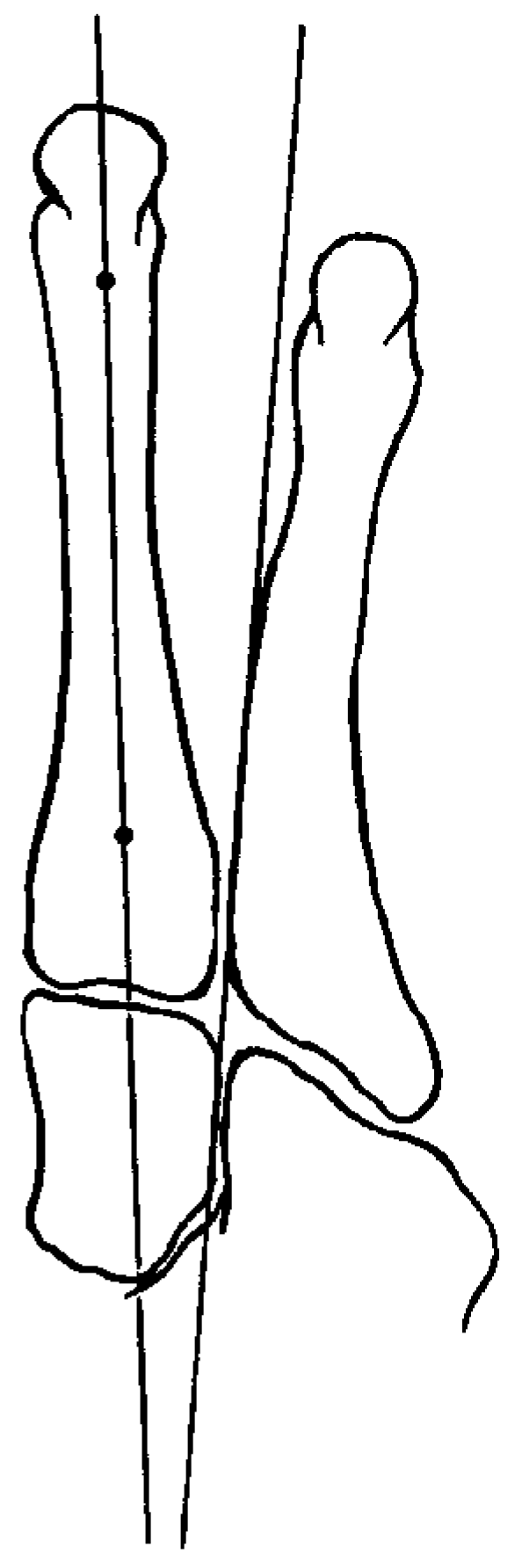
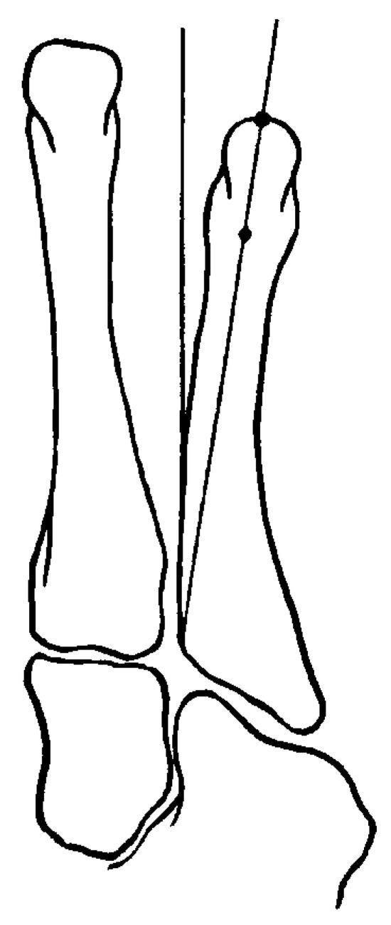
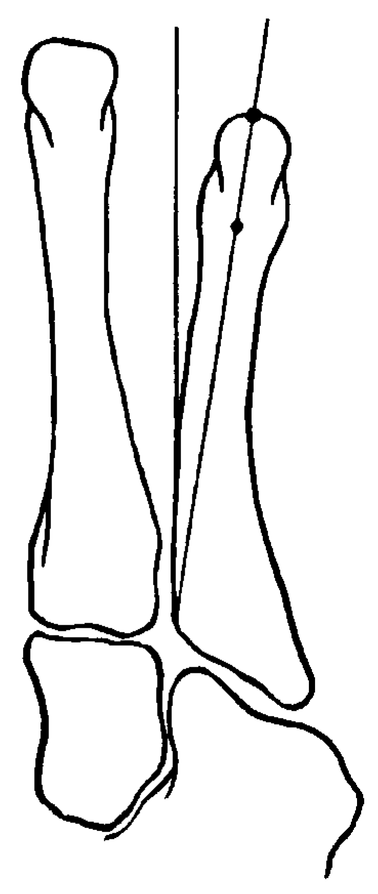
In 1980, Fallat and Buckholz [1] wrote the definitive article on the use of radiographic measurements to analyze and classify tailor’s bunion deformity. They established techniques for measuring the fourth intermetatarsal angle and lateral deviation angle in order to quantify the degree of deformity (Figure 1 and Figure 2). They determined that the average intermetatarsal angle was 6.47° with a range of 3° to 11°. In patients with tailor’s bunion deformities, the average intermetatarsal angle was 8.71° with a range of 3° to 15°. These results are consistent with those of Catanzariti et al [3] who found an average intermetatarsal angle of patients with tailor’s bunions of 9.11° ± 2.08°. This suggests that tailor’s bunions secondary to an increase in intermetatarsal angle may be a positional or functional deformity, rather than a structural deformity, a condition that would not be affected by changes in foot position or the orientation of more proximal joint complexes.
Figure 1.
Technique for measuring the fourth and fifth intermetatarsal angle (adapted from Fallat LM, Buckholz J: An analysis of the tailor’s bunion by radiographic and anatomical display. JAPA 70: 597, 1980).
Figure 2.
Technique for measuring the lateral bowing angle of the fifth metatarsal (adapted from Fallat LM, Buckholz J: An analysis of the tailor’s bunion by radiographic and anatomical display. JAPA 70: 597, 1980).
The lateral deviation angle is a measurement that quantifies the amount of lateral bowing of the distal third of the metatarsal shaft. The average lateral deviation angle in the normal foot was determined to be 2.64° with a range of 0° to 7°. The lateral deviation angle in patients with tailor’s bunions was determined to be 8.05°, with a range of 0° to 16° [1]. Fallat and Buckholz [1] determined that the lateral bowing of the fifth metatarsal was not affected by the position of the foot and, therefore, was a structural rather than functional deformity.
Catanzariti et al [3] described a third radiographic parameter, the sagittal deviation angle of the fifth metatarsal head. This was used to quantitate the amount of dorsal displacement of the capital fragment secondary to the surgical correction of the bunionette.
History of Surgical Treatments
Numerous surgical procedures have been advocated for the treatment of tailor’s bunions. The literature is replete with reviews that summarize these procedures, either chronologically or based on the location of the procedure in relation to the fifth metatarsal. Osteotomies of the metatarsal head were described by Davies [7], Dickson and Diveley [8], and DuVries [13]. LeLievre [12] advocated resection of both the lateral aspect of the fifth metatarsal head along with base of the proximal phalanx of the fifth toe.
The first distal transposition osteotomy for treatment of tailor’s bunions was described by Hohmann in 1951 [6]. Neither this transverse osteotomy described by Hohmann, the step-down osteotomy described by Leach and Igou [17], or the oblique distal osteotomies described by Sponsel [10] or Keating et al [24] used any form of internal fixation to stabilize the osteotomy. Steinke and Boll [25] adapted the Hohmann technique with peg-and-hole fixation described by Thompson in order to fixate the osteotomy. Throckmorton and Bradlee [26] described a transverse sliding V-osteotomy where they recommended a medial transposition of the capital fragment of up to 2.0 mm. Mercado [27] believed that when a medially displaced facet was present, a situation analogous to an increased proximal articular set angle seen in hallux valgus, a subcapital reverse-Reverdin osteotomy was most appropriate. [3] Habner and Kraft [28] advocated using a crescentic osteotomy at the metatarsal neck, with the convex side facing distally, for the treatment of tailor’s bunions with an associated plantar lesion. Yu et al [29] described an oblique closing wedge osteotomy of the metatarsal neck fixated with an intramedullary Kirschner wire. The base of the wedge faces medially, and the lateral cortex is left intact. This is used to treat tailor’s bunions when a dumbbell-shaped metatarsal head with lateral bowing of the distal portion of the fifth metatarsal is present.
Many of the authors mentioned above described their surgical procedures without using fixation. None of these authors stated that the use of fixation had been considered and subsequently, omitted. Davidson [30], White [31], and Zvijac et al [22], however, definitively stated that fixation was not necessary for a successful surgical outcome. The results from follow-up studies by Keating et al [24] and Catanzariti et al [3], both of which exhibited a significant occurrence of secondary transfer lesions, refute the claims made by the aforementioned authors. Hansson [32] reported on a sliding oblique neck osteotomy fixated with absorbable sutures. Recently, Castle et al [4] and Frankel et al [5] describe distal osteotomies of the fifth metatarsal with cortical screw fixation. Friend et al [33] advocate the use of absorbable pin fixation with an L-osteotomy of the metatarsal head. In each of these cases, the authors stated that fixation was necessary to achieve and maintain the desired degree of correction.
Other procedures have been reported. Resection of the fifth metatarsal head has been described by Harris [34] and Weisberg [35]. Addante and Kaufmann [36] and Petrich and Dull [37] supplemented resection of the fifth metatarsal head with the insertion of a silicone sphere implant in order to prevent digital retraction.
Many procedures performed at the metatarsal base have been described. Gerbert et al [38] advocated using a closing base wedge osteotomy with stainless steel suture fixation. A similar procedure was described by Rappaport [39]. LeLievre [12] described a proximal osteotomy in conjunction with tightening of the capsule to decrease the intermetatarsal angle. Mercado [27] used closing base wedge osteotomies to correct tailor’s bunions secondary to lateral deviation of the metatarsal shaft. Buchbinder [40] advocated the use of the derotational-angulational-transpositional osteotomy of the fifth metatarsal, 2 to 3 cm distal to the metatarsal base. Diebold and Bejjani [41] described a basal V-osteotomy with the apex oriented proximally. In this procedure, the shaft is transposed medially and fixated with Kirschner wires.
Surgical Classification
Based on the various etiologic components, radiographic criteria, and surgical procedures, in 1990, Fallat [6] described a classification system for the surgical management of tailor’s bunions.
1) Type 1: Enlargement of the lateral surface ofthe fifth metatarsal head. The enlargement could be secondary to exostosis, prominent lateral condyles, or a round or dumbbell-shaped metatarsal head. Conservative treatment, as discussed above, is most successful in these cases. If surgical intervention is necessary, resection of the lateral surface of the fifth metatarsal head only is the indicated procedure of choice.
2) Type 2: Lateral bowing of the distal fifth metatarsal. This occurs without hypertrophy of the metatarsal head. In this case, a distal osteotomy, preferably at the point of bowing, is the procedure of choice. Conservative treatment will be effective only if the amount of bowing is minimal.
3) Type 3: Increase in the intermetatarsal angle.This deformity is typically seen in splayfoot. Conservative treatment will be ineffective. Surgical correction may include a distal osteotomy for a mild increase in the intermetatarsal angle and a proximal osteotomy for more severe deformities.
4) Type 4: Combination of deformities. This involves two or more of the components listed above. These are frequently seen in the rheumatoid foot and are resistant to conservative treatment. Surgical correction should be directed toward the most severe or proximal component of the deformity.
Problem Statement
A controversy exists concerning the necessity of fixation for tailor’s bunion surgery. Cortical screw, Kirschner wire, absorbable pin and steel suture fixations have all been reported.[4,5,29,38,39,41] Beginning with the first distal transpositional osteotomy described by Hohmann [6] in 1951, several investigators have used procedures without fixations. [10,17,24,25] Based on successful surgical outcomes, Davidson [30], White [31], and Zvijac et al [22] advocated that fixation was not necessary. Coupled with the original biomechanical etiologies described by Hicks [19] and Root et al [20], it is unclear if the appropriate foot position and, hence, function can be maintained without fixation. As a result of this controversy and lack of objective measures to support one approach or the other, the following question was developed: Does fixation alter radiographic biomechanical parameters and clinical outcome in a retrospective assessment of tailor’s bunion procedures?
To answer the aforementioned question, the following hypothesis has been formulated:
Hypothesis 1
If a retrospective assessment of tailor’s bunion procedures is conducted, then no statistically significant differences will result between those cases with fixation and those cases without fixation for the following biomechanical parameters and clinical outcomes: lateral bowing angle, dorsal displacement, intermetatarsal angle, metatarsal length, time to shoes, and consolidation time
Materials and Methods
The data from this retrospective study were collected from 47 patients (56 feet), who presented to the Foot and Ankle Institute at the Pennsylvania College of Podiatric Medicine from 1985 to 1994. All subjects had tailor’s bunions with or without one or more of the following pathologies: pain associated with tailor’s bunion, plantarflexed fifth metatarsal, metatarsalgia or hyperkeratotic lesions in relation to the fifth metatarsal head, and bursitis.
The patient’s symptoms were recalcitrant to conservative treatment. All patients underwent one of four distal osteotomies of the fifth metatarsal with or without other surgical procedures to the forefoot. Wilson osteotomies were obliquely oriented, Hohmanns were transversely oriented, Austins were V-osteotomies in the transverse plane, and distal closing wedges were performed with the base of the wedge oriented medially. The postoperative course was standardized and all patients were weightbearing in a surgical shoe by the first week postoperatively.
Data were collected by reviewing the charts and preoperative and postoperative radiographs. All patients in the study had a complete medical record and at least three radiographic views preoperatively and postoperatively in the angle and base of gait. The preoperative films were not taken prior to 8 weeks from the date of the surgery. Some patients had other forefoot surgical procedures performed. Patients who had other lateral column procedures such as a Dwyer osteotomy and patients who were casted for other surgical procedures were excluded.
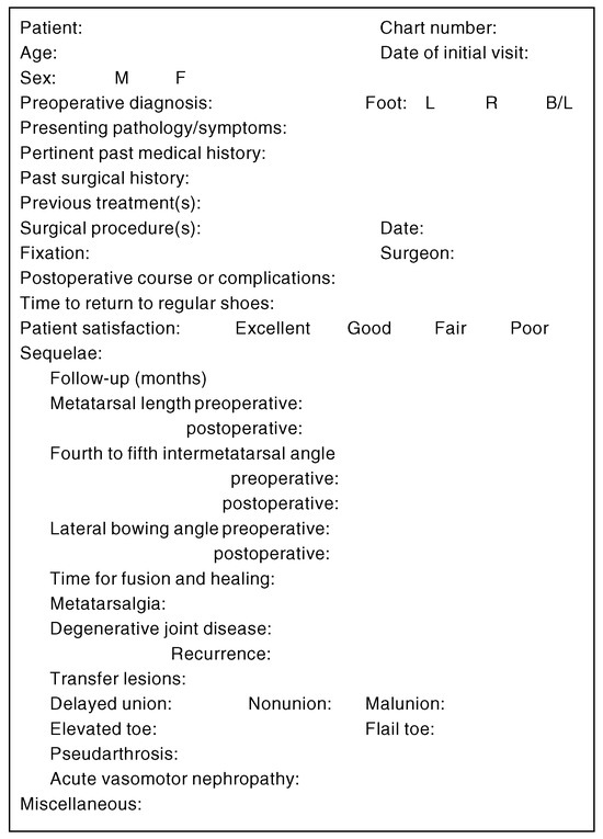
A standard form was used to collect biographical, subjective, and objective data (Figure 3). The preoperative data included presenting symptoms, diagnosis, pertinent past medical and surgical history, previous treatment to the affected ray, any documented abnormalities in biomechanics or foot type, and presence of an associated adducto varus fifth digit on the ipsilateral foot. The type of surgical procedure and fixation, if used, and postoperative course were recorded. Postoperative data included the length of follow-up care, time to return to regular shoes, and the development of any negative sequelae. For the purposes of this study, sequelae were defined as any negative occurrence presenting as a result of the surgical procedure. This included any dysfunction associated with the fixation devices used, such as pain at the pin site, or premature loosening or migration of the pin. Sequelae were differentiated from complications in that the latter did not include any pin dysfunction. The types of negative sequelae recorded included the development of lesser metatarsalgia, transfer lesions, delayed unions or nonunions, recurrence of the initial presenting pathology, and any dysfunction associated with the fixation device, including pain at the pin site and premature loosening or migration of the pin. Postoperative radiographs were evaluated to determine the time for consolidation of the osteotomy. The criteria for determining consolidation included presence of external bone callus and trabeculae crossing the osteotomy site.
Figure 3.
Data collection for fifth metatarsal osteotomy.
Various measurements were collected from preoperative and postoperative radiographs, including the intermetatarsal angle between the fourth and fifth metatarsals, the lateral bowing angle of the fifth metatarsal, the length of the fifth metatarsal, and dorsal displacement of the capital fragment. Any sagittal plane rotary angle of the distal segment relative to the proximal segment about the dorsal cortex of the proximal segment was visually noted and was excluded from the data set. The intermetatarsal and lateral bowing angles were measured according to the criteria described by Fallat and Buckholz [1] and outlined in the section on radiographic evaluation.
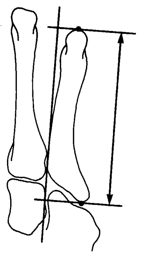
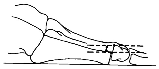
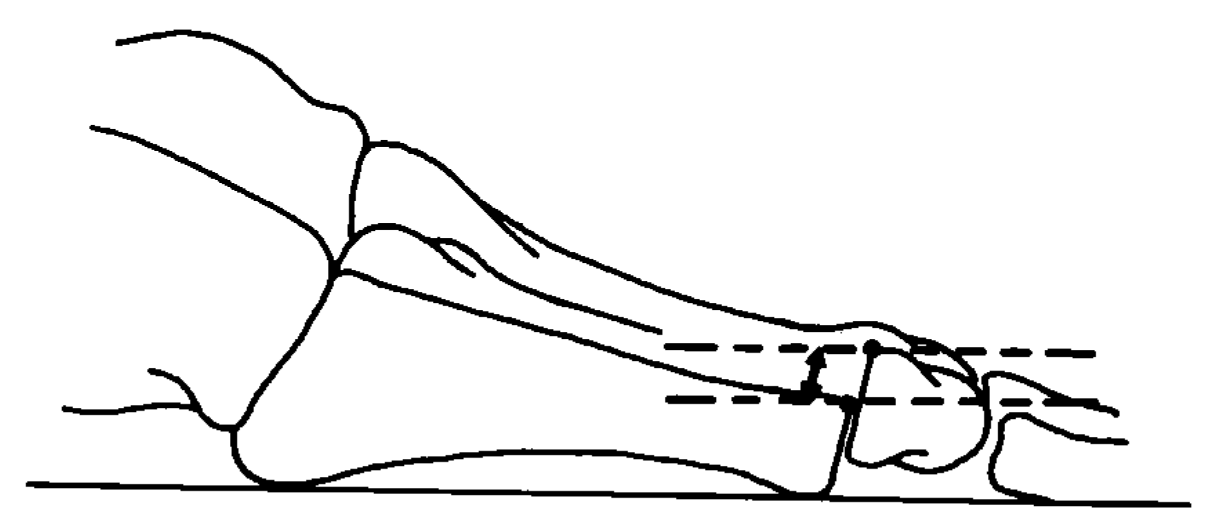
In order to measure the length of the fifth metatarsal, a previously undescribed method was used. On a dorsoplantar radiograph, a line parallel to the proximal medial shaft of the fifth metatarsal as described by Fallat and Buchkholz [1] was used as the axis for that ray. Two lines were drawn perpendicular to this axis; one tangent to the most proximal point on the styloid process, and the other tangent to the most distal point on the metatarsal head. The distance between these two lines was measured with a digital caliper, and determined to be the length of the meta-tarsal (Figure 4). In postoperative radiographs where dorsal displacement of the capital fragment was apparent, the distance between the dorsal cortices of the proximal and distal fragments on lateral views was measured with a digital caliper (Figure 5).
Figure 4.
Technique used for measuring the fifth metatarsal length.
Figure 5.
Technique for measuring dorsal displacement of the fifth metatarsal head.
Differences in measured values were determined by subtracting postoperative from preoperative values of each parameter. All data were statistically analyzed comparing fixated versus nonfixated groups with respect to the above-mentioned criteria. Any statistically significant differences between groups were determined with an unpaired t-test.
Results
Forty-seven patients (56 feet) who were treated at the Foot and Ankle Institute over a 9-year period were evaluated. Twenty-two feet had fixated osteotomies to correct tailor’s bunions, while 34 feet had no fixation. The surgical procedures were as follows: 31 Wilson osteotomies, 15 Hohmann osteotomies, 8 Austin osteotomies, and 2 distal closing wedge.
Ninety-six percent of the patients were female. The patients’ average age was 37.3 years, with a range of 15 to 64 years. Tailor’s bunion deformity occurred 16% bilaterally, and there was practically equal distribution between right and left feet (27 patients—right foot, 29 patients—left foot). The average follow-up care was 20 weeks, with a range of 2 weeks to 2 years. The ethnic distribution was as follows: 71% black, 20% white, 7% Hispanic, and 2% Asian. All patients were in good health and no specific consideration was made with respect to tobacco use.
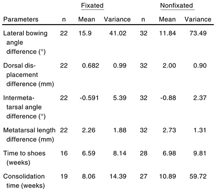
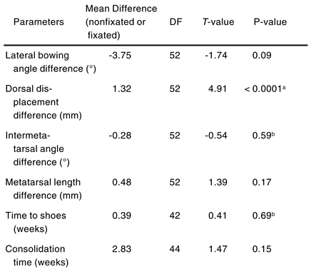
Four radiographic biomechanical parameters as described in the methodology were used to assess proper foot position: lateral bowing angle (°), dorsal displacement (mm), intermetatarsal angle (°), and metatarsal length (mm). To explore the effects of fixation, difference parameters were calculated by subtracting the preoperative from postoperative values. Two additional postoperative parameters were included to assess clinical outcome: time to return to shoes (weeks) and consolidation time (weeks). A descriptive summary of the mean values of these parameters and their variability is shown in Table 1.
Table 1.
Summary of Tailor’s Bunion Outcome Parameters.
The radiographic biomechanical parameters were extracted from each patient’s x-ray by manual measurement techniques as described in the methodology. This experiment design was not biased by errors from multiple raters and hence obviated the problem of establishing inter-rater reliability. Still, if the single rater was not consistent, then intra-rater reliability could be poor and corrupt the data. To determine the intra-rater reliability for the particular rater used in this research, the inter-class correlation coefficient was calculated. The rater took a subset of the subject pool (n = 10) and performed the radiographic measurements on three separate occasions for each of the ten subjects within a 24-hr time frame.
In accordance with the procedures reported by Shrout and Fleiss [45], the intra-class correlations were calculated and reported in Table 2. For the single rater of the tailor’s bunion radiographic measurements, there was consistency and hence reliability.
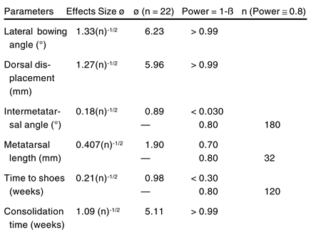
Table 2.
Parameter Effect Size Calculation for α = 0.05.
Prior to performing the unpaired t-tests for each parameter, the effect size calculations were conducted (Table 2). In this way, assuming a 0 level of significance equal to 0.05 to guard against the type I error, the power (ie,=1-ß) was calculated for n equal 22 subjects. If the experiment power was greater than 0.8, then the parameter was considered powerful enough for the study. If the experiment power was less than 0.8, then the increased sample size required for a power of 0.8 was determined with standard power function tables. As shown in Table 2, the intermetatarsal angle and time to shoe parameters would require an n of 180 and 120 subjects respectively, and hence are inconclusive with a group size on the order of 22 subjects. The metatarsal length parameter with a power of 0.7 is considered marginally acceptable.
The unpaired t-test results are shown in Table 3. Note that only the dorsal displacement parameter was statistically significant for differences between the fixation and nonfixation groups. The intermetatarsal angle difference and time to return to shoes parameters were inconclusive because of inadequate statistical power. The consolidation time, metatarsal length difference, and lateral bowing angle difference were not statistically significant at the α =0.05 level. Note that the p-value for the lateral bowing angle difference was actually to 0.0876, which is nearly significant.
Table 3.
Tailor’s Bunion Parameter T-Test Results.
Hypothesis 1, would be rejected from the perspective of the dorsal displacement parameter. In all other parameters, hypothesis 1 would be accepted.
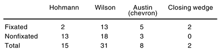
When examining the distribution of cases by surgical method and fixation, it is clear that the Hohmann, Austin, and closing wedge procedures have insufficient numbers of subjects in one or both groups to allow for surgical technique comparisons (Table 4).
Table 4.
Distributation of Cases by Surgical Procedure and Fixation.
After conducting the unpaired t-test for the Wilson procedure similar to the pooled results, only the dorsal displacement parameter was statistically significant (p = 0.0003) between fixated and nonfixated groups.
Discussion
The authors evaluate the effectiveness of using fixation when performing fifth metatarsal distal metaphyseal osteotomies for the correction of tailor’s bunion. The following parameters were evaluated preoperatively and postoperatively: metatarsal length, intermetatarsal angle, lateral bowing angle, and dorsal displacement for both the fixated and nonfixated groups. Postoperative complications and negative sequelae were also evaluated independently for each group.
It took an average of 5 weeks to get back to shoes for those patients who had fixation, and approximately 2 weeks longer for the nonfixated group. This was related to greater postoperative edema in the nonfixated group making it more difficult for those patients to get back into soft shoes.
Consolidation Time
The time required for consolidation of fifth metatarsal osteotomies has not been adequately documented. [42] According to the authors’ data, it took an average of 8.06 weeks for the osteotomy to consolidate in the fixated group, while the nonfixated group averaged 10.82 weeks. This data must be interpreted with the study’s limitations in mind. The patients did not have radiographs taken at standardized time periods postoperatively. Some patients may have consolidated prior to the next available radiograph. Some patients were excluded from the study, particularly some of the nonfixated patients, because they never consolidated radiographically and were discharged on the basis of being asymptomatic. Some were simply lost for follow-up care. This is significant because the patients that were discharged prior to radiographic consolidation often projected increased consolidation time. This would have changed the data and revealed a much longer consolidation time for those patients who were nonfixated.
A delayed union was defined as any osteotomy that took 3 months or longer to consolidate. White [31] reported a 5% nonunion rate with nonfixated osteotomies, while other authors consider a nonunion rare. [3] Seven patients of the nonfixated group had delayed or nonunions, while only one of the fixated group had this complication. Six of this group of eight were delayed unions. Of these, three were not consolidated by 4 months and then were lost for follow-up care. One patient was considered a nonunion and still did not radiographically consolidate at 1 year and was lost for follow-up care after that.
Interestingly, none of the seven patients had clinical signs of a delayed or nonunion, and they were all asymptomatic and considered a successful result by the surgeons. None were treated for the delayed union or nonunion, by immobilization or bone stimulation. There was only one delayed union in the fixated group, and it consolidated at 4 months.
Metatarsal Length
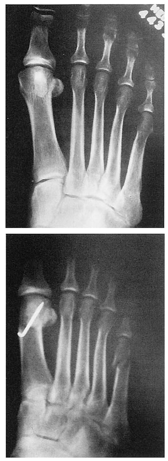
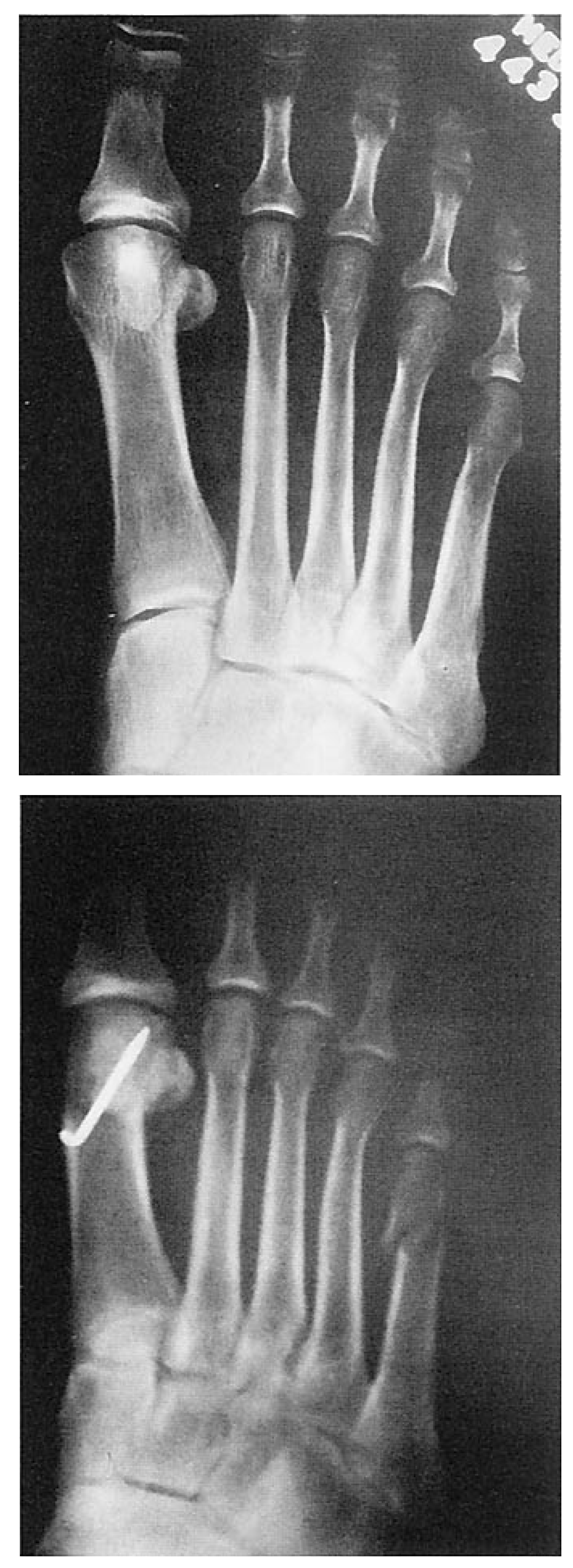
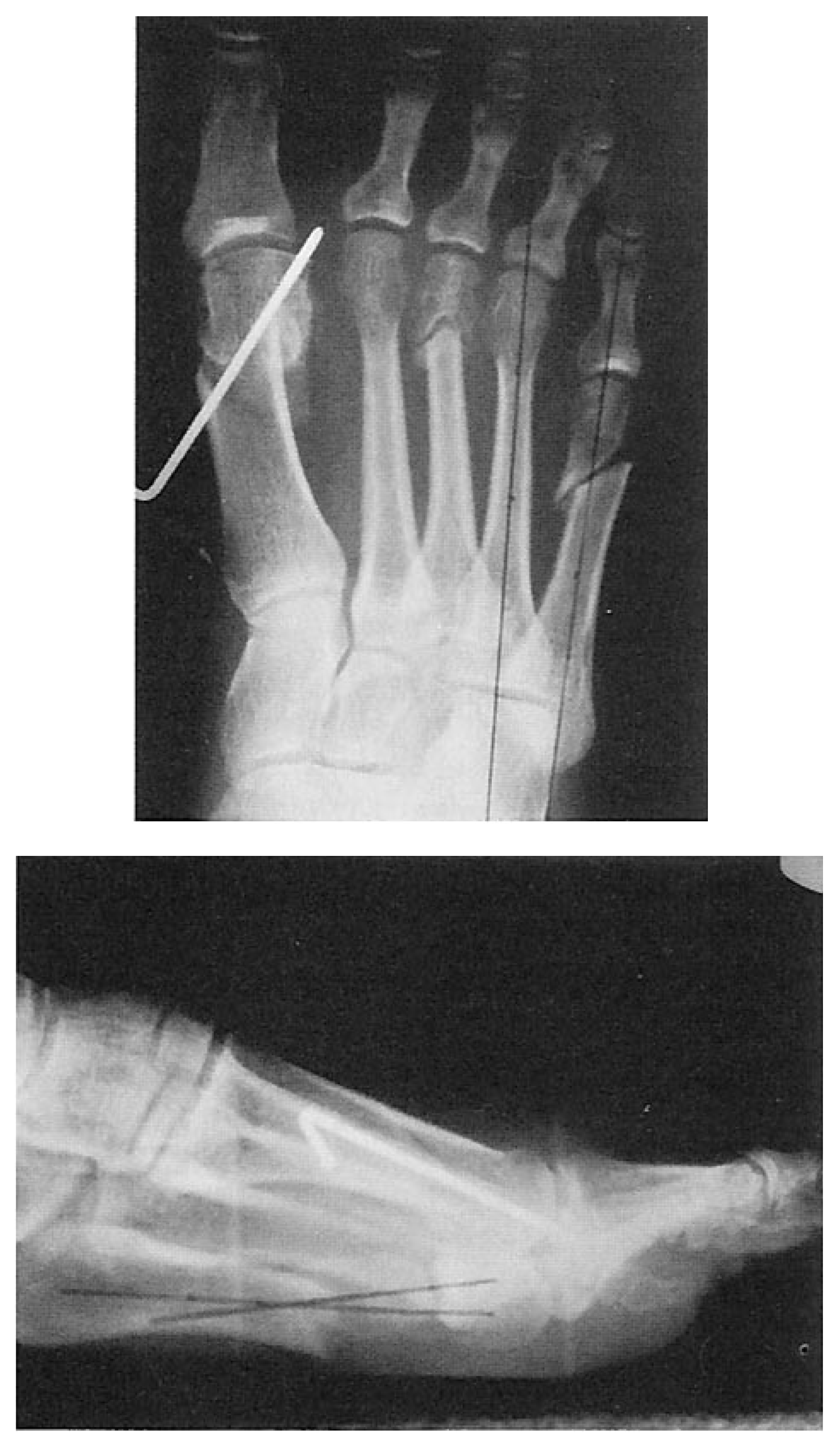
The fifth metatarsal shortened an average of 2.26 mm in the fixated group and 2.7 mm in the nonfixated group. The nonfixated osteotomies shortened by 0.45 mm more than the nonfixated group. Values of approximately 2 mm are consistent with other studies [22,43]. Nonfixated oblique osteotomies produced the most shortening. Figure 6 represents a patient who underwent a nonfixated oblique osteotomy showing excessive shortening of the fifth metatarsal.
Figure 6.
Anteroposterior radiographs showing excessive shortening following a nonfixated oblique osteotomy of the fifth metatarsal.
Angular Relationships
The fourth and fifth intermetatarsal angle was measured according to a method described by Fallat and Buckholtz [1]. Based on how this measurement is drawn, one would not expect it to change postoperatively, since it measures a proximal deformity between the fourth and fifth metatarsals. By performing a distal metaphyseal osteotomy, the angle is not affected based on how it is drawn. The authors’ data reflect a negative number for the difference in the angle and is reflective of the degree of error in measuring the angle. In most cases, measurements obtained preoperatively and postoperatively were within 1° to 2° of each other, with the postoperative measurement often being higher. Therefore, when subtracting the postoperative value from the preoperative value, a negative value was obtained. The fact that the difference was less than 1° shows that the intermetatarsal angle is not affected by distal osteotomies.
The average difference in lateral bowing in the fixated group was 15.68° while the average difference for the nonfixated group was 11.84°, ie, the fixated group had a higher amount of lateral bowing angle correction of 3.84°. The differences in lateral bowing angle may be attributed to the fact that the fixated group had more significant maintenance of correction than the nonfixated group. Perhaps fixation allowed for a greater amount of correction by the surgeon without the fear of malalignment, dislocation, or delayed union. All malalignments were excluded. A malalignment was considered to be any rotated, angulated, or dislocated capital fragment. This prevented miscalculations based on those patients who could have had a rotational component, for example, thereby falsely elevating the postoperative value.
Dorsal Displacement
Clearly, all plantar radiographic measurements are confounded by rotations. Note that all displacements were quantified with a digital linear caliper which has a resolution of 0.01 mm. Although some rotations may be present in these results and the magnitude of the dorsal displacements are generally small (< 3 mm), the difference between the fixated (xf = 0.682 mm) and nonfixated (xnf = 2.000 mm) groups is highly significant (p < 0.0001). This finding is unlikely if the errors caused by rotations are significant.
Excessive dorsal displacement has been correlated in the literature with an increase in transfer lesions, metatarsalgia, and recurrence of fifth metatarsal lesion.[3,5,24,43,44]
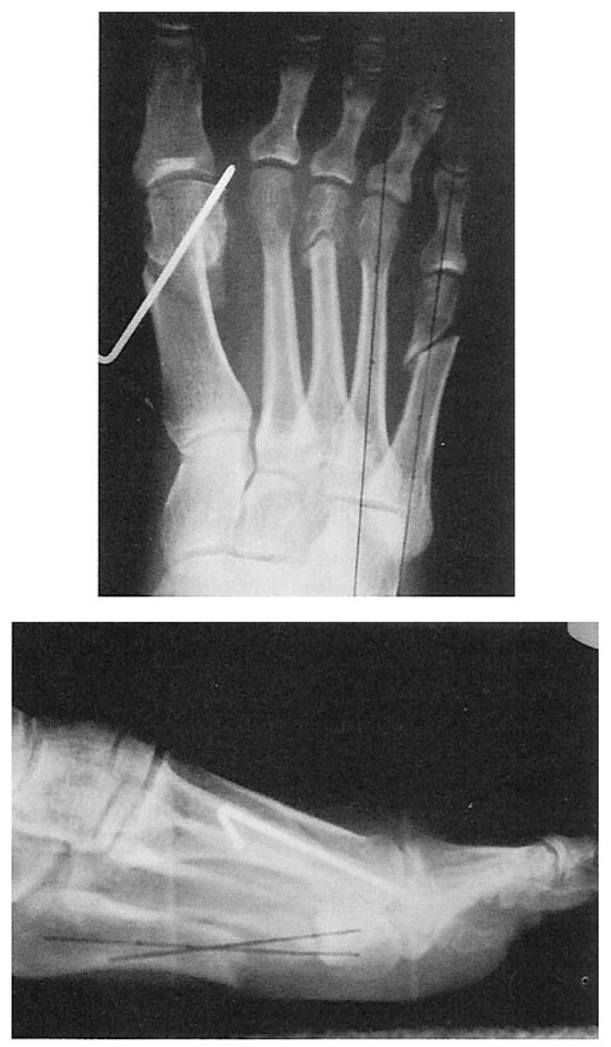
Two patients who had oblique nonfixated osteotomies showed a minimal amount of dorsal displacement (1 mm) postoperatively, but with an increase in weightbearing and time, displayed a progressive increase in the amount of dorsal displacement of an additional 2 mm. An example is shown in Figure 7.
Figure 7.
Anteroposterior and lateral radiographs taken one month after surgery showing excessive dorsal displacement following a nonfixated oblique fifth metatarsal osteotomy.
The authors observed three documented cases of recurrent lesions under the fifth metatarsal distally. All three patients had nonfixated osteotomies resulting in significant dorsal displacement and shortening of the metatarsal. As previously reported by Heckman et al [43], those lesions that reoccurred did so at the apex of the osteotomy and not at the metatarsal head level.
Metatarsalgia was also related to both excess dorsal displacement and shortening of the metatarsal. All those patients who had nonfixated osteotomies and resultant metatarsalgia had excess dorsal displacement and shortening. Of the fixated group, one patient was fixated in an excessively dorsiflexed position resulting in metatarsalgia. The second patient was actually fixated in a plantarflexed position, which probably resulted in a transfer in weight off of the fifth ray to the lesser metatarsals.
Of those eight patients who had chevron osteotomies, no dorsal displacement resulted. This osteotomy appeared to provide more stability in the sagittal plane and had no incidence of transfer lesions, metatarsalgia, or recurrence of lesion under the fifth metatarsal.
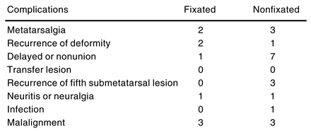
The overall complication rate is shown in Table 5. Overall, more complications were noted in the nonfixated group.
Table 5.
Complication Rate.
Fixation Time
The average fixation time was 4 weeks. All patients were fixated with a single Kirschner wire, either 0.045 or 0.062. The range of fixation time was 2 weeks to 7 1/2 weeks. Forty-one percent of those fixated with a Kirschner wire had minor negative sequelae. Two thirds had pin migration or pin loosening, while one third had pain at the pin site caused by irritation. These sequelae may be caused in part to the independent range of motion of the fifth ray. Proper bandaging to pad off the wire may help, and immobilization postoperatively with a soft or hard cast, particularly during the first 2 weeks after surgery.
Pin migration or loosening was most often seen in those patients fixated with a 0.045 Kirschner wire rather than those fixated with a 0.062 Kirschner wire. A larger Kirschner wire may allow for a more stable fixation of the osteotomy and may be affected less by weightbearing.
Other forms of fixation are also available such as screws, monofilament wire, intramedullary nailing, staple, or suture. Kirschner wire fixation is the most common form of fixation used and offers splintage and some compression of the osteotomy site. No pin tract infections were noted; this complication probably would be rare in fifth metatarsal head osteotomies.
Recurrence Rate
Tailor’s bunions recurred in 5.4% of the authors’ patients. This included one patient with a nonfixated oblique osteotomy and two patients of the fixated group, one with an oblique osteotomy and the other with a distal closing wedge osteotomy. In these cases, an adducto varus fifth digit was apparent radiographically and was not surgically corrected. Associated fifth toe deformities such as an adducto varus fifth digit may contribute to the malposition of the fifth metatarsal. It may produce a retrograde force of the fifth metatarsal head causing an increase in the intermetatarsal splaying between metatarsals four and five, thereby increasing the tailor’s bunion deformity.
Even though an adducto varus fifth toe may not be symptomatic, if it is severe enough that it is contributing to the tailor’s bunion deformity, surgical correction should be considered at the time of the tailor’s bunion correction.
Infection
Only one postoperative soft tissue infection was noted postoperatively. This patient had a nonfixated oblique osteotomy and was treated successfully with a short course of an oral antibiotic.
Neuritis
Two patients had postoperative neuritis. One patient had neuritis preoperatively from excessive pressure on the lateral aspect of the fifth metatarsophalangeal joint compressing the proper digital nerve. This patient had a nonfixated oblique osteotomy. The neuritis persisted postoperatively. The second patient had postoperative neuritis following a fixated chevron osteotomy. This was not relieved by surgery and was treated postoperatively with physical therapy, nonsteroidal anti-inflammatory drugs, and padding. It did not resolve by her last visit, 6 months postoperatively.
Operative Technique
The operative reports of all patients were reviewed to examine what, if any, techniques might account for a more desirable result.
Minimal dissection is recommended. Minimal stripping of the soft tissue attachments surrounding the joint will allow for osseous union to occur in a normal fashion and prevent interruption of the vascularity to the area. The soft tissue can also be used as a stabilizing factor and is particularly helpful in those osteotomies that are not fixated. The transverse metatarsal ligament should be preserved to stabilize the position of the fifth metatarsal head. This may be particularly important for nonfixated osteotomies (Harold Schoenhaus, DPM, personal communication, 1995).
For most tailor’s bunion osteotomies, dorsal displacement is not necessary. Fixation is therefore recommended for these osteotomies to prevent excessive dorsal displacement of the capital fragment. Otherwise, an increase in dorsal displacement can result in transferring more weight laterally, particularly in the fourth submetatarsal with possible lesion formation.
The capital fragment can be medially transposed approximately one third of the width of the metatarsal flare of the fifth metatarsal, approximately 2.5 mm. Excessive shifting can result in dislocation, malunion, and possible heloma molle formation if the capital fragment is too close in alignment to the lateral aspect of the fourth proximal phalanx base.
Resection of the lateral eminence may be necessary and should be performed only after the osteotomy has been transposed medially. Once the capital fragment has been transposed, more often the lateral aspect of the metatarsal head is not prominent and does not need resection.
With oblique osteotomies, the remaining spike of bone at the lateral distal aspect of the fifth metatarsal should be resected. This prominence can be a source of irritation postoperatively and an area where a lesion may develop secondary to shoe pressure.
Conclusion
Tailor’s bunions are not characterized by a high rate of complications. Nonetheless, complications may occur because of factors such as inappropriate procedure selection, excessive dorsal displacement, metatarsal shortening, and inadequate fixation.
This study shows that fixation may help control the postoperative dorsal displacement of the fifth metatarsal capital fragment (p < 0.0001) and produce less shortening of the metatarsal, resulting in fewer complications.
Fixation provides a more predictable healing time and prevents further displacement of the osteotomy with weightbearing, providing better maintenance of correction. Although nonfixated osteotomies may often produce successful results, a more predictable, successful result can be obtained by using fixation, not only to prevent excessive dorsal displacement at the osteotomy site, but to maintain the corrected position until early bone healing occurs. Fixation also provides better assurance that consolidation will take place.
Cast immobilization is not necessary but may be used as an adjunct in nonfixated osteotomies or when patient compliance is questioned.
When an adducto varus fifth toe is present, it should be evaluated to determine if it contributes to the tailor’s bunion deformity, and if so, surgically corrected at the same time as the tailor’s bunion. A postoperative functional orthosis may prevent or control transfer lesions or metatarsalgia when indicated.
References
- FALLAT LM, BUCKHOLZ J: An analysis of the tailor’s bunion by radiographic and anatomical display. JAPA 70: 597, 1980.
- TREPAL M: “Surgery of the Fifth Ray,” in Comprehensive Textbook of Foot Surgery, ed by ED McGlamry, AS Banks, MS Downey, Williams & Wilkins, Baltimore, 1992.
- CATANZARITI AR, FRIEDMAN C, DESTANZO J: Oblique osteotomy of the fifth metatarsal: a five year review. J Foot Surg 27: 316, 1988.
- CASTLE JE, COHEN AH, DOCKS G: Fifth metatarsal distal oblique wedge osteotomy utilizing cortical screw fixation. J Foot Surg 31: 478, 1992.
- FRANKEL JP, TURF RM, KING BA: Tailor’s bunion: clinical evaluation and correction by distal metaphyseal osteotomy with cortical screw fixation. J Foot Surg 28: 237, 1989.
- FALLAT LM: Pathology of the fifth ray. Clin Podiatr Med Surg 7: 689, 1990.
- DAVIES H: Metatarsus quintus valgus. BMJ 1: 664, 1949.
- DICKSON F, DIVELEY RL: “Hallux,” in Functional Disorders of the Foot, 3rd Ed, JB Lippincott, Philadelphia, 1953.
- BROWN JE: Functional and cosmetic correction of metatarsus latus. Clin Orthop 14: 166, 1959.
- SPONSEL KH: Bunionette correction by metatarsal osteotomy: preliminary report. Orthop Clin N Am 7: 809, 1976.
- WU KK: “Surgery of the Metatarsal Region,” in Surgery of the Foot, Lea & Febiger, Philadelphia, 1986.
- LELIEVRE J: Exostosis of the head of the fifth metatarsal bone, tailor’s bunion. Concours Med 78: 4815, 1956.
- DUVRIES HL: “Acquired Nontraumatic Deformities of the Foot,” in Surgery of the Foot, 4th Ed, CV Mosby, St Louis, 1978.
- YANCY HA: Congenital lateral bowing of the fifth metatarsal. Clin Orthop 62: 203, 1969.
- SGARLATO TE: Compendium of Podiatric Biomechanics, California College of Podiatric Medicine, San Francisco, 1971.
- NESTOR BJ, KITAOKA HB, STRUP D, ET AL: Radiologic anatomy of the painful bunionette. Foot Ankle 11: 6, 1990.
- LEACH RE, IGOU R: Metatarsal osteotomy for bunionette deformity. Clin Orthop 100: 171, 1974.
- GRAY H: Gray’s Anatomy, 36th Ed, WB Saunders, Philadelphia, 1980.
- HICKS JH: Mechanics of the foot: I. the joints. J Anat 87: 345, 1953.
- ROOT ML, ORIEN WP, WEED JH: Normal and Abnormal Function of the Foot: Clinical Biomechanics, Vol 2, Clinical Biomechanics, Los Angeles, 1977.
- SAKOFF M, LEVY AI, HANFT JS: Metaphyseal osteotomy for the treatment of tailor’s bunions. J Foot Surg 28: 537, 1989.
- ZVIJAC JE, JANECKI CJ, FREELING RM: Distal oblique osteotomy for tailor’s bunion. Foot Ankle 12: 171, 1991.
- THUL JR, HOFFMAN SJ: Neuromas associated with tailor’s bunion. J Foot Surg 24: 342, 1985.
- KEATING SE, DEVINCENTIS A, GOLLER WL: Oblique fifth metatarsal osteotomy: a follow-up study. J Foot Surg 71A: 423, 1989.
- STEINKE MS, BOLL KL. Holmann-Thompson metatarsal osteotomy for tailor’s bunion (bunionette). J Bone Joint Surg 71A: 423, 1989.
- THROCKMORTON JK, BRADLEE N: Transverse V sliding osteotomy: a new surgical procedure for the correction of tailor’s bunion deformity. J Foot Surg 18: 117, 1978.
- MERCADO OA: “Metatarsal-Phalageal Joint Surgery,” in An Atlas of Foot Surgery, Vol 1, Corolando Press, Oak Park, IL, 1979.
- HABNER JH, KRAFT J: Crescentic osteotomy osteotomies for fifth metatarsal head lesions. J Foot Surg 19: 66, 1980.
- YU GV, RUCH JA, SMITH TF: “Deformity and Surgery of the Fifth Ray,” in Comprehensive Textbook of Foot Surgery, ed by ED McGlamry, Williams & Wilkins, Baltimore, 1987.
- DAVIDSON MR: Non-stabilization metatarsal head oteotomies: a simple method for correcting second, third, fourth and fifth metatarsal head pathology. J Foot Surg 10: 121, 1971.
- WHITE DL: Minimal incision approach to osteotomies of the lesser metatarsals for treatment of intractable keratosis, metatarsalgia, and tailor’s bunion. Clin Podiatr Med Surg 8: 25, 1991.
- HANSSON G: Sliding osteotomy for tailor’s bunion: brief report. J Bone Joint Surg 71B: 324, 1989.
- FRIEND G, GRACE K, STONE HA: L-osteotomy with absorbable fixation correction of tailor’s bunion. J Foot Ankle Surg 32: 14, 1993.
- HARRIS MD: A surgical approach for digiti quinti varus. J Natl Assoc Chirop 47: 362, 1967.
- WEISBERG MH: Resection of the fifth metatarsal head in lateral segment problems. JAPA 57: 374, 1967.
- ADDANTE JB, KAUFMANN D: Repair of tailor’s bunion by means of fifth metatarsal head resection and insertion of a spherical silicone implant: a preliminary report of two cases. Arch Podiatr Med Foot Surg 4: 49, 1977.
- PETRICH RJ, DULL DD: Interpositional sphere implant in the fifth metatarsophalangeal joint. J Foot Surg 20: 93, 1981.
- GERBERT J, SGARLATO TE, SUBOTNICK IS: Preliminary study of a closing wedge osteotomy of the fifth metatarsal for correction of a tailor’s bunion deformity. JAPA 62: 212, 1972.
- RAPPAPORT MJ: “Wedge Osteotomy for Tailor’s Bunion,” in Reconstructive Surgery of the Foot and Leg, ed by ED McGlamry, Intercontinental Medical Book, New York, 1974.
- BUCHBINDER IJ: DRATO procedure for tailor’s bunion. J Foot Surg 21: 177, 1982.
- DIEBOLD PF, BEJJANI FJ: Basal osteotomy of the fifth metatarsal with intermetatarsal pinning: a new approach. Foot Ankle 8: 40, 1987.
- KITAOKA HA, LEVENTEN EO: Medial displacement metatarsal osteotomy for treatment of painful bunionette. Clin Orthop 243: 172, 1989.
- HECKMAN, JD, HARKLESS LB, HIGGINS KVZ: The Sponsel oblique fifth metatarsal osteotomy: evaluating with longterm follow-up. Foot 1: 37, 1991.
- DIEBOLD PF: Basal osteotomy of the fifth metatarsal for the bunionette. Foot Ankle 12: 74, 1991.
- SHROUT PE, FLEISS JL: Intra-class correlations: uses in assessing rater reliability. Psychol Bull 86: 420, 1979.
Additional References
- BELLACOSA RA, POLLACK RA: Complications of lesser metatarsal surgery. Clin Podiatr Med Surg 8: 383, 1991.
- KONRADSEN L, NIELSEN PT: Distal metatarsal osteotomy for bunionette deformity. J Foot Surg 27: 493, 1988.
- SCHABLER JA, TONEY M, HANFT JR, ET AL: Oblique metaphyseal osteotomy for the correction of tailor’s bunions: a three-year review. J Foot Surg 31: 79, 1992.
- SCHOENHAUS H, ROTMAN S, MESHON AL: A review of normal intermetatarsal angles. JAPA 63: 88, 1973.
- TREPAL MJ, HARKLESS LB, JULES KT, ET AL: “Tailor’s Bunion and Associated Fifth Metatarsal Conditions,” in Preferred Practice Guidelines, American College of Foot and Ankle Surgeons, Chicago, 1994.

© 1996 American Podiatric Medical Association