


Abstract
Calcaneal stress fracture is an uncommon osseous cause of heel and hindfoot pain. A 33-year-old woman with chronic underweight status (body mass index, 16.59 kg/m2) presented with a 2- to 3-week history of right lateral hindfoot pain that reportedly began during treadmill walking in association with a perceived twisting event. The medical record documented tenderness in the region of the calcaneofibular ligament and a negative talar tilt test, but heel-specific examination was incompletely documented. Magnetic resonance imaging (MRI) obtained at the initial evaluation was formally interpreted as showing a calcaneal tuberosity stress fracture and quadratus plantae strain, with no abnormality reported in the ankle ligament complexes. On retrospective MRI review, an incomplete, vertically oriented fracture line extending to the cortex without articular extension or osseous displacement was identified, with surrounding marrow signal abnormality meeting the imaging criteria for Nattiv grade 4. Weight-bearing radiographs obtained 4 days later showed a corresponding sclerotic band. This case illustrates MRI–radiographic correlation in a calcaneal tuberosity stress fracture presenting with lateral hindfoot pain and supports inclusion of osseous injury in the differential diagnosis.
1. Introduction
Musculoskeletal disorders represent a substantial burden in orthopedic outpatient practice [1]. Heel and hindfoot pain arise from a broad differential diagnosis that includes plantar fasciitis, heel fat pad pathology, Achilles and other tendinopathies, neuropathic disorders, inflammatory arthropathy, and osseous injury [2,3]. Calcaneal stress fracture is a recognized but relatively uncommon cause of heel pain and may be overlooked when symptoms develop gradually or resemble a soft-tissue disorder [3,4].
Stress fractures occur when repetitive loading produces microdamage that exceeds local repair capacity and have traditionally been categorized as fatigue fractures in normal bone and insufficiency fractures in weakened bone [4]. Published reports of calcaneal stress fracture have most often involved runners, military recruits, or other physically active adults, although civilian cases and delayed diagnoses have also been described [4,5,6,7,8,9]. In a nine-patient civilian series of magnetic resonance imaging (MRI)-confirmed calcaneal stress fractures, most patients reported pain over the lateral aspect of the heel, indicating that lateral pain localization is a recognized clinical presentation [9]. The calcaneus is exposed to repetitive ground-reaction forces and Achilles–plantar complex loading during gait, which may contribute to bone stress injury during repetitive loading [4,10].
Plain radiography is commonly used as the initial imaging examination for suspected stress fracture; however, early radiographs may be normal or nonspecific [3,4,11,12]. In a systematic review of suspected lower-extremity stress fractures, MRI showed the best overall diagnostic performance and was particularly useful when clinical suspicion persisted despite negative or indeterminate radiographs [11]. Low body mass may be a susceptibility factor for bone stress injury, but its contribution in an individual patient should be interpreted cautiously when contemporaneous nutritional, endocrine, and activity-related data are unavailable [10]. The present report describes a chronically underweight young woman with 2 to 3 weeks of lateral hindfoot pain that began during treadmill walking with a perceived twisting event. MRI obtained at the first clinical evaluation demonstrated a fracture line and surrounding marrow edema in the posterior calcaneal tuberosity, and weight-bearing radiographs obtained 4 days later showed a corresponding linear sclerotic band. Taken together, these findings highlight a clinically transferable diagnostic principle: calcaneal osseous injury should remain in the differential diagnosis of persistent lateral hindfoot pain even when the loading history is equivocal, and MRI and radiography may provide complementary information.
2. Case Report
A 33-year-old woman presented with right lateral ankle and hindfoot pain of 2 to 3 weeks’ duration. She reported that the pain began during treadmill walking in association with a perceived twisting event. The record did not document treadmill frequency or duration, recent activity changes, onset characteristics or preceding prodromal symptoms, or other relevant loading factors. Pain increased with weight bearing. She denied high-energy trauma, smoking, and alcohol use. No previous stress or fragility fracture was documented. Her height was 167.4 cm, and her weight was 46.5 kg, yielding a body mass index (BMI) of 16.59 kg/m2. Historical records showed chronic low body weight, with BMI values of 16.30 kg/m2 in 2021 and 15.4 kg/m2 in 2023.
No swelling or erythema was documented. The medical record described tenderness in the region of the right calcaneofibular ligament and a negative talar tilt test. Heel-specific and other relevant soft-tissue examinations, including the calcaneal squeeze test and localized calcaneal tuberosity tenderness, were incompletely documented; gait, weight-bearing tolerance, range of motion, and neurovascular status were also not documented. No definite provisional diagnosis of a lateral ankle ligament injury was recorded.
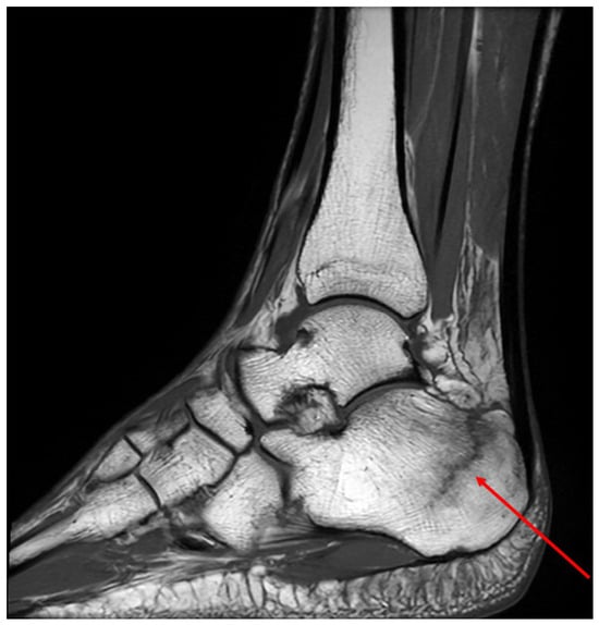
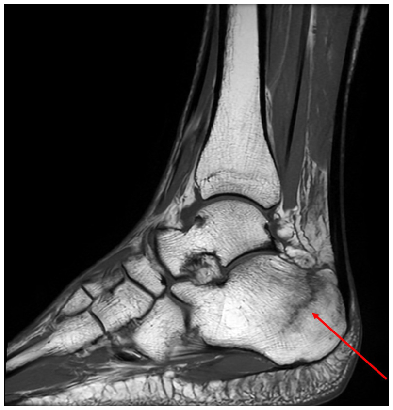
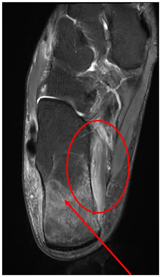
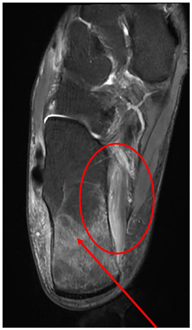
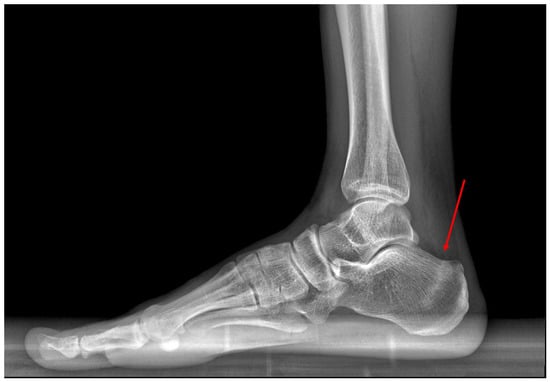
Magnetic resonance imaging of the right foot and ankle was obtained at the initial visit rather than plain radiography. The formal radiology report interpreted the osseous finding as a stress fracture at the calcaneal tuberosity and reported strain of the quadratus plantae muscle. No abnormality was reported in the lateral ankle ligaments, distal tibiofibular syndesmotic ligaments, deltoid ligament complex, or spring ligament. The MRI images were retrospectively reviewed by a fourth-year radiology resident with dedicated training in musculoskeletal imaging. The imaging findings were subsequently confirmed by a board-certified radiologist with experience in musculoskeletal imaging. On additional review of the MRI images, sagittal T1-weighted sequences demonstrated an incomplete, vertically oriented low-signal-intensity fracture line in the calcaneal tuberosity extending to the cortex, without articular extension or osseous displacement (Figure 1). Confluent marrow signal abnormality surrounding the fracture line was low on T1-weighted and high on fat-suppressed T2-weighted sequences. Axial T2-weighted fat-suppressed images also demonstrated the associated linear low-signal fracture line and adjacent increased signal in the quadratus plantae, consistent with muscle strain (Figure 2). Based on the marrow signal abnormality on both T1- and T2-weighted sequences and the visible fracture line, the lesion was retrospectively classified as grade 4 according to the Nattiv MRI grading system [13]. Although these findings characterized fracture morphology and MRI severity, the incomplete loading history did not permit confident determination of the precise loading mechanism or fatigue-versus-insufficiency subtype. Coronal fat-suppressed images showed no structural abnormality of the calcaneofibular ligament or deltoid ligament complex (Figure 3). Weight-bearing foot radiographs obtained 4 days later showed a linear sclerotic band along the posterior calcaneal tuberosity, corresponding to the lesion identified on MRI (Figure 4). Dual-energy X-ray absorptiometry (DXA) showed bone mineral density within the expected range for age (lumbar spine Z score, −0.6; total femur Z score, −0.6).
Figure 1.
Sagittal T1-weighted magnetic resonance image of the right ankle showing an incomplete, vertically oriented low-signal-intensity fracture line in the calcaneal tuberosity extending to the cortex without articular extension (arrow).
Figure 2.
Axial T2-weighted fat-suppressed magnetic resonance image of the right ankle showing confluent marrow edema centered at the calcaneal tuberosity with an associated linear fracture line (arrow) and adjacent quadratus plantae strain (circle).
Figure 3.
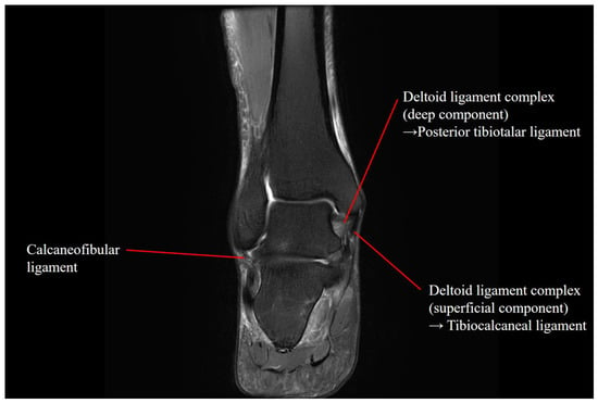
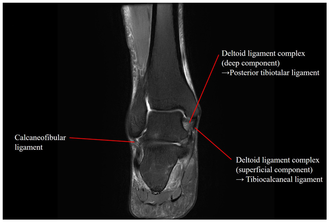
Coronal T2-weighted fat-suppressed magnetic resonance image of the right ankle showing no structural abnormality of the calcaneofibular ligament or deltoid ligament complex.
Figure 4.
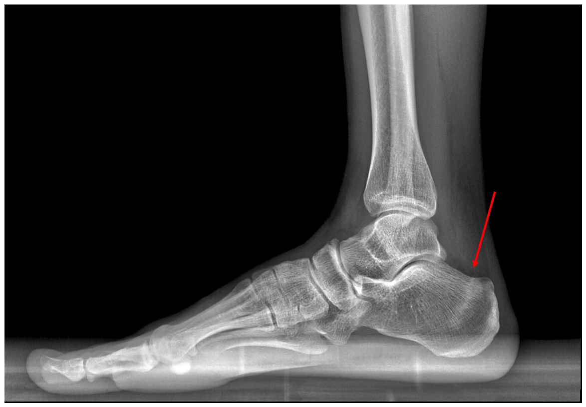
Weight-bearing lateral radiograph of the right foot obtained 4 days after magnetic resonance imaging, showing a linear sclerotic band in the posterior calcaneal tuberosity (arrow).
Historical chart review disclosed prior gynecologic evaluation for secondary amenorrhea/hypoestrogenism in 2021 and previous vitamin D deficiency, followed by a normal 25-hydroxyvitamin D level in 2023 (43.7 ng/mL). Low luteinizing hormone and estradiol values were also documented in 2023; however, because combined oral contraceptive therapy had been prescribed from 2021 and was ongoing at the time of fracture, these values may not reflect spontaneous ovarian function. Accordingly, spontaneous menstrual status at the time of injury could not be determined reliably. No complete blood count, ferritin/iron studies, or 25-hydroxyvitamin D measurement were available at the time of fracture. A walking boot was prescribed for 5 weeks; oral calcium, vitamin D, and analgesics were also prescribed. The record contained only a nonspecific notation of symptom improvement at an unspecified follow-up. Detailed treatment and follow-up data, including adherence, objective outcomes, repeat evaluation, and return-to-activity status, were unavailable; therefore, treatment effectiveness and long-term recovery could not be assessed.
3. Discussion
Previous civilian reports have established that calcaneal stress fracture may present with lateral heel pain, and delayed diagnosis after initial treatment for common soft-tissue disorders has also been described [8,9]. The clinical contribution of this report therefore lies not in a novel pain distribution, but in illustrating how calcaneal osseous injury can be characterized across imaging modalities in a patient with persistent lateral hindfoot pain and an equivocal loading history.
The imaging chronology requires cautious interpretation. MRI was obtained at the first clinical evaluation instead of plain radiography, and weight-bearing radiographs obtained 4 days later already demonstrated a corresponding linear sclerotic band in the posterior calcaneal tuberosity. Because no radiograph was obtained at the initial evaluation, this case cannot demonstrate initial radiographic false-negativity, earlier detection by MRI, or superiority of MRI over contemporaneous radiography. The value of the paired examinations therefore lies in cross-modality concordance rather than temporal comparison: MRI characterized the fracture line, cortical extension, and marrow response, whereas weight-bearing radiography demonstrated the corresponding sclerotic band. The broader literature indicates that early radiographs may be normal or nonspecific and that MRI has the best overall diagnostic performance when suspicion persists despite negative or indeterminate radiographs [3,4,11]. Plain radiography therefore remains the usual initial imaging examination, with MRI serving as a subsequent test when radiographs are negative or indeterminate or when clinical suspicion remains high [12].
The etiologic classification of the lesion remains uncertain. Stress fractures are generally attributed to repetitive loading that produces microdamage exceeding local repair capacity, with fatigue and insufficiency fractures distinguished according to the condition of the underlying bone [4,10]. In this patient, pain reportedly began during treadmill walking in association with a perceived twisting event, but activity exposure and onset characteristics were incompletely documented. The formal radiology report interpreted the lesion as a calcaneal tuberosity stress fracture, and additional image review characterized it as an incomplete, vertically oriented fracture with extension to the cortex but no articular extension or osseous displacement, corresponding to Nattiv MRI grade 4 [13]. The grade was applied retrospectively as a descriptor of MRI severity and does not determine the loading mechanism or the fatigue-versus-insufficiency subtype. Because activity exposure and onset characteristics were incompletely documented, etiologic classification remained uncertain, and an acute nondisplaced traumatic contribution could not be completely excluded.
The clinical examination was incompletely documented. The record noted tenderness in the region of the calcaneofibular ligament and a negative talar tilt test, whereas the formal MRI report identified a calcaneal tuberosity stress fracture and quadratus plantae strain without a reported structural ligament abnormality. Because heel-specific examination findings were incomplete and no provisional ligament diagnosis was recorded, it cannot be determined whether the presentation truly mimicked a lateral ligament injury or whether a diagnostic error occurred. These findings underscore that lateral localization alone should not be used to exclude calcaneal osseous injury [2,3,4,9]. Persistent hindfoot pain warrants assessment of the calcaneus as well as adjacent soft-tissue structures.
Chronic low BMI was the only clearly documented contemporaneous susceptibility factor [10]. Historical amenorrhea/hypoestrogenism and vitamin D deficiency were noted, but contemporaneous nutritional, menstrual, activity-related, and metabolic data were insufficient for syndromic assessment. Because the patient was not established to be an athlete, Relative Energy Deficiency in Sport and the Female Athlete Triad are mentioned only as contextual considerations and not as diagnoses or causal explanations [14,15]. DXA values were within the expected range for age, but the biological contribution of host factors could not be determined.
This report has several limitations. It is a single case with incomplete documentation of activity exposure, the characteristics of symptom onset and possible prodromal symptoms, heel-specific examination, and functional status. Retrospective MRI review allowed detailed morphologic grading, but the incomplete clinical history, unavailable contemporaneous metabolic and nutritional data, and limited follow-up precluded definitive mechanistic classification and assessment of treatment outcomes. Within these limitations, the case highlights several relevant diagnostic considerations for persistent hindfoot pain: symptom localization should guide but not determine tissue attribution, MRI and radiography may provide complementary information, and etiologic classification requires integration of imaging findings with loading history, trauma history, and host factors.
4. Conclusions
Calcaneal stress fracture should remain in the differential diagnosis of persistent hindfoot pain, even when symptoms are localized laterally and the loading history is equivocal. In this patient, MRI characterized the morphology of an incomplete calcaneal tuberosity fracture and the associated marrow signal abnormality, while weight-bearing radiography obtained 4 days later demonstrated a corresponding sclerotic band. Although the absence of baseline radiography and the incomplete exposure history preclude direct comparison of the imaging modalities and definitive classification as a fatigue or insufficiency fracture, the case highlights the complementary roles of MRI and radiography. Consistent with current imaging guidance, radiography remains the initial examination, followed by MRI when radiographs are negative or indeterminate and clinical suspicion persists.
Author Contributions
Conceptualization, H.B.; Formal analysis, H.H.; investigation, H.H., H.J.S., and C.H.L.; methodology, H.H.; software, H.H.; visualization, H.H.; data curation, H.H., H.J.S., and C.H.L.; resources, H.J.S.; project administration, C.H.L.; validation, C.H.L.; writing—original draft, H.H. and H.B.; writing—review and editing, M.-K.O., E.S.L., and H.B.; supervision, M.-K.O., E.S.L., and H.B.; funding acquisition, H.B. All authors have read and agreed to the published version of the manuscript.
Funding
This research was supported by the Gyeongsang National University Hospital Biomedical Research Institute Fund (Grant No. GNUHBRIF-2026-0004).
Institutional Review Board Statement
The Gyeongsang National University Hospital Institutional Review Board reviewed this single-patient case report and determined that it was exempt from IRB review (GNUH IRB No. 2026-07-018) because it involved no disclosure of personally identifiable information and posed no more than minimal risk. The report was prepared in accordance with the principles of the Declaration of Helsinki, and all reasonable measures were taken to protect patient privacy and confidentiality.
Informed Consent Statement
Written informed consent was obtained from the patient for publication of this report and accompanying images.
Data Availability Statement
The raw data supporting the conclusions of this article will be made available by the authors on request.
Conflicts of Interest
The authors declare no conflicts of interest.
References
- Ülgen, N.K.; Nazlıgül, A.S.; Yiğit, N.; Erginoğlu, S.E.; Akkurt, M.O. Epidemiologic patterns of musculoskeletal disorders by body region across 1.2 million orthopedic visits: A 15-year experience from a middle-income country. J. Eval. Clin. Pract. 2025, 31, e70230. [Google Scholar] [CrossRef] [Scilit] [PubMed]
- Yi, T.I.; Lee, G.E.; Seo, I.S.; Huh, W.S.; Yoon, T.H.; Kim, B.R. Clinical characteristics of the causes of plantar heel pain. Ann. Rehabil. Med. 2011, 35, 507–513. [Google Scholar] [CrossRef] [Scilit] [PubMed]
- Chang, C.D.; Wu, J.S. MR imaging findings in heel pain. Magn. Reson. Imaging. Clin. N. Am. 2017, 25, 79–93. [Google Scholar] [CrossRef] [Scilit] [PubMed]
- Weber, J.M.; Vidt, L.G.; Gehl, R.S.; Montgomery, T. Calcaneal stress fractures. Clin. Podiatr. Med. Surg. 2005, 22, 45–54. [Google Scholar] [CrossRef] [Scilit] [PubMed]
- Hopson, C.N.; Perry, D.R. Stress fractures of the calcaneus in women marine recruits. Clin. Orthop. Relat. Res. 1977, 128, 159–162. [Google Scholar] [CrossRef] [Scilit]
- Sormaala, M.J.; Niva, M.H.; Kiuru, M.J.; Mattila, V.M.; Pihlajamäki, H.K. Stress injuries of the calcaneus detected with magnetic resonance imaging in military recruits. J. Bone Jt. Surg. Am. 2006, 88, 2237–2242. [Google Scholar] [CrossRef] [Scilit] [PubMed]
- Niva, M.H.; Sormaala, M.J.; Kiuru, M.J.; Haataja, R.; Ahovuo, J.A.; Pihlajamaki, H.K. Bone stress injuries of the ankle and foot: An 86-month magnetic resonance imaging-based study of physically active young adults. Am. J. Sports Med. 2007, 35, 643–649. [Google Scholar] [PubMed]
- Imerci, A.; Incesu, M.; Bozoglan, M.; Canbek, U.; Ursavas, H.T. Bilateral calcaneal stress fractures: A case report. Ortop. Traumatol. Rehabil. 2012, 14, 477–481. [Google Scholar] [CrossRef] [Scilit] [PubMed]
- Labronici, P.J.; Pires, R.E.S.; Amorim, L.E. Calcaneal stress fractures in civilian patients: An epidemiological study. J. Foot Ankle 2021, 15, 54–59. [Google Scholar] [CrossRef] [Scilit]
- Hoenig, T.; Ackerman, K.E.; Beck, B.R.; Bouxsein, M.L.; Burr, D.B.; Hollander, K.; Popp, K.L.; Rolvien, T.; Tenforde, A.S.; Warden, S.J. Bone stress injuries. Nat. Rev. Dis. Primers 2022, 8, 26. [Google Scholar] [CrossRef] [Scilit]
- Wright, A.A.; Hegedus, E.J.; Lenchik, L.; Kuhn, K.J.; Santiago, L.; Smoliga, J.M. Diagnostic accuracy of various imaging modalities for suspected lower extremity stress fractures: A systematic review with evidence-based recommendations for clinical practice. Am. J. Sports Med. 2016, 44, 255–263. [Google Scholar] [PubMed]
- Morrison, W.B.; Deely, D.; Fox, M.G.; Blankenbaker, D.G.; Dodds, J.A.; French, C.N.; Frick, M.A.; Jawetz, S.T.; Khurana, B.; Kresin, M.; et al. ACR Appropriateness Criteria® Stress (Fatigue-Insufficiency) Fracture Including Sacrum Excluding Other Vertebrae: 2024 Update. J. Am. Coll. Radiol. 2024, 21, S490–S503. [Google Scholar] [CrossRef] [Scilit] [PubMed]
- Nattiv, A.; Kennedy, G.; Barrack, M.T.; Abdelkerim, A.; Goolsby, M.A.; Arends, J.C.; Seeger, L.L. Correlation of MRI grading of bone stress injuries with clinical risk factors and return to play: A 5-year prospective study in collegiate track and field athletes. Am. J. Sports Med. 2013, 41, 1930–1941. [Google Scholar] [PubMed]
- Angelidi, A.M.; Stefanakis, K.; Chou, S.H.; Valenzuela-Vallejo, L.; Dipla, K.; Boutari, C.; Ntoskas, K.; Tokmakidis, P.; Kokkinos, A.; Goulis, D.G.; et al. Relative energy deficiency in sport (REDs): Endocrine manifestations, pathophysiology and treatments. Endocr. Rev. 2024, 45, 676–708. [Google Scholar] [CrossRef] [Scilit] [PubMed]
- Williams, N.I.; De Souza, M.J.; Misra, M.; Nattiv, A.; Joy, E.; Barrack, M.; Ricker, E.A.; Gorrell, S.; Koltun, K.J.; O’Donnell, E.; et al. 2025 update to the Female Athlete Triad Coalition consensus statement Part 2: Clinical guidelines for screening, diagnosis, treatment, and return to play for adolescents and adults. Sports Med. 2026, 56, 375–427. [Google Scholar] [CrossRef] [Scilit] [PubMed]
Disclaimer/Publisher’s Note: The statements, opinions and data contained in all publications are solely those of the individual author(s) and contributor(s) and not of MDPI and/or the editor(s). MDPI and/or the editor(s) disclaim responsibility for any injury to people or property resulting from any ideas, methods, instructions or products referred to in the content. |
© 2026 by the authors. Published by MDPI on behalf of the American Podiatric Medical Association. Licensee MDPI, Basel, Switzerland. This article is an open access article distributed under the terms and conditions of the Creative Commons Attribution (CC BY) license.