


Abstract
Treatment of osteochondral lesions of the talus (OLTs) with bone grafting is an alternative option to osteochondral transplantation for larger OLTs. The purpose of this study is to evaluate treatment of OLTs with bone grafting with continuous functional outcome scores, return-to-play data, and meta-analyses of overall complication rates after bone graft treatment for OLT lesions as reported and best- and worst-case scenarios. A systematic review was conducted based on PRISMA guidelines using specific search terms on the PubMed, Embase and Cochrane Library databases. Level and quality of evidence were evaluated by The Journal of Bone & Joint Surgery and the Modified Coleman Methodology Score, respectively. Meta-analysis by fixed-effects models was performed if heterogeneity was low (I2 < 25%) and by random-effects models if heterogeneity was moderate to high (I2 > 25%). Eleven studies were included in this systematic review. The Mean American Orthopaedic Foot and Ankle Society score was 56.0 ± 10.1 (range 40.3–67.3) preoperatively to 88.7 ± 5.7 (range 80.8–95.0) points postoperatively. The mean 10 mm-VAS pain score was 6.5 ± 0.89 (range 5.6–7.7) preoperatively to 1.7 ± 1.1 (range 0.4–3.7) postoperatively. The overall pooled complication rate was 3.82% (95% CI: 0.05–10.92). Limited debilitating complications were noted in these studies, with one case of deep vein thrombosis noted. Autologous bone grafting offered satisfactory outcomes for pain relief, with notable improvement of functional outcomes scores. This review proposed that autologous bone grafting should be considered as the first line of treatment for lesion sizes >150 mm2. Studies with longer follow-ups are desirable to determine the longevity of bone grafting and evaluate the durability of bone grafting for larger OLT lesions.
1. Introduction
Osteochondral lesion of the talus (OLT) is an injury commonly suffered due to ankle trauma in athletes [1,2]. The treatment can be conservative or surgical. Conservative treatment is typically for small and/or asymptomatic lesions [3]. For symptomatic lesions, reparative or replacement techniques can be employed. Reparative techniques include bone marrow stimulation (BMS), and replacement techniques include autologous osteochondral transplantation (AOT) [4]. BMS has been indicated to be optimally employed for lesion sizes < 107.4 mm2; larger lesions are often transplanted with an osteochondral autograft [3]. The drawback with AOT has been donor-site morbidity. Notably, with large lesion sizes, the inherent need for a sizeable harvest can further predispose complications. Alternative replacement techniques for lesion sizes for which AOT harvesting is uncomfortable have included autologous bone grafting and allograft use. However, access to allografts can be logistically inconvenient and/or economically unfavourable.
Theoretically, autologous bone grafting to alleviate symptoms (pain) makes sense [5]. Larger lesion sizes inherently hold a considerable void that allows joint fluid to enter and generate rising pressures that stimulate the highly innervated subchondral bone to cause pain [1,6]. Filling this void negates fluid pressure generation and the associated pain. However, the long-term durability of autologous bone grafting for the treatment of OLT is questioned as the lack of a cartilaginous aspect challenges its physiological integration and robustness [7]. To date, multiple studies have evaluated autologous bone grafting for the treatment of OLT, with modest outcomes reported, but a comprehensive review of its efficacy remains to be performed [8,9,10].
The papers included in this study suggested varying techniques of autologous bone grafting. Typically, an incision to the respective lesion location followed by osteotomy is performed. A cannulated drill is then placed over a Kirschner wire to curette out the necrotic or sclerotic bone and/or cystic material with subsequent harvesting. Bone grafting is harvested carefully using a bone extractor from the desired donor site, with some procedures using the defect as a template to guide the harvest [11]. The autologous bone graft would be placed in a press-fit manner, fixating it to the level of the subchondral plate [9]. The operation is then concluded, ranging from reduction and internal fixation of the osteotomy and screw fixation with a splint to no cast or ankle bracing [9,10,12].
Therefore, the purpose of the study was to evaluate the outcomes following autologous bone grafting for the treatment of OLT. The hypothesis was that an autologous bone graft can provide symptomatic relief translated as favourable functional outcomes. However, the inherent physiological disparity of an osteochondral construct may suffer in the long term, resulting in symptomatic recurrence that may affect function.
1.1. What Is Known About This Subject?
Conservative and surgical treatment methods for OLT are chosen by the clinician mainly based on the lesion sizes and symptoms that patients experience accompanying the lesion. Given that bone marrow aspiration is ideal for lesion sizes less than 107.4 mm2 and the occurrence of donor-site morbidity is associated with autologous osteochondral transplantation, bone grafting is therefore brought into light, questioning its ability and sustainability for the treatment of larger OLT lesion sizes.
1.2. What Does This Study Add to Existing Knowledge?
Previous studies have been extensively done on bone marrow stimulation and autologous osteochondral transplantation and have demonstrated the optimal OLT lesion sizes for bone marrow aspiration. To the best of our knowledge, prior studies were done to evaluate the use of bone grafting, but comprehensive review studies that investigate the efficacy of bone grafting for treatment of OLT are scarce. Therefore, this systematic review plays a role in evaluating the outcomes following bone grafting and aids surgeons/physicians in determining the appropriateness of adopting autologous bone grafting for the treatment of OLTs with larger lesion sizes.
2. Materials and Methods
2.1. Study Design
A systematic review was performed according to the Preferred Reporting Items for Systematic Reviews and Meta-Analyses (PRISMA) guidelines (Supplementary File S1). Retrospective OSF registration was done, which can be accessed via the link https://doi.org/10.17605/OSF.IO/7CM82. Two authors searched the PubMed, Embase, and Cochrane Library databases independently from inception to 23 May 2022 [13]. Titles, abstracts, and full texts were screened in that order. Further screening was performed on the references of selected full-text studies to include any additional studies that met the eligibility criteria. Studies were included based on the consensus of both authors, with any disputes resolved by the opinion of an additional author.
2.2. Search Strategy/Terms
(Graft OR autograft OR allograft) AND (cartilage OR chondral OR osteochondral OR osteochondritis dissecans OR transchondral) AND (ankle OR talar OR talus).
2.3. Eligibility Criteria
The inclusion criteria were (1) clinical studies that reported complication rates, continuous functional outcome scores and/or RTP data following autologous bone grafting for the treatment of OLT; (2) full-text studies; (3) studies published in peer-reviewed journals; and (4) studies written in English. The exclusion criteria were (1) animal studies, (2) cadaver studies, (3) case reports, (4) in vitro studies, and (5) reviews.
2.4. Assessment of Level of Evidence
The level of evidence (LoE) was assessed using criteria published by Marx et al. in The Journal of Bone & Joint Surgery [14]. LoE I included randomised controlled trials, LoE II included prospective cohort studies, LoE III included retrospective cohort studies and case–control studies, and LoE IV included case series.
2.5. Assessment of Quality of Evidence
The quality of evidence (QoE) of the included studies was assessed using the Modified Coleman Methodology Score (MCMS). The Coleman Methodology Score was initially described by Coleman et al. to evaluate the quality of studies in the treatment of patella tendinopathy [15]. This was subsequently modified by Jakobsen et al. to assess cartilage repair [16]. Ramponi et al. then further modified it to assess the QoE of studies on the treatment of OLT [3]. Studies were graded as having excellent quality for scores 85 to 100, good quality for scores 70 to 84, fair quality for scores 55 to 69, and poor quality for scores < 55.
2.6. Definitions
- ▪
- Osteochondral lesion of the talus: damage to or minor fractures of the talus, with variable involvement of the subchondral bone and cartilage.
- ▪
- As protocol: the total number of patients that completed treatment with loss to follow-up not yet accounted for.
- ▪
- Per protocol: the total number of patients that completed treatment with loss to follow-up accounted for.
- ▪
- Return to play: patients that returned to sporting activities.
- ▪
- Return to pre-injury level status: patients that returned to sporting activities at the same level as before sustaining an OLT injury.
2.7. Data Extraction
Data were extracted onto a Microsoft® Excel datasheet (Microsoft® Excel, Redmond, WA, USA). The study and patient characteristics extracted were treatment protocol, rehabilitation protocol, donor site, lesion location, OLTs as protocol, OLTs per protocol, mean age, mean lesion size, and mean follow-up. In addition, the study outcomes extracted were three-fold: complications, continuous function outcomes scores, and RTP data.
2.8. Statistical Analysis
The statistical analysis was performed using R version 4.2.2 (R Foundation for Statistical Computing, Vienna, Austria), supported by the meta package tool [17]. Descriptive statistics were calculated for all continuous and categorical variables. A meta-analysis of proportions was conducted, with the restricted maximum likelihood method following the Freeman–Tukey double-arcsine transformation [18]. “Best-case scenario” and “worst-case scenario” meta-analyses were also performed. The best- and worst-case scenarios assumed that all patients lost to follow-up did not or did have complications, respectively. Heterogeneity was determined using the I2 statistic. Fixed effects models were employed for analyses with low heterogeneity, defined as I2 < 25%. Random effects models were employed for analyses with moderate to high heterogeneity, defined as I2 ≥ 25% [19,20]. A p-value < 0.05 was deemed statistically significant.
3. Results
3.1. Literature Search
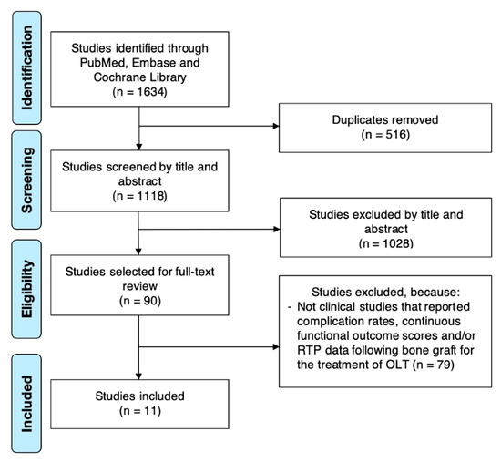
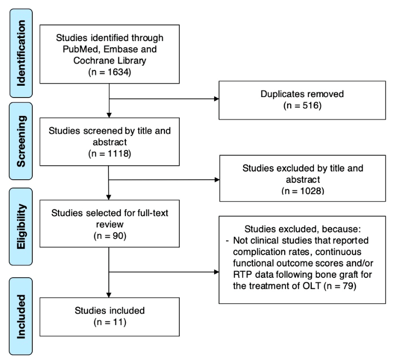
A literature search based on the search strategy yielded 1634 studies for review. Once duplicates were removed, 1118 studies were screened by title and abstract. Subsequently, there were 90 studies selected for full-text review. Only 11 studies met the eligibility criteria and were included in the systematic review (Figure 1) [8,9,10,11,12,21,22,23,24,25,26].
Figure 1.
Preferred Reporting Items for Systematic Reviews and Meta-Analyses Flow Diagram.
3.2. Study and Patient Characteristics
The included studies were published between 2000 and 2021, with ten studies with LoE IV (90.9%) and one with LoE II (9.09%) [8]. The mean QoE was 57.09 ± 7.88 (range 46–71), with one classified as “Good” (9.10%), five classified as “Fair” (45.45%) and five classified as “Poor” (45.45%). Rehabilitation protocols were reported in nine out of eleven studies. Partial weightbearing initiation varied from immediately to 4 weeks postoperatively; range of motion varied from immediately to 6 weeks postoperatively. The progression to full weightbearing was seen the earliest at week 4. The source of the bone graft differed; it was mainly taken from the autologous medial calcaneus, tibia, and iliac crest. Six out of the eleven studies reported the same number of patients pre- and postoperatively with follow-up, with one study observing a loss of two patients [23], and four studies did not report the OLT per protocol [9]. The mean age observed was 37.1 ± 6.6 (range 31.5 to 57.0). Studies predominantly reported medially located lesions with mean lesion sizes of 171.2 ± 109.5 mm2 (range 63.6 to 310.0) and mean follow-up of 35.2 ± 20.0 (range 6.2 to 84.0). A summary of study characteristics and patient demographics is illustrated in Table 1. A summary of study outcomes is illustrated in Table 2.
Table 1.
Study and Patient Characteristics.
Table 2.
Study Outcomes.
3.3. Complications
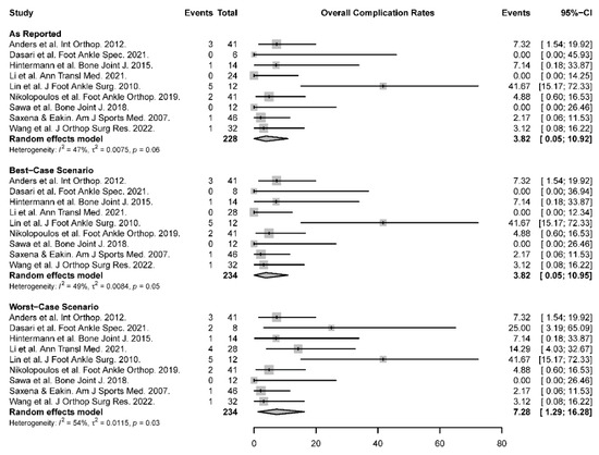
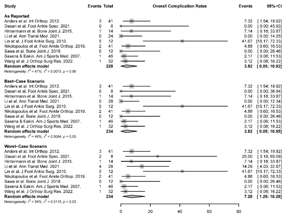
The overall pooled complication rate was 3.82% (95% CI: 0.05 to 10.92). Six out of the nine studies reported the overall complication breakdown. Among the six studies, the revision rate and infection rate were zero, with one case of DVT recorded [22]. One notable complication reported was a case of severe cicatricial diathesis; the scar gradually increased following healing of the surgical incision, resulting in medial soft tissue contraction and limited movement of the foot valgus. The patient received extracorporeal shock wave therapy; however, the ankle mobility was still mildly limited to a mobility angle of 28.1 degrees [11]. Based on the random-effects models in the meta-analysis, the overall complication rate was 3.82% (95% CI: 0.05 to 10.95) in the best-case scenario and 7.28% (95% CI: 1.29 to 16.28) in the worst-case scenario. One of the studies reported two cases of reduced sensation around the wound out of the four cases, and it was excluded from the forest plot as an outlier and will be further elaborated in the discussion [26]. The forest plot of overall complication rates is illustrated in Figure 2. A summary of complications is illustrated in Table 2.
Figure 2.
Forest Plot of Overall Complication Rates [8,9,10,11,12,22,23,24,25].
3.4. Functional Outcome Scores
Functional outcome scores were reported in all eleven studies, with a total of 12 unique scoring systems, including AOFAS, 10 mm VAS pain scores, and other scoring systems that were recorded under “Other Functional Outcome Scores”. The mean American Orthopaedic Foot and Ankle Society (AOFAS) score was 56.0 ± 10.1 (range 40.3 to 67.3) preoperatively to 88.7 ± 5.7 (range 80.8 to 95.0) postoperatively, which was reported in nine studies. The mean 10 mm VAS pain score was 6.5 ± 0.9 (range 5.6 to 7.7) preoperatively to 1.7 ± 1.1 (range 0.4 to 3.7) postoperatively, which was reported in six studies.
3.5. Return to Play and Return to Pre-Injury Level Status
Return to play was discussed in two of the included studies. Hintermann et al. reported a mean time of 5.2 months for the resumption of sports after surgery [25], whilst Saxena recorded the return to play to be 19.6 ± 5.9 weeks for bone grafting and 15.1 ± 4.0 weeks for microfracture [8]. Two other studies mentioned that a return to sport activities was allowed after six months but did not specify the mean time of RTP or return to pre-injury level status [10,12].
4. Discussion
Autologous bone grafting can be employed for larger OLT lesions to minimise donor site morbidity and the relative ease of obtaining the donor graft autologously for larger lesions. This systematic review highlighted the optimistic clinical outcomes produced by autologous bone grafting with tolerable complication rates in short to medium durations for larger OLT lesions as opposed to other treatment strategies. Demonstrable improvement in the functional outcome scores was seen for autologous bone grafting, particularly in the AOFAS and 10 mm VAS pain scores postoperatively. Heterogenous functional outcome scores with varying postoperative follow-up duration were utilised to monitor the durability of autologous bone grafting for larger OLT lesions. A low complication rate was observed from the included studies, with the complications mostly self-limiting. Currently, BMS remains the gold standard of treatment for OLT lesion sizes of less than 150 mm2 [3,27]. No consensus has been reached regarding the gold standard of treatment for OLT lesion sizes of more than 150 mm2. AOT is still the most commonly used technique with the caveat of donor site morbidity. With evidence from this study, autologous bone grafting demonstrated its ability to serve as an alternative treatment for larger OLT lesions, achieving symptomatic relief with favourable clinical outcomes and the possibility of omitting donor site morbidity as one of the major complications. Clinically, autologous bone grafting should be taken into consideration as a viable treatment choice for larger OLTs with evidence of encouraging outcomes from this study.
Best- and worst-case analyses of complications in OLT were performed in previous studies. In a meta-analysis looking into the knee-to-talus donor site morbidity following AOT, the best- and worst-case analyses were 6.7% (95% CI: 2.8 to 11.8), assuming no patients lost to follow-up had donor-site morbidity, and 10.8% (95% CI: 4.8 to 18.3), assuming all patients lost to follow-up experienced donor-site morbidity, respectively, with a short- to medium-term follow-up [18]. In this study, no complications related to donor site morbidity were noted, with an overall complication rate of 3.82% (95% CI: 0.05 to 10.95) in the best-case and 7.28% in the worst-case scenario (95% CI: 1.29 to 16.28). Although no direct comparisons could be made as the outcome of interest varied, the studies have comparable rigorous pooling methods. Autologous bone grafting has demonstrated its advantage of low, self-limiting overall complication rates as opposed to donor site morbidity in AOT, suggesting its superiority in treating OLT with preservation of the donor site.
BMS, on the other hand, was known to produce ideal outcomes for OLT lesion sizes less than 150 mm2 [3,27]. In the study by Ramponi et al., an improvement of the weighted mean AOFAS scores from 62.4 ± 7.9 to 83.9 ± 9.2 was seen over a mean follow-up of 54.1 months. Interestingly, comparable AOFAS scores of 56.0 ± 10.1 (range 40.3 to 67.3) preoperatively to 88.7 ± 5.7 (range 80.8 to 95.0) postoperatively were produced in this study within a shorter mean follow-up period of 35.2 ± 20.0 (range 6.2 to 84.0). Autologous bone grafting, exhibiting a remarkable improvement in the functional outcome score analogous to the gold standard of treatment for lesion sizes less than 107.4 mm2, is a promising indication to adopt the technique in treating larger lesion sizes. Although autologous bone grafting is an alternative treatment method that has not been adequately explored, with only eleven studies appraising its potential, there have been promising outcomes seen in this systematic review, which suggests its comparability to BMS and AOT in terms of a relatively low complication rate and optimistic functional outcome scores within a modest follow-up duration. More comparative studies are indicated to demonstrate the ability of autologous bone grafting for the treatment of larger OLT lesions.
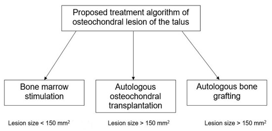
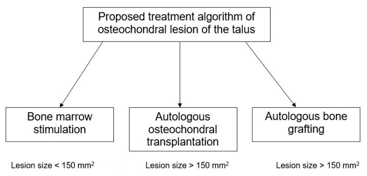
Treatment using AOT poses the risk of donor-site morbidity due to the release of proinflammatory cytokines, resulting in pain as the main symptom [18]. Donor site morbidity, as defined by Shimozono et al. [18], refers to any disability at the knee donor site following AOT surgery for OLT. In a systematic review of 578 AOT procedures that utilised the ipsilateral knee as the donor site, 34.8% of patients experienced complications, of which 4.1% were related to the knee joint. Despite good reported clinical outcomes, the high risk of donor site morbidity in AOT could eventually worsen postoperative clinical outcomes [28]. BMS is currently indicated for lesion sizes < 150 mm2, while AOT is reserved for lesions ≥ 150 mm2 or those that have failed BMS [28,29,30]. While AOT has been employed for lesions up to 249 mm2, overall complication rates of up to 5.3% and revision rates of 71.4% have been reported [31,32]. It nonetheless remains uncertain if larger lesion sizes are a poor prognostic factor of outcomes following AOT, and evidence pertaining to the maximum size of lesions that can be treated by AOT is lacking [33,34]. Within this meta-analysis, autologous bone grafting showed low overall complication and revision rates up to a mean lesion size of 391 mm2, which suggests that autologous bone grafting may be considered for large lesions not amenable to BMS or AOT [9]. While a subgroup analysis to compare outcomes between studies with mean lesion size < 150 mm2 and mean lesion size ≥ 150 mm2 was attempted, there were no significant differences in overall complication rates between subgroups. This could have arisen from the limited number of reported studies pertaining to the use of autologous bone grafting, particularly for various lesion sizes. Studies within the literature were also inconsistent in the reporting of lesion sizes. Therefore, further studies evaluating outcomes following autologous bone grafting are warranted to determine the efficacy of this procedure for varying lesion sizes. This proposed treatment algorithm is outlined in Figure 3.
Figure 3.
Suggested treatment algorithm for OLT [3,27].
OLT injuries are commonly caused by an ankle sprain due to rotational, shear-type forces along with a compression component that causes the talus to slide against the fibula or tibial plafond. With no direct muscle attachments on the talar dome, the cartilage on the talus is known to be 18–37% less stiff than the tibia and thus is more prone to traumatic injuries at the site [35]. Although the current systematic review observed satisfactory outcomes of treatment of larger OLT lesions with autologous bone grafting, the longevity of autologous bone grafting for the treatment of OLT is in question, as the lack of a cartilaginous aspect challenges its physiological integration and robustness. In an autologous bone graft study done on rat models with femoral bone defects, good bone union was seen in autologous cancellous bone grafting at 8 weeks over the control group with no implant, which had no signs of bone fusion. Autologous bone grafts are easily revascularised and could be rapidly incorporated into host sites as the large surface area is covered with dormant and active osteoblasts [7,36]. In addition, the benefits of autologous bone grafting in alleviating immediate symptoms such as pain are evident. Treatment using autologous bone grafting could fill the void and negate the fluid pressure generation without stimulating the highly innervated subchondral bone, thus eliminating OLT-associated pain [11,37]. For OLT treatment using AOT, transplantation of both bone and cartilage is involved, and type-II collagen could be maintained with the potential regeneration of original hyaline articular cartilage at the lesion site [38,39]. Yet, autologous bone grafting lacks “like with like” replacement and may not be as ideal as AOTs in this respect [4]. Subsequently, the lack of the inherent physiological disparity of an osteochondral construct may lead to complications in the long term, translated as symptomatic recurrence that may affect talar function. Further studies with a longer follow-up are suggested to observe the long-term outcomes of autologous bone grafting treatment alongside alleviating immediate symptoms.
There are some limitations to the study. First, the literature search was limited to PubMed, Embase databases, and Cochrane Library, with only full-text studies published in English, which may predispose the study to selection bias. In addition, the heterogenous donor sites and lesion sizes investigated in the eleven studies could be potential confounders affecting the autologous bone graft treatment outcomes. Additionally, the 50% complication rate found by Tanaka et al. was considered as an outlier, where two out of the four patients experienced complications of reduced sensations around the wound [26]. The absurdly high complication rate could be attributed to the small patient cohort of four patients, the highest mean patient age and the largest mean lesion size with large cysts, leading to exclusion of their complication rate from this study. Moreover, we observed the lack of reports on the patients’ sporting background, extending to poor reporting on RTP and return to pre-injury level status, which were noted as OLT is a common result of sporting injury [40]. Only Saxena et al. reported the sporting background of their cohort with fifteen runners and an additional twelve who were involved in either soccer or basketball [8]. RTP was only reported in two out of the eleven studies [8,25]. The modest follow-up duration in this study, furthermore, may not be reflective of the actual potential of autologous bone graft, and its physiological variation may eventually result in deterioration of the functional outcome scores. Future studies are warranted to elicit long-term outcomes of patients who received autologous bone grafting treatment.
5. Conclusions
This current systematic review demonstrated that autologous bone grafting is a satisfactory alternative treatment for OLTs of larger lesion sizes, with notable improvement in the functional outcome scores and low complication rates in the short- to medium-term follow-up. Autologous bone grafting should be considered as the first line of treatment for lesion sizes > 150 mm2, as demonstrated by satisfactory clinical outcomes, low complication rates, and notably eliminating the risk of donor site morbidity. However, due to the lack of cartilaginous aspect, further delineation is required to determine the sustainability of autologous bone grafts with a longer follow-up.
Supplementary Materials
The following supporting information can be downloaded at https://www.mdpi.com/article/10.3390/japma116050058/s1: File S1: the PRISMA checklist [41].
Author Contributions
Conceptualisation, K.H.K.L. and D.S.; data curation, K.H.K.L. and D.S.; methodology, K.H.K.L. and D.S.; Validation, K.H.K.L. and D.S.; formal analysis, K.H.K.L., J.H.K. and D.S.; investigation, K.H.K.L. and D.S.; Writing—original draft preparation, K.H.K.L.; writing—review and editing, K.H.K.L. and D.S.; supervision, D.S.; project administration, D.S. All authors have read and agreed to the published version of the manuscript.
Funding
This research received no external funding.
Institutional Review Board Statement
Not applicable.
Informed Consent Statement
Not applicable.
Data Availability Statement
All data generated or analyzed during this systematic review are included in this published article and its Supplementary Materials. The dataset consists of data extracted from the original primary studies, which are publicly available.
Conflicts of Interest
The authors declare no conflicts of interest.
References
- van Dijk, C.N.; Reilingh, M.L.; Zengerink, M.; van Bergen, C.J. Osteochondral defects in the ankle: Why painful? Knee Surg. Sports Traumatol. Arthrosc. 2010, 18, 570–580. [Google Scholar] [CrossRef] [Scilit] [PubMed]
- Badekas, T.; Takvorian, M.; Souras, N. Treatment principles for osteochondral lesions in foot and ankle. Int. Orthop. 2013, 37, 1697–1706. [Google Scholar] [CrossRef] [Scilit] [PubMed]
- Ramponi, L.; Yasui, Y.; Murawski, C.D.; Ferkel, R.D.; DiGiovanni, C.W.; Kerkhoffs, G.; Calder, J.D.F.; Takao, M.; Vannini, F.; Choi, W.J.; et al. Lesion Size Is a Predictor of Clinical Outcomes After Bone Marrow Stimulation for Osteochondral Lesions of the Talus: A Systematic Review. Am. J. Sports Med. 2017, 45, 1698–1705. [Google Scholar] [CrossRef] [Scilit] [PubMed]
- Kennedy, J.G.; Murawski, C.D. The Treatment of Osteochondral Lesions of the Talus with Autologous Osteochondral Transplantation and Bone Marrow Aspirate Concentrate:Surgical Technique. CARTILAGE 2011, 2, 327–336. [Google Scholar] [CrossRef] [Scilit] [PubMed]
- Chew, K.T.; Tay, E.; Wong, Y.S. Osteochondral lesions of the talus. Ann. Acad. Med. Singap. 2008, 37, 63–68. [Google Scholar] [CrossRef] [Scilit]
- Aspenberg, P.; Van der Vis, H. Migration, particles, and fluid pressure. A discussion of causes of prosthetic loosening. Clin. Orthop. Relat. Res. 1998, 352, 75–80. [Google Scholar]
- Pape, H.C.; Evans, A.; Kobbe, P. Autologous bone graft: Properties and techniques. J. Orthop. Trauma 2010, 24, S36–S40. [Google Scholar] [CrossRef] [Scilit] [PubMed]
- Saxena, A.; Eakin, C. Articular talar injuries in athletes: Results of microfracture and autogenous bone graft. Am. J. Sports Med. 2007, 35, 1680–1687. [Google Scholar] [PubMed]
- Nikolopoulos, D.; Sergides, N.; Safos, G.; Moustakas, K.; Safos, P.; Moutsios-Rentzos, A. Large Osteochondral Lesions of the Talus Treated With Autologous Bone Graft and Periosteum Transfer. Foot Ankle Orthop. 2019, 4, 2473011419874039. [Google Scholar] [CrossRef] [Scilit] [PubMed]
- Anders, S.; Lechler, P.; Rackl, W.; Grifka, J.; Schaumburger, J. Fluoroscopy-guided retrograde core drilling and cancellous bone grafting in osteochondral defects of the talus. Int. Orthop. 2012, 36, 1635–1640. [Google Scholar] [CrossRef] [Scilit] [PubMed]
- Wang, X.; Zhang, D.; Zhang, F.; Jin, L.; Shi, D.; Hou, Z. Effect analysis of iliac bone autografting for Hepple V osteochondral lesions of the talus. J. Orthop. Surg. Res. 2022, 17, 33. [Google Scholar] [CrossRef] [Scilit] [PubMed]
- Sawa, M.; Nakasa, T.; Ikuta, Y.; Yoshikawa, M.; Tsuyuguchi, Y.; Kanemitsu, M.; Ota, Y.; Adachi, N. Outcome of autologous bone grafting with preservation of articular cartilage to treat osteochondral lesions of the talus with large associated subchondral cysts. Bone Jt. J. 2018, 100-B, 590–595. [Google Scholar] [CrossRef] [Scilit] [PubMed]
- Liberati, A.; Altman, D.G.; Tetzlaff, J.; Mulrow, C.; Gotzsche, P.C.; Ioannidis, J.P.; Clarke, M.; Devereaux, P.J.; Kleijnen, J.; Moher, D. The PRISMA statement for reporting systematic reviews and meta-analyses of studies that evaluate health care interventions: Explanation and elaboration. PLoS Med. 2009, 6, e1000100. [Google Scholar] [CrossRef] [Scilit] [PubMed]
- Marx, R.G.; Wilson, S.M.; Swiontkowski, M.F. Updating the assignment of levels of evidence. J. Bone Jt. Surg. Am. 2015, 97, 1–2. [Google Scholar] [CrossRef] [Scilit] [PubMed]
- Coleman, B.D.; Khan, K.M.; Maffulli, N.; Cook, J.L.; Wark, J.D. Studies of surgical outcome after patellar tendinopathy: Clinical significance of methodological deficiencies and guidelines for future studies. Scand. J. Med. Sci. Sports 2000, 10, 2–11. [Google Scholar] [CrossRef] [Scilit] [PubMed]
- Jakobsen, R.B.; Engebretsen, L.; Slauterbeck, J.R. An analysis of the quality of cartilage repair studies. J. Bone Jt. Surg. Am. 2005, 87, 2232–2239. [Google Scholar] [CrossRef] [Scilit] [PubMed]
- Balduzzi, S.; Rücker, G.; Schwarzer, G. How to perform a meta-analysis with R: A practical tutorial. Evid. Based Ment. Health 2019, 22, 153–160. [Google Scholar] [CrossRef] [Scilit] [PubMed]
- Shimozono, Y.; Seow, D.; Yasui, Y.; Fields, K.; Kennedy, J.G. Knee-to-Talus Donor-Site Morbidity Following Autologous Osteochondral Transplantation: A Meta-Analysis with Best-case and Worst-case Analysis. Clin. Orthop. Relat. Res. 2019, 477, 1915–1931. [Google Scholar] [CrossRef] [Scilit] [PubMed]
- Higgins, J.P.; Thompson, S.G.; Deeks, J.J.; Altman, D.G. Measuring inconsistency in meta-analyses. BMJ 2003, 327, 557–560. [Google Scholar] [CrossRef] [Scilit] [PubMed]
- Haidich, A.B. Meta-analysis in medical research. Hippokratia 2010, 14, 29–37. [Google Scholar] [PubMed]
- Draper, S.D.; Fallat, L.M. Autogenous bone grafting for the treatment of talar dome lesions. J. Foot Ankle Surg. 2000, 39, 15–23. [Google Scholar] [CrossRef] [Scilit] [PubMed]
- Lin, J.S.; Andersen, L.B.; Juliano, P.J. Effectiveness of composite bone graft substitute plugs in the treatment of chondral and osteochondral lesions of the talus. J. Foot Ankle Surg. 2010, 49, 224–231. [Google Scholar] [CrossRef] [Scilit] [PubMed]
- Dasari, S.P.; Langer, T.M., 3rd; Parshall, D.; Law, B. Open Treatment of Osteochondral Lesions of the Talus With Bone Grafting and Particulated Juvenile Cartilage Allografting. Foot Ankle Spec. 2021, 17, 39–48. [Google Scholar] [CrossRef] [Scilit] [PubMed]
- Li, S.; Lu, R.; Zhang, J.; Tao, H.; Hua, Y. Outcomes of arthroscopic bone graft transplantation for Hepple stage V osteochondral lesions of the talus. Ann. Transl. Med. 2021, 9, 884. [Google Scholar] [CrossRef] [Scilit] [PubMed]
- Hintermann, B.; Wagener, J.; Knupp, M.; Schweizer, C.; Schaefer, D.J. Treatment of extended osteochondral lesions of the talus with a free vascularised bone graft from the medial condyle of the femur. Bone Jt. J. 2015, 97-B, 1242–1249. [Google Scholar] [CrossRef] [Scilit] [PubMed]
- Tanaka, Y.; Omokawa, S.; Fujii, T.; Kumai, T.; Sugimoto, K.; Takakura, Y. Vascularized bone graft from the medial calcaneus for treatment of large osteochondral lesions of the medial talus. Foot Ankle Int. 2006, 27, 1143–1147. [Google Scholar] [CrossRef] [Scilit] [PubMed]
- Flynn, S.; Ross, K.A.; Hannon, C.P.; Yasui, Y.; Newman, H.; Murawski, C.D.; Deyer, T.W.; Do, H.T.; Kennedy, J.G. Autologous Osteochondral Transplantation for Osteochondral Lesions of the Talus. Foot Ankle Int. 2015, 37, 363–372. [Google Scholar] [CrossRef] [Scilit] [PubMed]
- Ferreira, C.; Vuurberg, G.; Oliveira, J.M.; Espregueira-Mendes, J.; Pereira, H.; Reis, R.L.; Ripoll, P.L. Good clinical outcome after osteochondral autologous transplantation surgery for osteochondral lesions of the talus but at the cost of a high rate of complications: A systematic review. J. ISAKOS 2016, 1, 184–191. [Google Scholar] [CrossRef] [Scilit]
- Zengerink, M.; Struijs, P.A.; Tol, J.L.; van Dijk, C.N. Treatment of osteochondral lesions of the talus: A systematic review. Knee Surg. Sports Traumatol. Arthrosc. 2010, 18, 238–246. [Google Scholar] [CrossRef] [Scilit] [PubMed]
- Choi, W.J.; Park, K.K.; Kim, B.S.; Lee, J.W. Osteochondral lesion of the talus: Is there a critical defect size for poor outcome? Am. J. Sports Med. 2009, 37, 1974–1980. [Google Scholar] [PubMed]
- Nguyen, A.; Ramasamy, A.; Walsh, M.; McMenemy, L.; Calder, J.D.F. Autologous Osteochondral Transplantation for Large Osteochondral Lesions of the Talus Is a Viable Option in an Athletic Population. Am. J. Sports Med. 2019, 47, 3429–3435. [Google Scholar] [CrossRef] [Scilit] [PubMed]
- Winkler, P.W.; Geyer, S.; Walzl, D.; Woertler, K.; Paul, J.; Siebenlist, S.; Imhoff, A.B.; Achtnich, A. Favorable long-term clinical and radiologic outcomes with high survivorship after autologous osteochondral transplantation of the talus. Knee Surg. Sports Traumatol. Arthrosc. 2023, 31, 2166–2173. [Google Scholar] [CrossRef] [Scilit] [PubMed]
- Haleem, A.M.; Ross, K.A.; Smyth, N.A.; Duke, G.L.; Deyer, T.W.; Do, H.T.; Kennedy, J.G. Double-Plug Autologous Osteochondral Transplantation Shows Equal Functional Outcomes Compared With Single-Plug Procedures in Lesions of the Talar Dome: A Minimum 5-Year Clinical Follow-up. Am. J. Sports Med. 2014, 42, 1888–1895. [Google Scholar] [PubMed]
- Kim, Y.S.; Park, E.H.; Kim, Y.C.; Koh, Y.G.; Lee, J.W. Factors Associated With the Clinical Outcomes of the Osteochondral Autograft Transfer System in Osteochondral Lesions of the Talus: Second-Look Arthroscopic Evaluation. Am. J. Sports Med. 2012, 40, 2709–2719. [Google Scholar] [CrossRef] [Scilit] [PubMed]
- Athanasiou, K.A.; Niederauer, G.G.; Schenck, R.C., Jr. Biomechanical topography of human ankle cartilage. Ann. Biomed. Eng. 1995, 23, 697–704. [Google Scholar] [CrossRef] [Scilit] [PubMed]
- Hamada, T.; Matsubara, H.; Hikichi, T.; Shimokawa, K.; Tsuchiya, H. Rat model of an autologous cancellous bone graft. Sci. Rep. 2021, 11, 18001. [Google Scholar] [CrossRef] [Scilit] [PubMed]
- Schmidt, A.H. Autologous bone graft: Is it still the gold standard? Injury 2021, 52, S18–S22. [Google Scholar] [CrossRef] [Scilit] [PubMed]
- Giannini, S.; Buda, R.; Grigolo, B.; Vannini, F. Autologous chondrocyte transplantation in osteochondral lesions of the ankle joint. Foot Ankle Int. 2001, 22, 513–517. [Google Scholar] [CrossRef] [Scilit] [PubMed]
- Krause, F.; Anwander, H. Osteochondral lesion of the talus: Still a problem? EFORT Open Rev. 2022, 7, 337–343. [Google Scholar] [CrossRef] [Scilit] [PubMed]
- Tol, J.L.; Struijs, P.A.; Bossuyt, P.M.; Verhagen, R.A.; van Dijk, C.N. Treatment strategies in osteochondral defects of the talar dome: A systematic review. Foot Ankle Int. 2000, 21, 119–126. [Google Scholar] [CrossRef] [Scilit] [PubMed]
- Page, M.J.; McKenzie, J.E.; Bossuyt, P.M.; Boutron, I.; Hoffmann, T.C.; Mulrow, C.D.; Shamseer, L.; Tetzlaff, J.M.; Akl, E.A.; Brennan, S.E.; et al. The PRISMA 2020 statement: An updated guideline for reporting systematic reviews. BMJ 2021, 372, n71. [Google Scholar] [CrossRef] [Scilit] [PubMed]

Disclaimer/Publisher’s Note: The statements, opinions and data contained in all publications are solely those of the individual author(s) and contributor(s) and not of MDPI and/or the editor(s). MDPI and/or the editor(s) disclaim responsibility for any injury to people or property resulting from any ideas, methods, instructions or products referred to in the content. |
© 2026 by the authors. Published by MDPI on behalf of the American Podiatric Medical Association. Licensee MDPI, Basel, Switzerland. This article is an open access article distributed under the terms and conditions of the Creative Commons Attribution (CC BY) license.