Technical Note

Allograft Tendon Pretensioning Technique Guide
by
Joseph R. Brown
1,* and
Robert W. Mendicino
2
1
Department of Foot and Ankle Surgery, OhioHealth Grant Medical Center, Columbus, OH 43215, USA
2
St. Clair Medical Group Orthopedic Surgery, Pittsburgh, PA 15423, USA
*
Author to whom correspondence should be addressed.
J. Am. Podiatr. Med. Assoc. 2026, 116(5), 59; https://doi.org/10.3390/japma116050059
Submission received: 11 June 2024 / Revised: 1 December 2024 / Accepted: 5 January 2025 / Published: 26 August 2026

Abstract

In foot and ankle surgery, allograft tendons are frequently used in both primary and revision cases to enhance stability and function. However, postoperative complications, such as creep, can compromise the intended correction and stability over time. This paper presents a practical guide on pretensioning techniques prior to implantation, aiming to reduce creep and improve long-term outcomes. Using two common allograft specimens, we demonstrate pretensioning methods with specific focus on tendon stitching, techniques for securing the allograft to the graft board, optimal tensioning and time parameters. We recommend pretensioning at 80 N for approximately 30 min to effectively decrease creep and maintain correction. This technique guide offers foot and ankle surgeons streamlined methods for tendon preparation to improve efficiency and consistency in the operating room.

1. Introduction

Allograft tendons play a critical role in foot and ankle procedures to assist with restoring function and stability. Although the majority of research on tendon tensioning revolves around anterior cruciate ligament (ACL) reconstruction [1,2,3,4,5], there is a growing interest in utilizing allografts in various foot and ankle procedures, such as lateral ankle ligament reconstruction [6,7]. Achilles tendon repair [8,9,10], tibialis anterior tendon repair [11], peroneus longus and brevis repair [12], deltoid ligament reconstruction [13], and flatfoot reconstruction [14]. However, there remains limited literature on how allografts are prepared prior to implantation in foot and ankle surgery.
Scant literature is available in foot and ankle literature on pretensioning protocols. The majority of reports utilizing allograft tendons for reconstruction either did not pretension their allografts or failed to mention tensioning protocols [8,9,10,12,14,15]. In their technique on lateral ankle stabilization, Wang and Xu reported after soaking and trimming the allograft to an appropriate length, they utilized a nonabsorbable #2 braided polyester suture which was secured to each end of the graft. The graft was then pretensioned for 15 min prior to implantation [6]. In contrast, another report on utilizing allografts for lateral ankle stabilization reported they were inconsistent with their pretensioning prior to implantation due to the controversial necessity of this step [7]. Haddad et al. described their technique of fashioning an allograft for deltoid reconstruction. After the tendon ends were tubularized and suture buttons were placed at each end of the tendon, the graft was pretensioned to minimize creep and loosening following reconstruction [13].
Creep, or graft elongation, is a known concern when fixating soft tissue grafts [4]. Loss of tension over time may occur due to stress relaxation or an increase in temperature, such as when grafts are thawed or during implantation into warmer temperatures within the human body [5]. Howard et al. tested 153 grafts and found a mean increase in length of 6.0 mm or 14.0% in their in vivo models, and a mean increase in length of 4.30 mm or 10.12% in their in vitro models. Their findings supported that pretensioning should be performed to eliminate the elastic deformation and minimize graft creep over time [16]. Ciccone et al. reported grafts lose approximately 50% and 80% of tension and stiffness, respectively, following stress relaxation. They recommended keeping tendon grafts warm during preconditioning to prevent the decrease in stiffness and tension [5]. In their biomechanical study, Fujii et al. reported the majority of graft elongation occurred at the stitched portion and recommended focusing on removing any slack during stitching of the graft ends [17]. Yasuda et al. prospectively evaluated outcomes in three groups of ACL reconstruction patients based on initial graft tension (20 N, 40 N, and 80 N). They noted significantly less postoperative laxity in patients who had initial graft tensioning of 80 N. They concluded that there is a significant correlation between the degree of initial graft tensioning and the amount of postoperative laxity and recommended a high tension of 80 N to reduce postoperative laxity [18].
To the best of our knowledge, no allograft tendon pretensioning guides for foot and ankle surgery are described in the literature. Herein, we present techniques and tips to pretension allografts prior to implantation. We hope these simplified technical tips will help to increase surgeon efficiency in the operating room.

2. Surgical Technique

Two allograft specimens, a peroneus longus and semitendinosus tendon, were donated for this technique guide. The graft board utilized in this technique guide was provided by the surgery department at our institution.

2.1. OR Setup

The graft board is placed on the back table or Mayo stand. Multiple tissue clamps and attachment devices are provided within the kit. Attachments can be used interchangeably, depending on surgeon preference (Figure 1).

2.2. Tendon Stitching

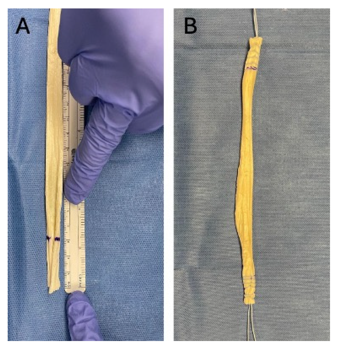
The choice of allograft tendon depends on surgeon preference and may include: semitendinosus, peroneus longus, peroneus brevis, gracilis, tibialis anterior, or Achilles tendons. The allograft should be measured and cut to an appropriate length for the procedure being performed. A sizing device can be utilized prior to securing the tendon to the graft board, or during tension, depending on surgeon preference. Following sizing of the allograft tendon, a whipstitch should be performed at both ends of the tendon. The distal end of each tendon should be marked out utilizing a measuring device. The distal 1.5 to 3.0 cm of the allograft tendon ends should be secured with a suture material in a whipstitch type fashion (Figure 2). Utilizing a suture with a suture button may assist with securing to the tensioner attachment, although this is not necessarily required if sutures are wrapped around and tied securely to the attachment device. Alternatively, surgeons may wish to weave suture material throughout the entirety of the tendon for additional structural support.
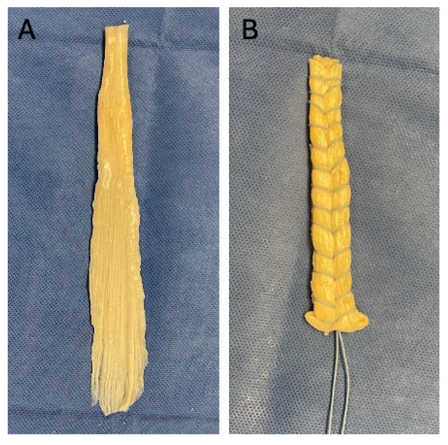
Some tendons, such as the Achilles tendon or semitendinosus, may be flattened at one end of the allograft. If this is the case, consideration should be made for tubularizing the tendon back onto itself and securing the tendon with suture material, again in a whipstitch type fashion (Figure 3).

2.3. Securing Tendon and Tensioning

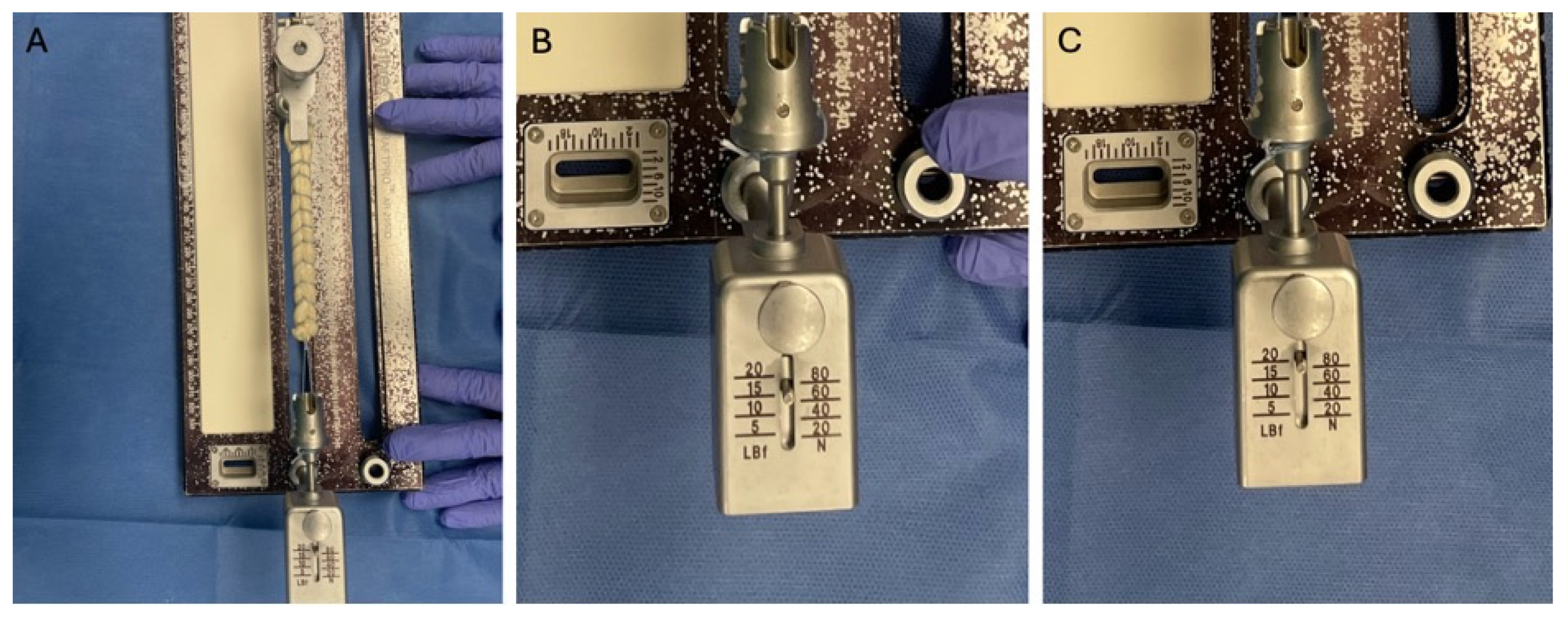
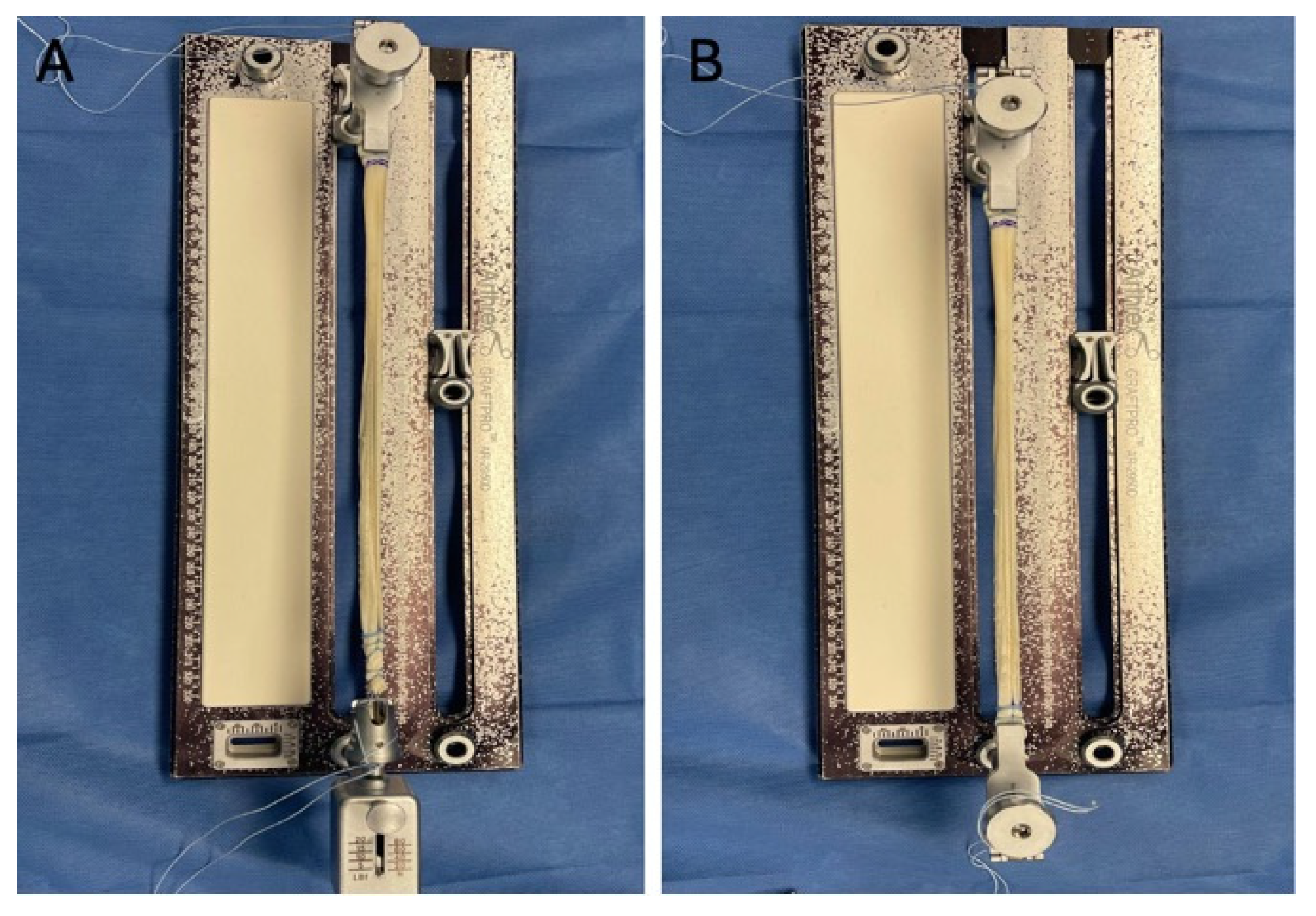
Following suturing, the tendon should be secured to the graft board. There are multiple variations to securing the tendon to the graft board. The tendon can be secured to the tensioner attachment by wrapping the suture around the attachment and utilizing a hand-tie technique. Alternatively, some graft boards allow for a suture button slot to easily attach to the tensioner. One end of the tendon is secured to the tensioner; the opposite end of the tendon can be secured to the clamp attachment. Most of these clamps are atraumatic and provide for an easy loading option of the allograft (Figure 4). The next step is to provide tension to the graft. This is accomplished by pulling the clamp attachment in the opposite direction of the tensioner. The clamp will click into place as the surgeon continues to pull. The amount of tension applied to the graft can be read on the tensioner device. We prefer to apply 80 N of tension to the graft. This tension should be applied for approximately 30 min to minimize the amount of creep. Often, one may see the tensioner device drop below 80 N as the creep occurs. If this happens, you may be required to re-tension several times until the elongation is complete and the tension is constant (Figure 5).
An alternative technique to securing the tendon allograft is to utilize two clamp attachments, one for each end of the tendon. This allows the surgeon to easily secure the tendon to the graft board. Again, the surgeon should pull the clamp attachment along the graft board sliders. It is important to note that the amount of tension applied will not be recorded with this technique.
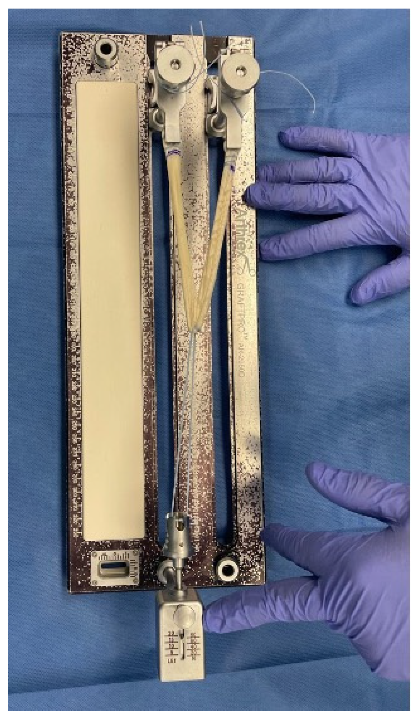
Some grafts may need to be implanted in a V- or Y-shaped pattern. If this is the case, there are two techniques the surgeon can utilize to simultaneously tension both limbs of the graft. First, the graft should be measured and marked at the central point. If the graft board has dual tracks, two clamp attachments should be inserted into the sliders. Each end of the graft should be placed into the clamp attachments. At the central point of the graft, a wide-based suture material, such as FiberWire (Arthrex, Naples, FL, USA) or Ethibond (Johnson & Johnson, New Brunswick, NJ, USA), should be wrapped around and secured to the tensioning or clamp attachment, as described above (Figure 6). Alternatively, if the graft board kit comes with a post attachment, this may be utilized on one of the slide tracks to wrap the graft around prior to securing to the tensioner or clamp attachment.
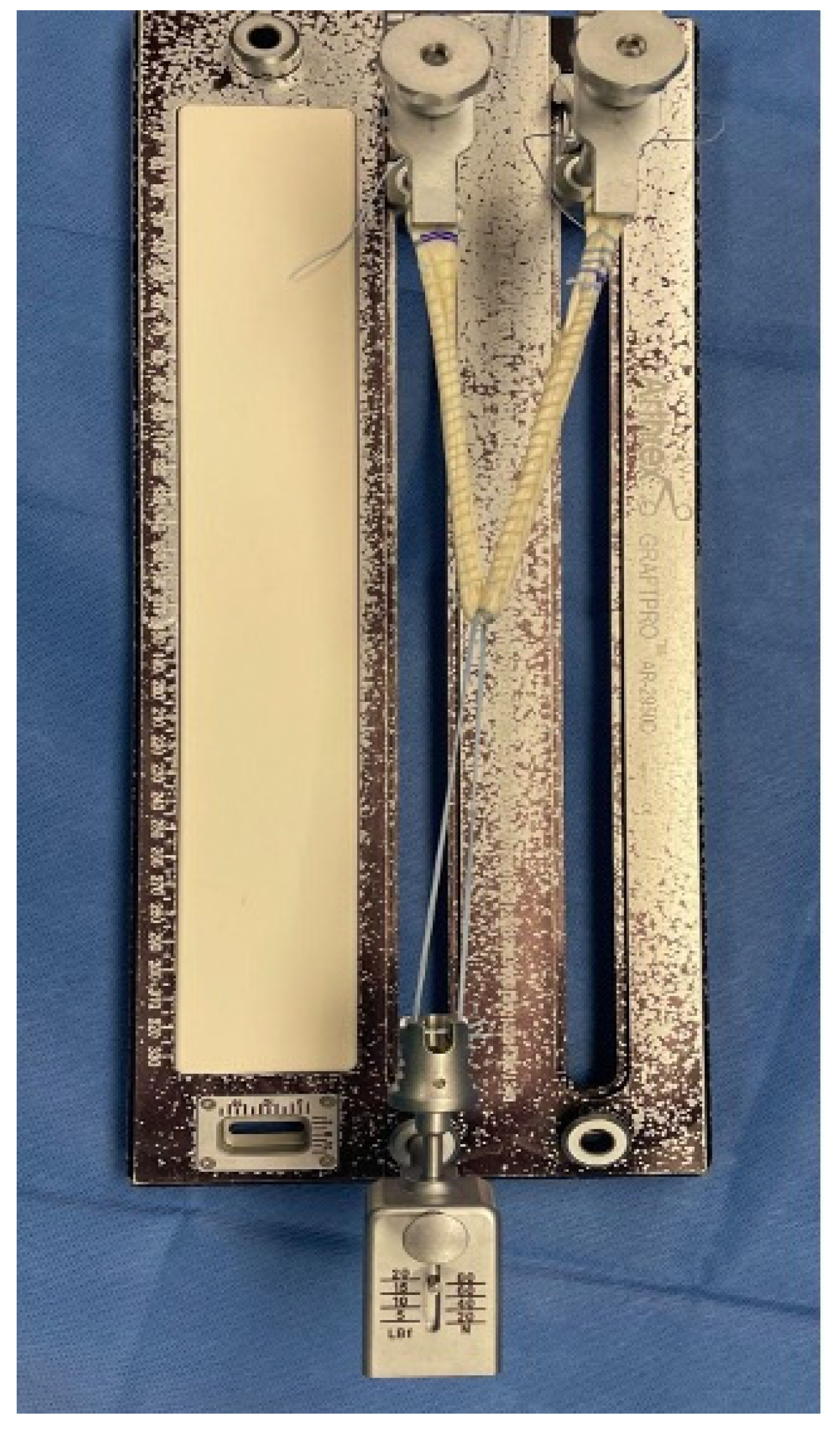
During the tensioning procedure, surgeons should consider performing a continuous running type suture to provide additional structural support and ideally maintain the tensioned positioning of the allograft. We prefer to utilize a stronger suture material, such as Fiberwire or Ethibond, for this step (Figure 7). Following the tensioning protocol, tendons are prepared for implantation and may be utilized for a variety of procedures.

3. Discussion

Foot and ankle surgeons commonly utilize allograft tendons during reconstructive procedures to address various pathologies. However, minimal data is available on how allografts are prepared prior to implantation in the foot and ankle literature. A stepwise technique guide on pretensioning is beneficial to decrease the amount of creep and to maintain the tension and correction over time.
Creep is a well-documented issue in soft tissue grafts due to the inherent viscoelastic properties of tendon tissue and is a concern for elongation over time [4,13]. In their cadaveric study, Pilia et al. [1] reported that pretensioning or preconditioning resulted in increased residual tension and significantly higher retained tension over time than either technique alone. They recommended surgeons precondition and pretension tendons prior to final tibial fixation during ACL reconstruction. In their systematic review, Jisa et al. [4] found the majority of studies utilized a pretensioning or preconditioning protocol around 80 N. Although there are potential benefits of pretensioning and preconditioning, including decreased graft elongation and greater residual tension over time, no consensus on an optimal tensioning protocol has been reported. Furthermore, the necessity of pretensioning allografts prior to implementation has been questioned. Nurmi et al. [19] compared no preconditioning to cyclic and isometric preconditioning protocols. They found a decrease in tension over time and questioned the applicability of preconditioning protocols as they were unable to eliminate the intrinsic viscoelasticity of soft tissue grafts. We believe there is benefit to pretensioning or preconditioning to decrease the amount of creep and elongation, which can ultimately compromise the end surgical result over time. Additionally, there are no studies that show a harmful result from pretensioning grafts.
The amount of tension applied and length of tensioning time are two important factors to consider for a tensioning protocol. Tensioning protocols in the literature have ranged from tensions of 20 to 88 N for between 10 and 30 min [5,6,19,20,21,22,23,24]. Koga et al. [20] evaluated the minimum amount of tension required for restoring knee stability in ACL reconstruction. They recommended that a minimum tension of 25 N was required. Cunningham et al. [25] reported that 80 N of force is within the range that a surgeon can physically apply during hamstring tendon preparation for ACL reconstruction. We believe this amount of force is reasonable and corresponds to the maximum force on most tensioner attachments. In our protocol, we attempt to maintain this amount of force over a 30 min period.
Some studies have evaluated whether to pretension (on the graft board) or precondition (after initial fixation of single end). Pilia et al. [1] performed tensile mechanical testing utilizing four different pretensioning and/or preconditioning protocols. They utilized tendons with an initial 80 N pull for tensioning as a control. Other groups utilized 88 N of pretensioning for 20 min and/or cyclic tensioning for 20 cycles at 10–80 N. Although all groups experienced elongation, the pretensioning and cyclic tensioning groups showed greater stiffness than the control. Their results show that pretensioning or preconditioning provides higher residual tension and that combining techniques provides more retained tension than each individual technique. They recommended against simply pulling the graft prior to fixation, as this may lead to a higher likelihood of failure. They recommended that surgeons should precondition prior to implantation, or pretension prior to final fixation, to maintain tension in the graft. In foot and ankle reconstruction, pretensioning is a more ideal approach and has been briefly described by Haddad et al. [13] for deltoid reconstruction. Our protocol allows foot and ankle surgeons to easily follow a stepwise approach to pretension grafts prior to implantation.
In conclusion, foot and ankle surgeons should be aware of tensioning protocols when using allografts for reconstruction. By following the techniques outlined in this guide, surgeons can reduce the risk of postoperative graft creep and optimize the stability and functionality of their reconstructions. We recommend applying a consistent tension of 80 N for 30 min. This guide bridges a critical gap in the literature by providing practical and reproducible methods for allograft preparation by simplifying pretensioning techniques into a stepwise approach that can be incorporated prior to implantation. Future studies should focus on the clinical outcomes of pretensioning allografts in foot and ankle reconstructions.

Author Contributions

Conceptualization, J.R.B. and R.W.M.; methodology, J.R.B. and R.W.M.; software, J.R.B.; validation, J.R.B. and R.W.M.; formal analysis, J.R.B.; investigation, J.R.B. and R.W.M.; resources, J.R.B.; data curation, J.R.B. and R.W.M.; writing—original draft preparation, J.R.B.; writing—review and editing, R.W.M.; visualization, J.R.B. and R.W.M.; supervision, R.W.M.; project administration, J.R.B. and R.W.M. All authors have read and agreed to the published version of the manuscript.

Funding

This research received no external funding.

Institutional Review Board Statement

Not applicable.

Informed Consent Statement

Not applicable.

Data Availability Statement

The raw data supporting the conclusions of this article will be made available by the authors on request.

Conflicts of Interest

The authors declare no conflicts of interest.

References

  1. Pilia, M.; Murray, M.; Guda, T.; Heckman, M.; Appleford, M. Pretensioning of Soft Tissue Grafts in Anterior Cruciate Ligament Reconstruction. Orthopedics 2015, 38, e582–e587. [Google Scholar] [CrossRef] [Scilit] [PubMed]
  2. Cruz, A.I., Jr.; Fabricant, P.D.; Seeley, M.A.; Ganley, T.J.; Lawrence, J.T. Change in Size of Hamstring Grafts During Preparation for ACL Reconstruction: Effect of Tension and Circumferential Compression on Graft Diameter. J. Bone Jt. Surg. Am. 2016, 98, 484–489. [Google Scholar] [CrossRef] [Scilit] [PubMed]
  3. Arneja, S.; McConkey, M.O.; Mulpuri, K.; Chin, P.; Gilbart, M.K.; Regan, W.D.; Leith, J.M. Graft tensioning in anterior cruciate ligament reconstruction: A systematic review of randomized controlled trials. Arthroscopy 2009, 25, 200–207. [Google Scholar] [PubMed]
  4. Jisa, K.A.; Williams, B.T.; Jaglowski, J.R.; Turnbull, T.L.; LaPrade, R.F.; Wijdicks, C.A. Lack of consensus regarding pretensioning and preconditioning protocols for soft tissue graft reconstruction of the anterior cruciate ligament. Knee Surg. Sports Traumatol. Arthrosc. 2016, 24, 2884–2891. [Google Scholar] [PubMed]
  5. Ciccone, W.J.; Bratton, D.R.; Weinstein, D.M.; Elias, J.J. Viscoelasticity and temperature variations decrease tension and stiffness of hamstring tendon grafts following anterior cruciate ligament reconstruction. J. Bone Jt. Surg. Am. 2006, 88, 1071–1078. [Google Scholar] [CrossRef] [Scilit]
  6. Wang, W.; Xu, G.H. Allograft tendon reconstruction of the anterior talofibular ligament and calcaneofibular Ligament in the treatment of chronic ankle instability. BMC Musculoskelet. Disord. 2017, 18, 150. [Google Scholar] [CrossRef] [Scilit] [PubMed]
  7. Miller, A.G.; Raikin, S.M.; Ahmad, J. Near-anatomic allograft tenodesis of chronic lateral ankle instability. Foot Ankle Int. 2013, 34, 1501–1507. [Google Scholar] [CrossRef] [Scilit] [PubMed]
  8. Huang, X.; Huang, G.; Ji, Y.; Ao Rg Yu, B.; Zhu, Y.L. Augmented Repair of Acute Achilles Tendon Rupture Using an Allograft Tendon Weaving Technique. J. Foot Ankle Surg. 2015, 54, 1004–1009. [Google Scholar] [CrossRef] [Scilit] [PubMed]
  9. Benny, A.; Balg, F.; Svotelis, A.; Vézina, F. Reconstruction of Overlengthening After Gastrocnemius Recession with an Achilles Tendon Allograft: Case Report. Foot Ankle Int. 2016, 37, 1249–1254. [Google Scholar] [CrossRef] [Scilit] [PubMed]
  10. So, E.; Consul, D.; Holmes, T. Achilles Tendon Reconstruction with Bone Block Allograft:Long-Term Follow-Up of Two Cases. J. Foot Ankle Surg. 2019, 58, 779–784. [Google Scholar] [CrossRef] [Scilit] [PubMed]
  11. Burton, A.; Aydogan, U. Repair of Chronic Tibialis Anterior Tendon Rupture with a Major Defect Using Gracilis Allograft. Foot Ankle Spec. 2016, 9, 345–350. [Google Scholar] [CrossRef] [Scilit] [PubMed]
  12. Pellegrini, M.J.; Adams, S.B.; Parekh, S.G. Reversal of Peroneal Tenodesis with Allograft Reconstruction of the Peroneus Brevis and Longus: Case Report and Surgical Technique. Foot Ankle Spec. 2014, 7, 327–331. [Google Scholar] [CrossRef] [Scilit] [PubMed]
  13. Haddad, S.L.; Dedhia, S.; Ren, Y.; Rotstein, J.; Zhang, L.Q. Deltoid ligament reconstruction: A novel technique with biomechanical analysis. Foot Ankle Int. 2010, 31, 639–651. [Google Scholar] [CrossRef] [Scilit] [PubMed]
  14. Dominick, D.R.; Catanzariti, A.R. Posterior Tibial Tendon Allograft Reconstruction for Stage II Adult Acquired Flatfoot: A Case Series. J. Foot Ankle Surg. 2020, 59, 821–825. [Google Scholar] [CrossRef] [Scilit] [PubMed]
  15. Diniz, P.; Pacheco, J.; Flora, M.; Quintero, D.; Stufkens, S.; Kerkhoffs, G.; Batista, J.; Karlsson, J.; Pereira, H. Clinical applications of allografts in foot and ankle surgery. Knee Surg. Sports Traumatol. Arthrosc. 2019, 27, 1847–1872. [Google Scholar] [CrossRef] [Scilit] [PubMed]
  16. Howard, M.E.; Cawley, P.W.; Losse, G.M.; Johnston, R.B. Bone-patellar tendon-bone grafts for anterior cruciate ligament reconstruction: The effects of graft pretensioning. Arthroscopy 1996, 12, 287–292. [Google Scholar] [CrossRef] [Scilit] [PubMed]
  17. Fujii, M.; Furumatsu, T.; Miyazawa, S.; Tanaka, T.; Inoue, H.; Kodama, Y.; Masuda, K.; Seno, N.; Ozaki, T. Features of human autologous hamstring graft elongation after pre-tensioning in anterior cruciate ligament reconstruction. Int. Orthop. 2016, 40, 2553–2558. [Google Scholar] [CrossRef] [Scilit] [PubMed]
  18. Yasuda, K.; Tsujino, J.; Tanabe, Y.; Kaneda, K. Effects of initial graft tension on clinical outcome after anterior cruciate ligament reconstruction. Autogenous doubled hamstring tendons connected in series with polyester tapes. Am. J. Sports Med. 1997, 25, 99–106. [Google Scholar] [PubMed]
  19. Nurmi, J.T.; Kannus, P.; Sievänen, H.; Järvelä, T.; Järvinen, M.; Järvinen, T.L. Interference screw fixation of soft tissue grafts in anterior cruciate ligament reconstruction: Part 2: Effect of preconditioning on graft tension during and after screw insertion. Am. J. Sports Med. 2004, 32, 418–424. [Google Scholar] [PubMed]
  20. Koga, H.; Muneta, T.; Yagishita, K.; Watanabe, T.; Mochizuki, T.; Horie, M.; Nakamura, T.; Otabe, K.; Sekiya, I. Effect of Initial Graft Tension on Knee Stability and Graft Tension Pattern in Double-Bundle Anterior Cruciate Ligament Reconstruction. Arthroscopy 2015, 31, 1756–1763. [Google Scholar] [CrossRef] [Scilit] [PubMed]
  21. Edgar, C.M.; Zimmer, S.; Kakar, S.; Jones, H.; Schepsis, A.A. Prospective comparison of auto and allograft hamstring tendon constructs for ACL reconstruction. Clin. Orthop. Relat. Res. 2008, 466, 2238–2246. [Google Scholar] [CrossRef] [Scilit] [PubMed]
  22. Austin, J.C.; Phornphutkul, C.; Wojtys, E.M. Loss of knee extension after anterior cruciate ligament reconstruction: Effects of knee position and graft tensioning. J. Bone Jt. Surg. Am. 2007, 89, 1565–1574. [Google Scholar]
  23. Brady, M.F.; Bradley, M.P.; Fleming, B.C.; Fadale, P.D.; Hulstyn, M.J.; Banerjee, R. Effects of initial graft tension on the tibiofemoral compressive forces and joint position after anterior cruciate ligament reconstruction. Am. J. Sports Med. 2007, 35, 395–403. [Google Scholar] [CrossRef] [Scilit] [PubMed]
  24. Kuhn, M.A.; Ross, G. Allografts in the treatment of anterior cruciate ligament injuries. Sports Med. Arthrosc. Rev. 2007, 15, 133–138. [Google Scholar] [CrossRef] [Scilit] [PubMed]
  25. Cunningham, R.; West, J.R.; Greis, P.E.; Burks, R.T. A survey of the tension applied to a doubled hamstring tendon graft for reconstruction of the anterior cruciate ligament. Arthroscopy 2002, 18, 983–988. [Google Scholar] [CrossRef] [Scilit] [PubMed]
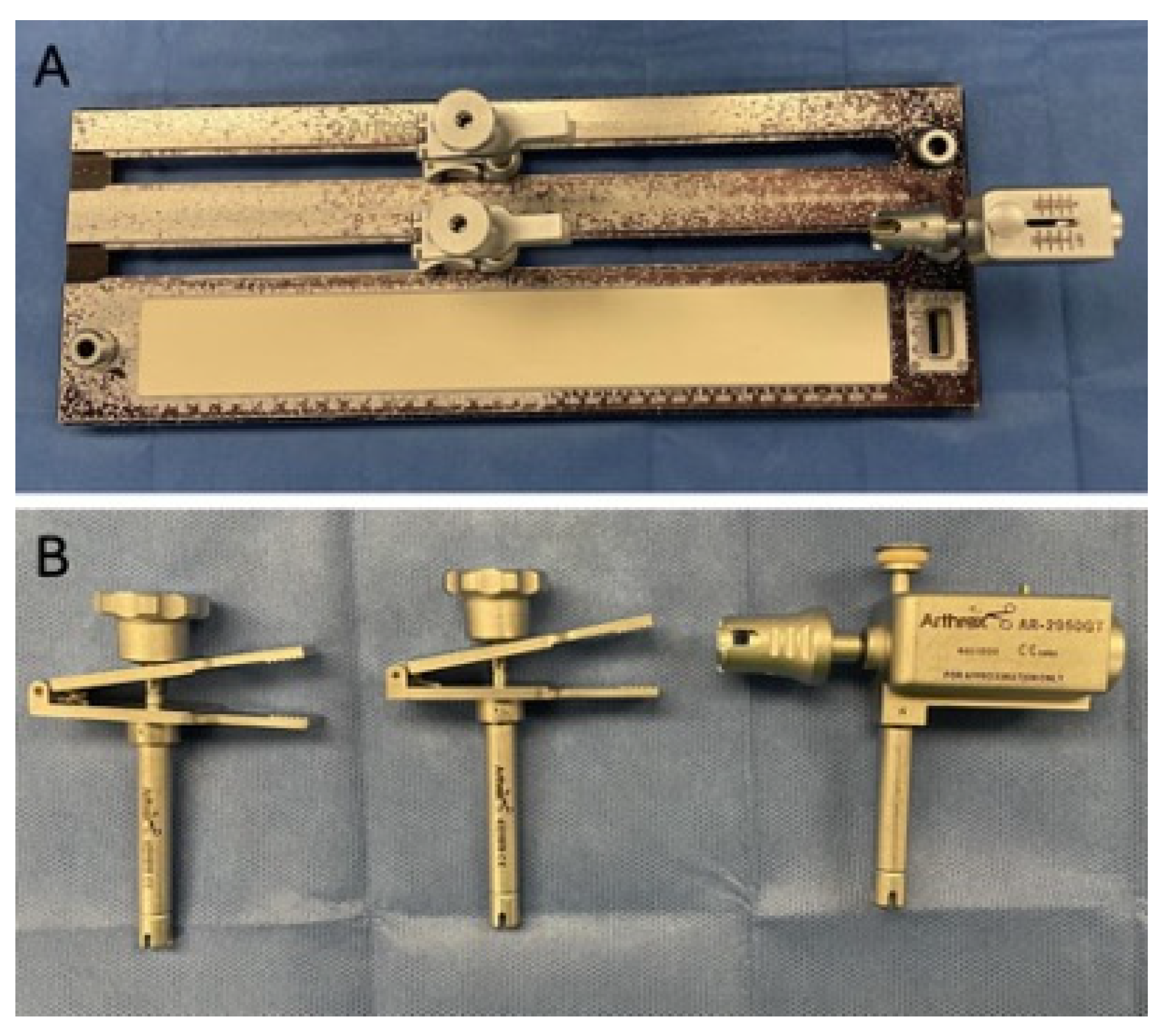
Figure 1. (A) Graft tensioning board. (B) Clamp and tensioner attachments.
Japma 116 00059 g001
Figure 2. Stitching tendon allograft ends. (A) Marking out 1.5–3.0 cm from the distal tip of each end of the tendon. (B) Whipstitch secured to the distal 1.5–3.0 cm of tendon ends.
Japma 116 00059 g002
Figure 3. Tubularizing tendon. (A) Semitendinosus allograft with flattened end. (B) The same semitendinosus allograft tubularized and reinforced with whipstitch.
Japma 116 00059 g003
Figure 4. Securing graft to graft board. (A) Tendon secured to tensioner and clamp attachments. (B) Tendon secured at both ends to clamp attachments.
Japma 116 00059 g004
Figure 5. Tensioning of graft. (A) Tendon from Figure 3 secured to tensioner and clamp attachments. Note the tensioner reads 80 N of force. (B) One can appreciate the loss in tension from 80 N to 60 N over a period of time. (C) Re-tensioning to 80 N of force during the course of graft preparation.
Japma 116 00059 g005
Figure 6. Securing tendon to graft board to simultaneously tension both limbs. Note the clamps are equidistant from the tensioner attachment, which is at 80 N of force.
Japma 116 00059 g006
Figure 7. The same tendon from Figure 5 with both limbs reinforced with a continuous running suture while under 80 N of tension. Note that this reinforcement technique under tension can be performed with a single limb tendon.
Japma 116 00059 g007
Disclaimer/Publisher’s Note: The statements, opinions and data contained in all publications are solely those of the individual author(s) and contributor(s) and not of MDPI and/or the editor(s). MDPI and/or the editor(s) disclaim responsibility for any injury to people or property resulting from any ideas, methods, instructions or products referred to in the content.