


Abstract
Background: We evaluated patients who were followed after acutely developing single-sided footdrop and improving with conservative management or spontaneously. Methods: In 2019 and 2020, ten patients were retrospectively evaluated for unilateral weakness of the lower extremity in the form of absent dorsiflexion at the ankle joint and were given a diagnosis of footdrop without etiologic cause. Patients were followed for 18 months. Patients were evaluated for acute footdrop of the affected extremity with electromyography, lumbar spine magnetic resonance imaging (MRI), knee MRI, peripheral MRI neurography, and noncontrast brain MRI. Each patient was evaluated for a history of COVID-19 infection during the past year. Patients with any identified cause were excluded. Results: Initial evaluation of muscle strength revealed 0/5 by the Medical Research Council muscle testing scale. In two patients, muscle strength was 3/5 at month 6 and in eight patients it was 4/5. Muscle strength of all of the patients improved to 5/5 at 1 year. Six patients were dispensed an ankle-foot orthosis, and nine patients performed physical therapy. Electromyography identified significant neuropathy at the level of the common peroneal at the fibular head in all of the patients. Compared with peroneal nerve stimulation below and above the fibular head in the lateral popliteal fossa, a 50% reduction in sensory amplitude and motor conduction slowing greater than 10 m/sec were present. Knee MRI revealed no masses, edema, or anatomical variations at the level of the fibular head. Conclusions: Spontaneous resolution of unilateral acute footdrop without an etiologic cause can occur within 1 year.
Unilateral footdrop is a condition that causes difficulty in walking, often due to peripheral causes. It occurs with loss of dorsiflexion of the ankle. In the etiology of footdrop, there are many causes in addition to fibular head entrapment neuropathy of the peroneal nerve, including L5 radiculopathy and upper motor neuron diseases. There are traumatic causes such as sciatic nerve lesions, polyneuropathy, lumbar plexus pathologies, penetrating injuries, space-occupying lesions, and external compressions that can develop due to tight knee bandages, leg crossing, or prolonged squatting.[1] Spontaneous footdrop is most commonly seen due to peroneal neuropathy caused by compression of the nerve at the level of the fibula head, where it is covered only by skin and subcutaneous tissue.[2] Gait problems, leg pain, and muscle strength and sensory deficits may occur with acute or chronic compression, stretching, or trauma on the peroneal nerve before it divides into superficial and deep branches.[3] In the present study, we showed that patients who were evaluated in an orthopedic outpatient clinic for footdrop and whose etiology could not be identified after conducting an evaluation and testing of the peroneal neuropathy would completely recover with nonsurgical treatments.
Patients and Methods
The retrospective study was approved by the institutional review board of Prof. Dr. Cemil Taşcıoğlu City Hospital (Istanbul, Turkey) and was performed under the ethical standards laid out in the Declaration of Helsinki.
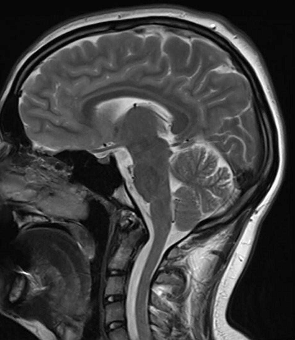
Between May 17, 2019, and June 20, 2020, ten patients were included in the study for the complaint of inability to raise the front part of the foot whose tibialis anterior muscled strength was determined as 0/5 by the Medical Research Council muscle testing scale.[4] These patients were aged 5 to 53 years (mean age, 23 years). Three patients were female and seven were male. The right side was affected in all of the patients. Four patients had a history of long-term drug use (Table 1). The patients were followed up for at least 18 months. Peripheral neuropathy was detected on electromyography (EMG) after acute footdrop in all of the patients. There was no pathology detected at the L5 plexus in the lumbar region magnetic resonance images (MRIs) (Fig. 1). No pathology was detected in the region of the fibular head of the fibular nerve in the knee MRIs of the affected extremity, and the morphology of the biceps muscle was normal throughout the course of the fibula (Fig. 2). Orthoroentgenography was performed in all of the patients, deformity analysis was performed, and no angular pathology was found (Fig. 3). All of the patients were evaluated for acute footdrop with EMG, lumbar and knee MRI, peripheral MRI neurography of the affected extremity (Fig. 4), and noncontrast brain MRI (Fig. 5). To identify the etiology, the posture and sitting positions of the patients were examined, and no pathology was detected. Detailed anamnesis was questioned, and no diagnosed autoimmune or metabolic disease was detected. Patients were checked for a history of COVID infection by looking at COVID-19 polymerase chain reaction results during the past 1 year. In all of the patients, the history of trauma was questioned and excluded. The use of an ankle-foot orthosis and the process of physical therapy rehabilitation were evaluated. Patients with an identified cause, such as pathologies pressing on the fibular nerve or cranial or lumbar pathology, and those who had surgical treatment as the cause were excluded from the study.

Table 1.
Data of the Ten Study Participants
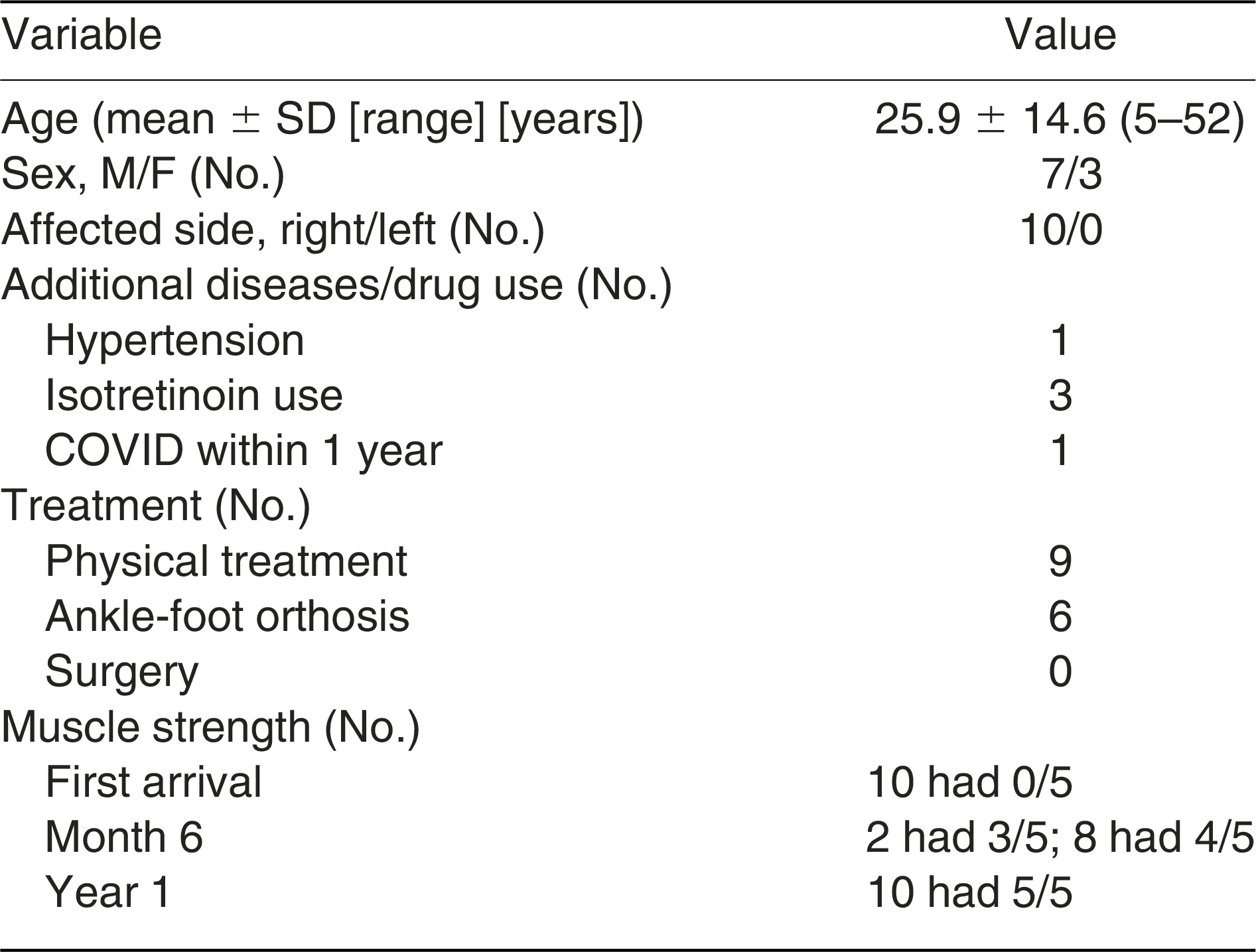
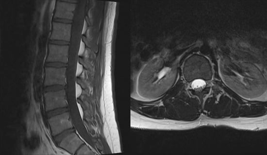
Figure 1.
Lumbar magnetic resonance images in the sagittal and axial sections indicate that there is no pathology at the L5 plexus.
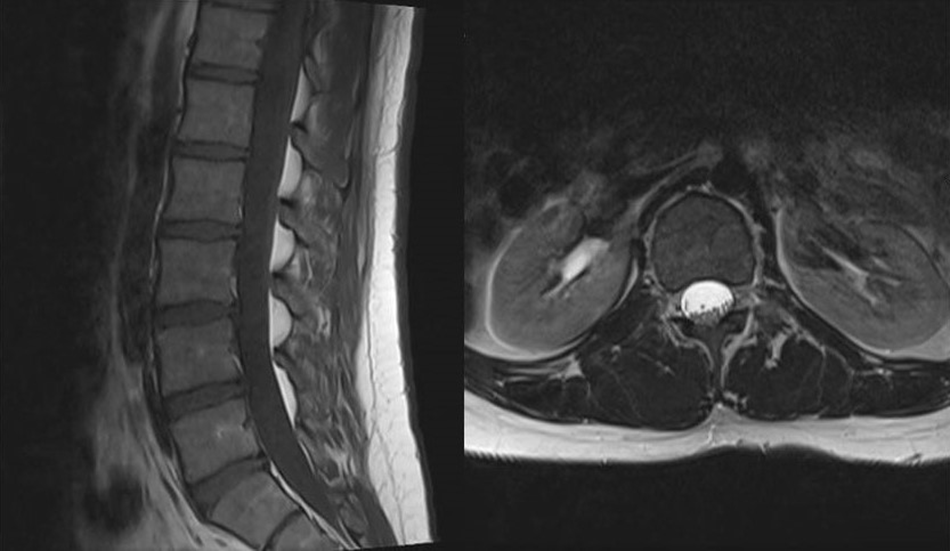
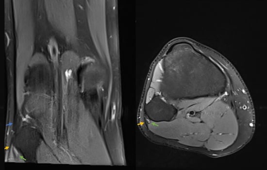
Figure 2.
Knee magnetic resonance images show that the common peroneal (fibular) nerve coursing through the popliteal fossa toward naturally the head of the fibula (green arrows), the normal anatomy of the fibular insertion of the biceps femoris tendon (blue arrow), and the origin of the peroneus longus tendon, which forms the fibular tunnel, did not cause compression or irritation (yellow arrows).
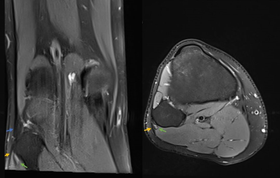
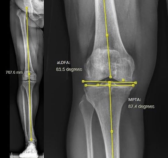
Figure 3.
Lower-extremity deformity analysis evaluated as normal.
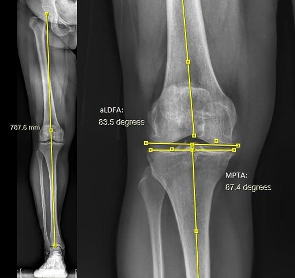
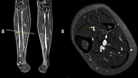
Figure 4.
Peripheral magnetic resonance neurography scan shows that there are no pathologies such as impingement or edema along the peroneal nerve trace. The deep peroneal nerve is indicated by yellow arrows and the posterior tibial nerve is indicated by green arrows.
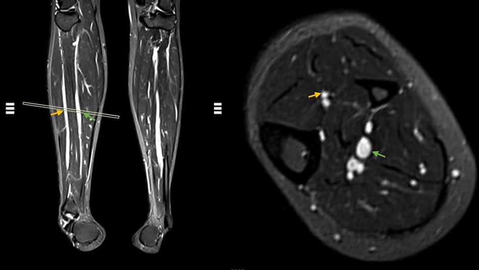
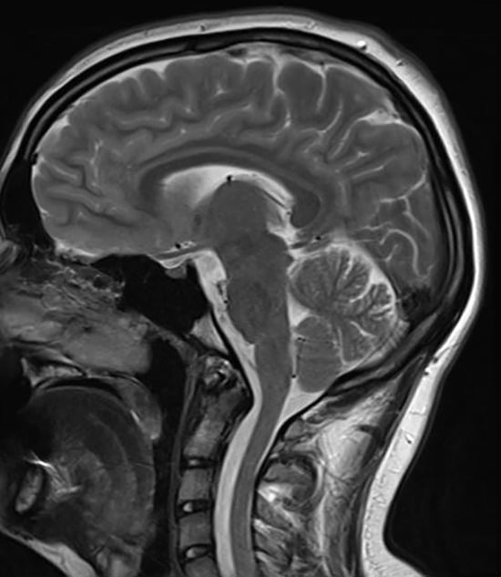
Figure 5.
Cranial magnetic resonance image evaluated as normal.
Results
The muscle strength at the initial application of all of the patients was 0/5 according to the Medical Research Council muscle testing scale. In two patients, the muscle strength was 3/5 at month 6, and in the other eight patients it was 4/5 at month 6. The muscle strength improved to 5/5 in 1 year at the latest. Three patients were using isorethionine, a vitamin A derivative, for acne treatment. One patient was under treatment for hypertension. An ankle-foot orthosis was applied to six patients, and nine patients underwent physical therapy. One patient rejected physical therapy, but muscle strength improved spontaneously at month 6. Vitamin B and nonsteroidal anti-inflammatory drug (NSAID) treatment were given to nine of the patients. Evaluation of EMG results identified significant neuropathy at the level of the common peroneal at the fibular head in all of the patients. Compared with peroneal nerve stimulation below the fibular head (BFH) and above the fibular head (AFH) in the lateral popliteal fossa, a 50% reduction in sensory amplitude (BFH: mean ± SD: 4.9 ± 0.9 μV and AFH: 18.3 ± 2.32 μV) and motor conduction slowing greater than 10 m/sec (BFH: mean ± SD: 33.1 ± 2.79 m/sec and AFH: 59.3 ± 6.92 m/sec) were present. Electrodiagnostic data collected included deep fibular nerve conduction block/slowing, extensor digitorum brevis/tibialis anterior compound muscle action potential amplitude and axon loss estimate, superficial fibular nerve sensory nerve action potential amplitude and axon loss estimate, and needle EMG neurogenic changes in the muscles supplied by the deep versus superficial branches of the common fibular nerve. In all of the patients, normal knee MRI findings for the peroneal nerve were observed at the level of the head of the fibula, and no mass that would cause any compression or edema was found. No anatomical variations were detected in the knee MRIs of the patients. Knee MRI identified three of the patients with meniscal degeneration and edema of the anterior cruciate ligament fibers. Nine of the patients were diagnosed as having a COVID infection during the past year. The tenth patient, who was 5 years old, did not have a polymerase chain reaction performed; however, his family members were diagnosed as having COVID-19 during the past year. Patients using isotretinoin were found to have mildly elevated liver enzyme levels. After performing a thorough physical and diagnostic evaluation of each patient for unilateral footdrop, no etiologic cause could be identified other than each patient had been exposed to the COVID-19 virus. These patients continued their social lives during follow-up.
The EMG was not repeated, and no additional radiologic investigations were performed on the patients, whose muscle strength improved. During follow-up, the patients did not develop new foot complaints.
Footdrop Rehabilitation
Patients were treated conservatively. The main goals of treatment were to have a pain-free, stable foot that can dorsiflex voluntarily in a functional gait cycle.
All of the patients were informed about the prognosis and management of their individual condition. Patients in the acute phase were advised about precautions such as avoiding leg crossing, kneeling, and squatting. Patients with acute injury were immobilized and treated with oral or local NSAIDs. Lifestyle modifications were encouraged to reduce the risk of falling. Flat shoes with ankle support or an ankle-foot orthosis was prescribed depending on the patient’s muscle strength.
All of the patients were referred to physiotherapy and rehabilitation. For patients with low muscle strength, neuromuscular electrical stimulation was administered to prevent muscle atrophy. The rehabilitation program consisted of active or passive daily range of movement and stretching exercises for the ankle to prevent development of contractures, and rigidity isometric or resistive strengthening exercises were instructed. An experienced physiotherapist provided gait and stair training to each patient to achieve safe ambulation. Some of the patients were dispensed a cane to help with ambulation and stability.
Discussion
Throughout the literature, several authors have identified many etiologic causes of footdrop. Upton and McComas proposed the theory of double crush in nerve entrapment syndromes in 1973, and this proposal is still supported by many authors.[5,6] So far, this theory of double crushing is still supported by several authors.[6] However, experience from clinical practice reveals that many patients have additional or competing causes for footdrop.[7] Weakness or loss of dorsiflexion at the ankle is known as footdrop. This may be caused by upper or lower motor neuron deficits. It is most commonly caused by lower motor neuron deficits and mostly due to peroneal nerve neuropathy. Common peroneal nerve compression around the head of the fibula is the most common site of peroneal nerve injury. Other causes of lower motor neuropathy that cause footdrop include sciatic mononeuropathy, lumbosacral plexopathy, polyneuropathy, or severe L5 radiculopathy.[8] Cases of footdrop secondary to ischemia after squatting or crossing legs have been reported in the literature, and some of them were treated with conservative treatment. Pigott and Jefferson[9] also reported idiopathic cases that did not improve in 2 years of follow-up. In the present study, muscle strength was 4/5 in the 6-month period, and muscle strength returned to previous function after 1 year. Contrary to the literature, all of the patients recovered. We attribute this outcome to the rehabilitation process in the early period. Barton et al[10] mention that conditions affecting the central nervous system, such as COVID-19, cause footdrop. Also, in some case series, the peripheral neuropathy may have been determined to be pathogens such as COVID-19.[11] The common identifier in the present study was that each patient was either infected or exposed to COVID infection during the past year before developing unilateral footdrop. We believe that COVID infection, in rare cases, can cause footdrop.
It has been reported in some studies that the activity levels and sitting positions of the individuals cause peroneal nerve lesions.[7] Compression of the nerve in this region may lead to footdrop, which may lead to surgical intervention. In the present study group, we observed that patient activities such as sitting, standing, and sports participation had no involvement in injury to the peroneal nerve. Some authors have advocated for the use of surgical nerve decompression to decrease the length of time of idiopathic footdrop.[12,13]
When the nerve is managed conservatively, it takes time to fully recover muscle strength. In the present study, we observed that all of the patients regained their former muscle strength and function at the end of 1 year when following them conservatively.
There are several options in the conservative treatment of footdrop, including rest, NSAID treatment, corticosteroid treatment, and orthosis use.[14–16] In the present study, we used NSAIDs and orthoses in five patients with pain complaints, consistent with the literature. Four patients refused to use orthoses. Ultimately, muscle strength was seen as 5/5 in orthosis users and in those who did not use orthoses. Despite the small number of patients, we think that orthotic device treatment may not have to be used in patients whose etiology has not been identified. Use of vitamin B complex and NSAIDs was recommended to all of the patients. Contrary to the literature, no patient received oral or parenteral corticosteroid therapy. Despite the small number of patients, we believe that the use of orthotic devices may be unnecessary for the treatment of footdrop when associated with an unknown etiologic factor.
The most common cause of unilateral acute footdrop is lumbar pathology. This is mostly caused by L5 radiculopathy.[17] Lumbar region MRI was performed to rule out this pathology. In the present study, no pathology was found in the tests performed on the lumbar region. Although it is rare, footdrop due to anatomical variations has been mentioned in the literature. To exclude this, evaluation of MRIs has been recommended.[18,19] In the present study group, no variation was found in the MRIs.
The most important limitation of this study is the low number of patients. This number could have been increased if the study was multicentered. In addition, the EMG was not retaken because the complaints due to footdrop were resolved and the muscle strength was improved in the patients.
Conclusions
The response of footdrop to treatment varies depending on the etiology. Note that when there is no cause found in the investigation of unilateral acute footdrop that comes to the orthopedic outpatient clinic, functional muscle strength will be improved and peripheral neuropathy can be resolved without surgery within 1 year.
Financial Disclosure: None reported.
Conflict of Interest: None reported.
References
- Baima J, Krivickas L: Evaluation and treatment of peroneal neuropathy. Curr Rev Musculoskelet Med 1: 147, 2008.
- Marciniak C: Fibular (peroneal) neuropathy: electrodiagnostic features and clinical correlates. Phys Med Rehabil Clin N Am 24: 121, 2013.
- Anderson JC: Common fibular nerve compression: anatomy, symptoms, clinical evaluation, and surgical decompression. Clin Podiatr Med Surg 33: 283, 2016.
- Medical Research Council: Aids to the Investigation of the Peripheral Nervous System, Her Majesty’s Stationary Office, London, 1943.
- Upton AR, McComas AJ: The double crush in nerve entrapment syndromes. Lancet 2: 359, 1973.
- Matsumoto J, Isu T, Kim K, et al: Impact of additional treatment of paralumbar spine and peripheral nerve diseases after lumbar spine surgery. World Neurosurg 112: e778, 2018.
- Carolus AE, Becker M, Cuny J, et al: The interdisciplinary management of foot drop. Dtsch Arztebl Int 116: 347, 2019.
- Nori SL, Stretanski MF: “Foot drop,” in StatPearls [Internet], StatPearls Publishing, Treasure Island, FL, 2022.
- Pigott TJ, Jefferson D: Idiopathic common peroneal nerve palsy: a review of thirteen cases. Br J Neurosurg 5: 7, 1991.
- Barton EC, Crosbie G, Hobson S, et al: A critical care follow-up service evaluation: acquired peripheral nerve injury after admission with COVID-19 respiratory disease. J Intensive Care Soc 24: 230, 2023.
- Diamond KB, Weisberg MD, Ng MK, et al: COVID-19 peripheral neuropathy: a report of three cases. Cureus 13: e18132, 2021.
- Tarabay B, Abdallah Y, Kobaiter-Maarrawi S, et al: Outcome and prognosis of microsurgical decompression in idiopathic severe common fibular nerve entrapment: prospective clinical study. World Neurosurg 126: e281, 2019.
- Broekx S, Weyns F: External neurolysis as a treatment for foot drop secondary to weight loss: a retrospective analysis of 200 cases. Acta Neurochir 160: 1847, 2018.
- Bregman PJ, Schuenke MJ: A commentary on the diagnosis and treatment of superficial peroneal (fibular) nerve injury and entrapment. J Foot Ankle Surg 55: 668, 2016.
- Fortier LM, Markel M, Thomas BG, et al: An update on peroneal nerve entrapment and neuropathy. Orthop Rev (Pavia) 13: 24937, 2021.
- Morris TR, Keenan MA, Baldwin K: Peroneal nerve palsy: review article. Curr Orthop Pract 26: 1, 2014.
- Abdalla M, Mostofi A, Shtaya A, et al: Acute foot drop secondary to lumbar disc prolapse after seizure. Br J Neurosurg 36: 524, 2022.
- Park JH, Park KR, Yang J, et al: Unusual variant of distal biceps femoris muscle associated with common peroneal entrapment neuropathy: a cadaveric case report. Medicine 97: e12274, 2018.
- Van den Bergh FR, Vanhoenacker FM, De Smet E, et al: Peroneal nerve: normal anatomy and pathologic findings on routine MRI of the knee. Insights Imaging 4: 287, 2013.
© 2024 American Podiatric Medical Association