


Abstract
Several challenges exist for the foot and ankle surgeon when addressing the complications associated with ankle fracture repair. The risk of joint destruction, debilitation, and even limb loss may be amplified by the noncompliant patient. There is an abundance of literature documenting the management of fracture nonunions in the setting of infection with a variety of techniques proven successful in managing large osseous defects and eradicating infection. We present a particularly challenging case in which we modified an existing method of treatment to preserve the ankle joint of a failed reduction complicated by septic nonunion following placement of a fibular intramedullary nail.
The incidence of ankle fractures is reported at 100 to 184 fractures per 100,00 people each year with isolated fibular fractures accounting for 59% of the cases.1–5 Certain comorbid conditions increase the risk of postoperative complications in individuals undergoing operative management of ankle fractures.3 Complications of ankle fractures include wound dehiscence, deep tissue and bone infections, delayed union, nonunion or malunion, as well as post-traumatic arthritis.4,5
Although nonunion and delayed wound healing are fairly common complications encountered after surgical repair of ankle fractures, osteomyelitis is rare.6 When osteomyelitis is present after repair of a fracture, treatment options are limited. Prolonged antibiotic regimens may subdue the infection but may result in systemic complications including renal and hepatic toxicity, pseudomembranous colitis, and increased risk for antibiotic resistance.7,8 Surgical resection remains the recommended management of choice and according to the Infectious Disease Society of America guidelines, surgical resection should be considered in circumstances where it would be tolerated and cause no functional deficit.9 However, complete resection often results in large osseous defects that may present other functional challenges. Several approaches have been described in the literature regarding managing osseous defects including bone transport, the Masquelet technique with cement spacers and grafting, bulk allograft or autograft, and vascularized free or pedicled grafts.10–18
We present a case of a septic nonunion of an isolated fibular fracture attributed to tobacco use and weightbearing noncompliance with salvage of the ankle joint using an antibiotic spacer.
Case Report
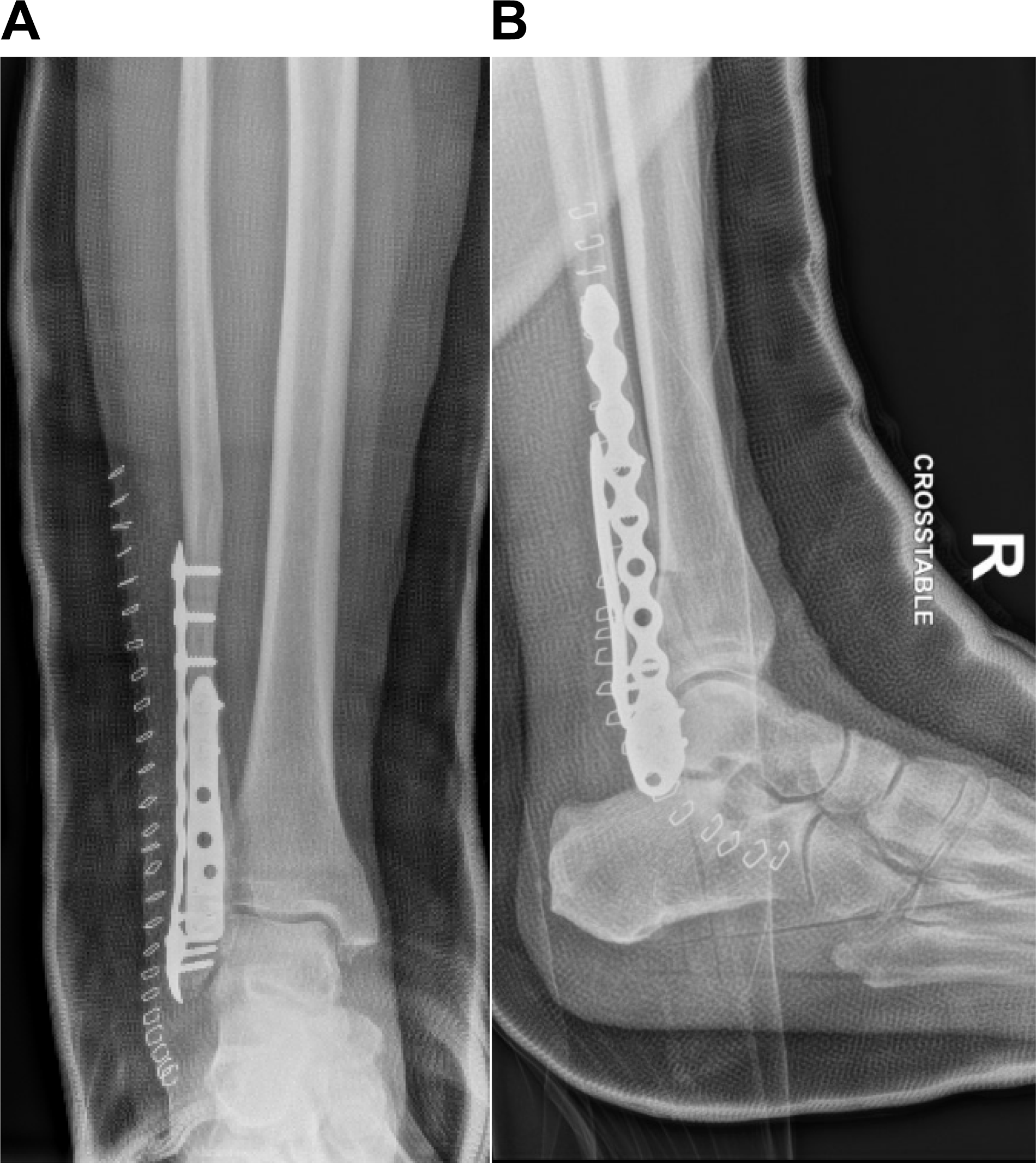
In January 2019, a 59-year-old female with a past medical history significant for bipolar disorder, schizophrenia, asthma, seizures, high cholesterol, and ovarian cancer in remission presented to the emergency department after sustaining a fall at home with an isolated injury to the right ankle and without loss of consciousness. Radiographs and computed tomography demonstrated a 3-mm posteriorly displaced isolated Weber B fibular fracture. The patient's social history was significant for 46 pack-years of smoking. Following medical optimization, the patient underwent minimally invasive open reduction of the fibular fracture with use of an intramedullary nail without complication by the attending on call at the time (Fig. 1). It is uncertain if any other methods of treatment were considered, such as cast immobilization or external fixation. The patient followed-up with the surgeon and was counseled on smoking cessation and instructed to remain nonweightbearing to the surgical limb. At 8 weeks post repair, the patient presented with pain to the right ankle and an inability to walk. Radiographs revealed displacement of the distal fibula with fracturing through the intramedullary nail. The patient continued to smoke and bear weight against medical advice (Fig. 2). A revision of the ankle fracture was performed using multiplanar plating of the fibula (Fig. 3). The postoperative course progressed uneventfully with initial nonweightbearing in a short leg cast, transitioning to weightbearing in controlled ankle motion (CAM) boot with an assistive device at 8 weeks.
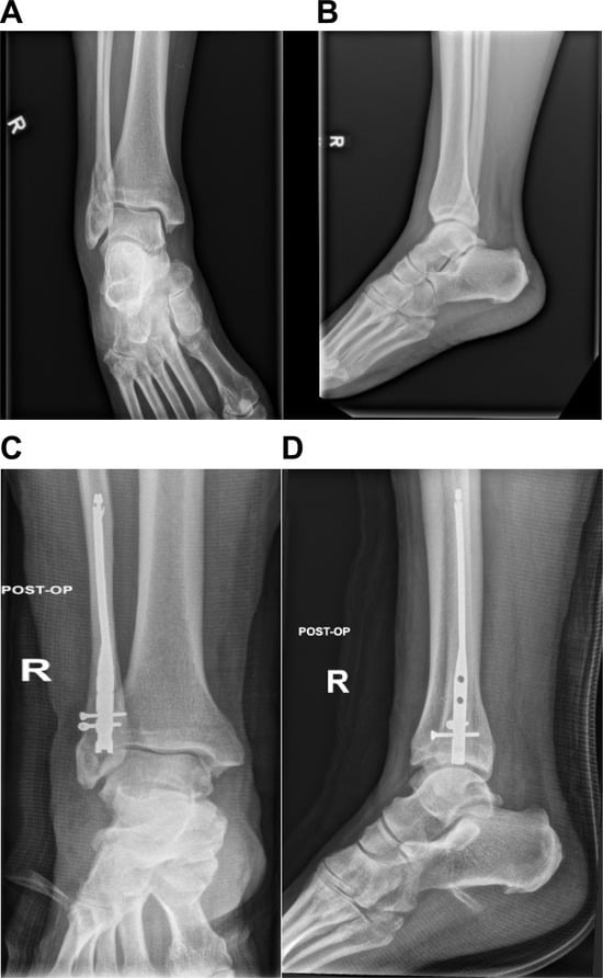
Figure 1.
A, Initial nonweightbearing anteroposterior radiograph of the right ankle revealing a Weber B fibular fracture with subsequent reduction. B, Initial nonweightbearing lateral radiograph of the right ankle revealing a Weber B fibular fracture with subsequent reduction and showing 3 mm of posterior displacement. C, Anteroposterior and D, lateral radiographs after open reduction and internal fixation of isolated fibular fracture with intramedullary nail.
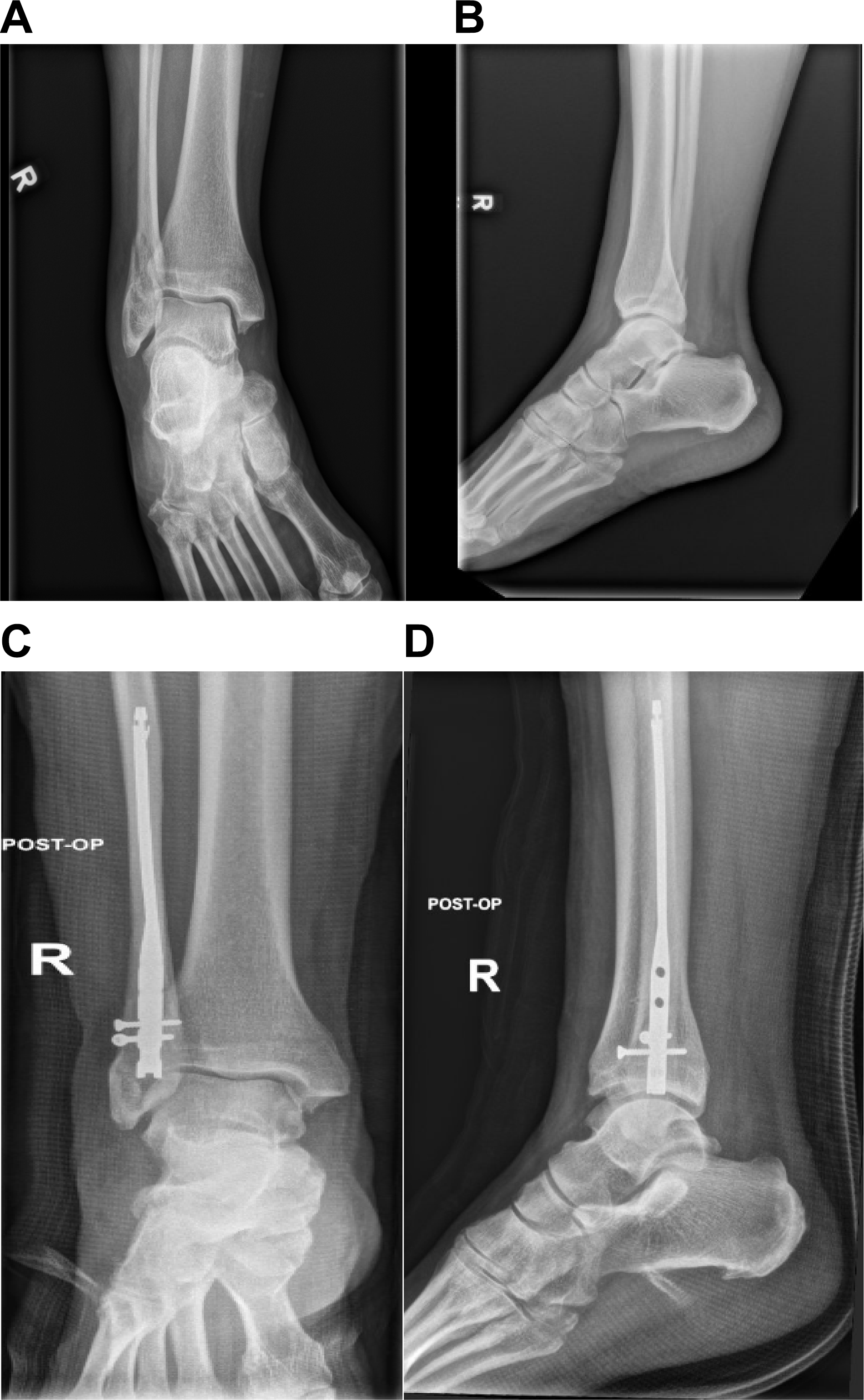
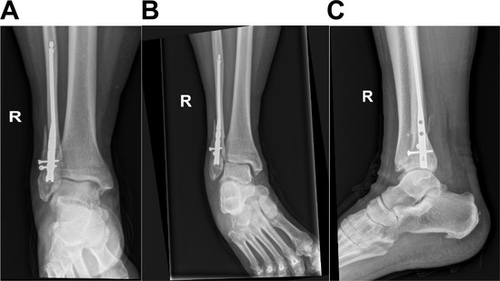
Figure 2.
Anteroposterior (A), mortise (B), and lateral (C) radiographs of the right ankle 8 weeks after repair, displaying a nonhealing fracture with displacement through the intramedullary nail.
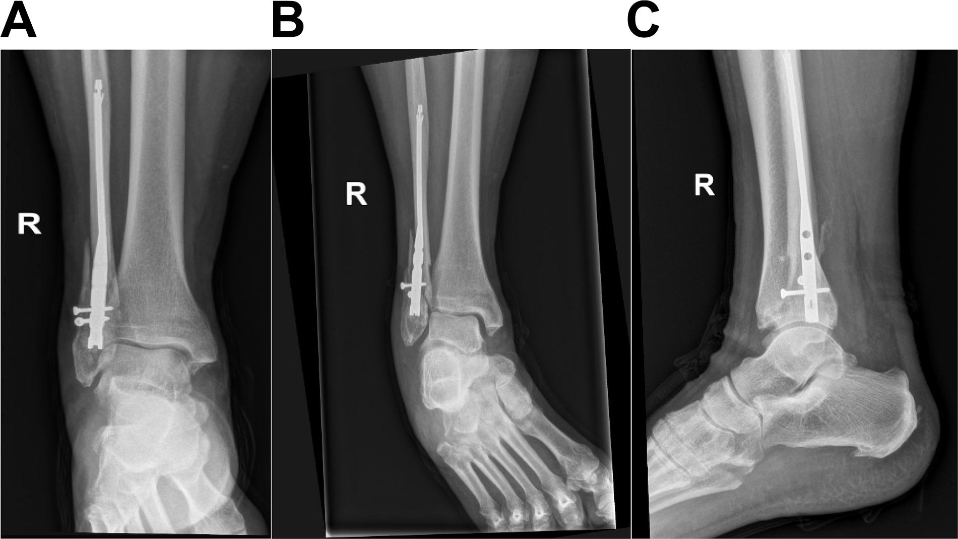
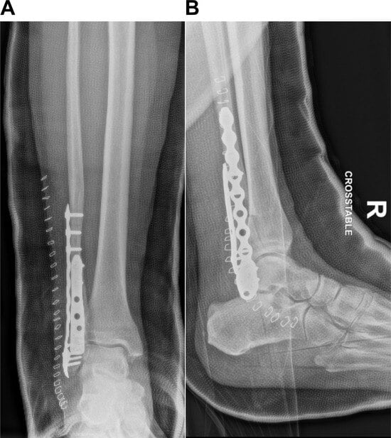
Figure 3.
Anteroposterior (A) and lateral (B) radiographs demonstrating revision of the fibular fracture with multiplanar plating.
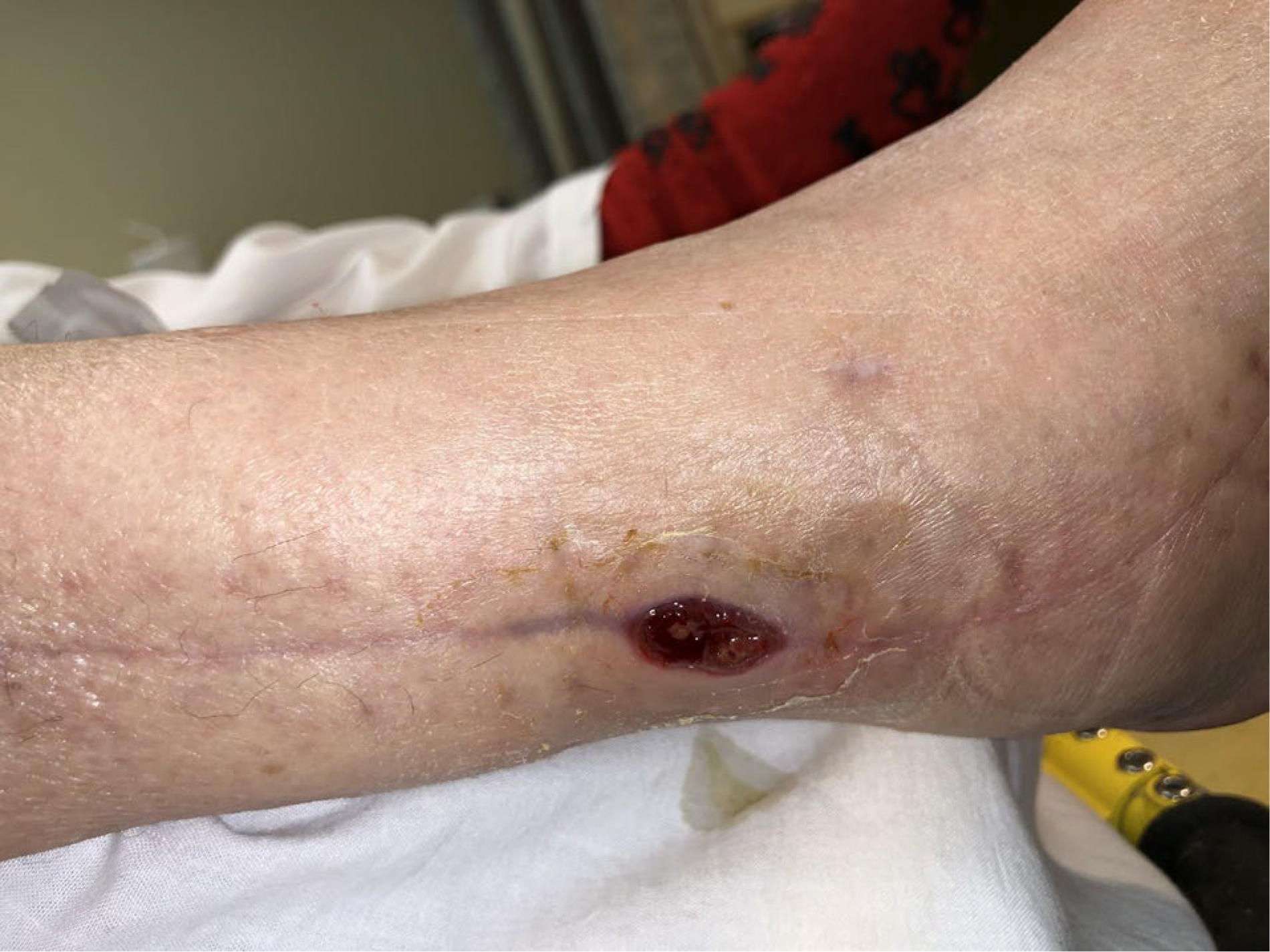
At 13 weeks, the patient presented with a new small malodorous wound along the previously healed surgical incision with depth to the level of hardware (Fig. 4). Serologic tests and inflammatory markers were within normal limits, white blood cell count (WBC) of 8.6 x 10³/uL and an erythrocyte sedimentation rate (ESR) of 6 mm/hr. The decision was made to retain the hardware for stability of fibular fracture in consultation with the department of infectious disease. Wound cultures demonstrated scant growth of methicillin-resistant Staphylococcus aureus and Enterobacter cloacae. An incision and drainage with insertion of vancomycin-impregnated calcium sulfate beads was performed for local infection control. The patient was subsequently discharged with 4 weeks of intravenous antibiotic therapy via a peripherally inserted central catheter (PICC) tailored toward wound cultures. Negative-pressure wound therapy was initiated to promote formation of granulation tissue and wound closure.
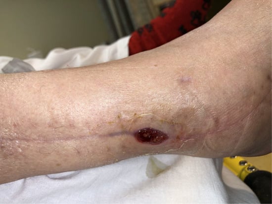
Figure 4.
Clinical presentation of a 1.5-cm circumferential malodorous wound along the previously healed surgical incision with purulent drainage extending to the hardware, 13 weeks after revisional surgery.
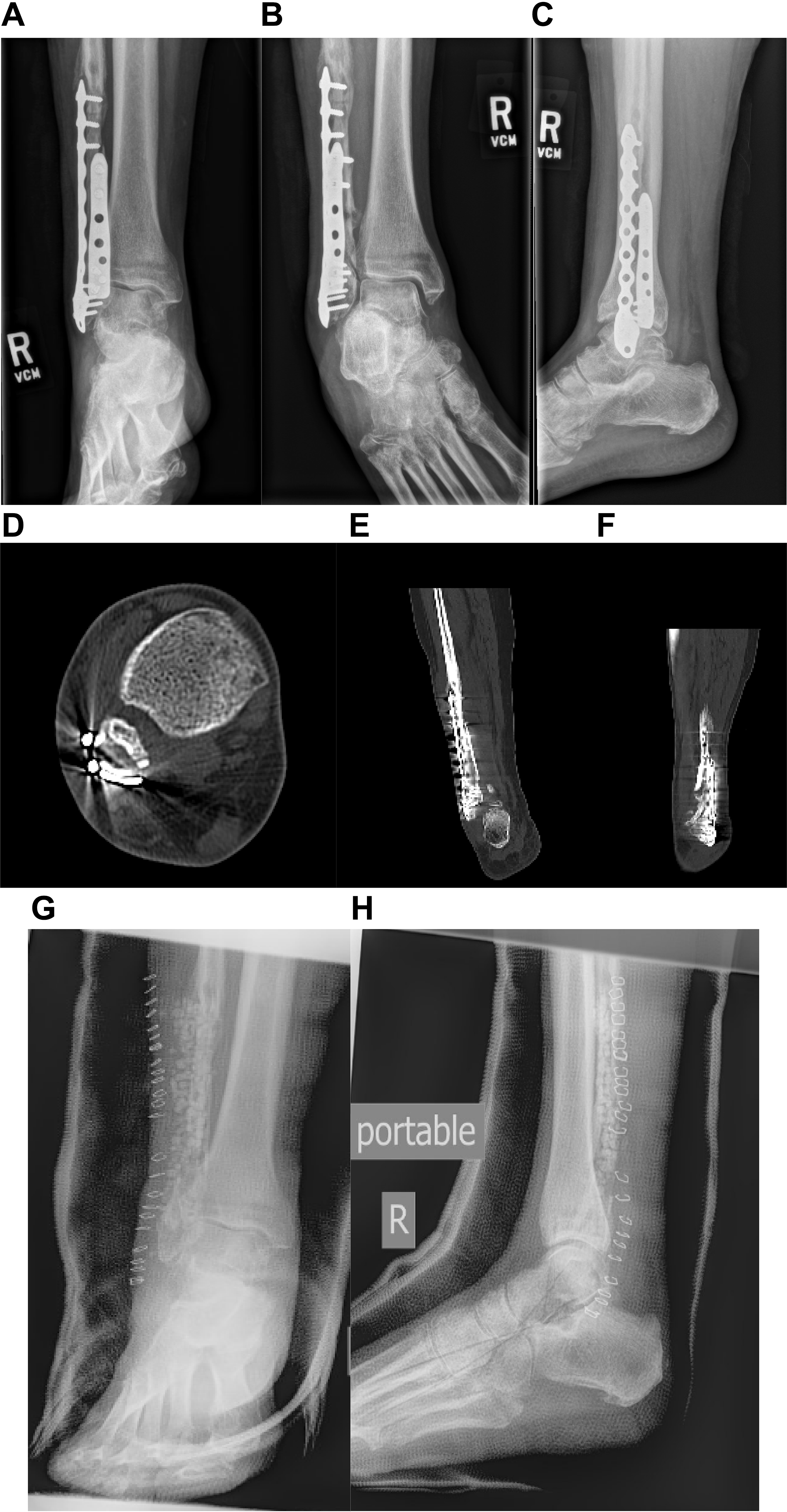
After 1 month, the wound remained contiguous with the hardware despite continued treatment of intravenous antibiotics and negative-pressure wound therapy with purulent, malodorous drainage present. Serologic and inflammatory markers were significant for leukocytosis (WBC of 14.6 x 10³/uL), ESR of 19mm/hr, and C-reactive protein (CRP) of 6mg/L, in addition to radiographic and computed tomography evidence of periosteal elevation with thickening highly suggestive of osteomyelitis. The decision was made to subsequently remove the hardware with tissue and bone specimens sent for microbiologic and pathologic examination. Vancomycin- and gentamicin-impregnated calcium sulfate beads were placed within the surgical site (Fig. 5). Soft-tissue and bone culture results were positive for moderate growth of E. cloacae, and histopathology reported acute and chronic inflammation consistent with osteomyelitis. The patient remained nonweightbearing with a bivalved cast and started on vancomycin and cefepime antibiotics.
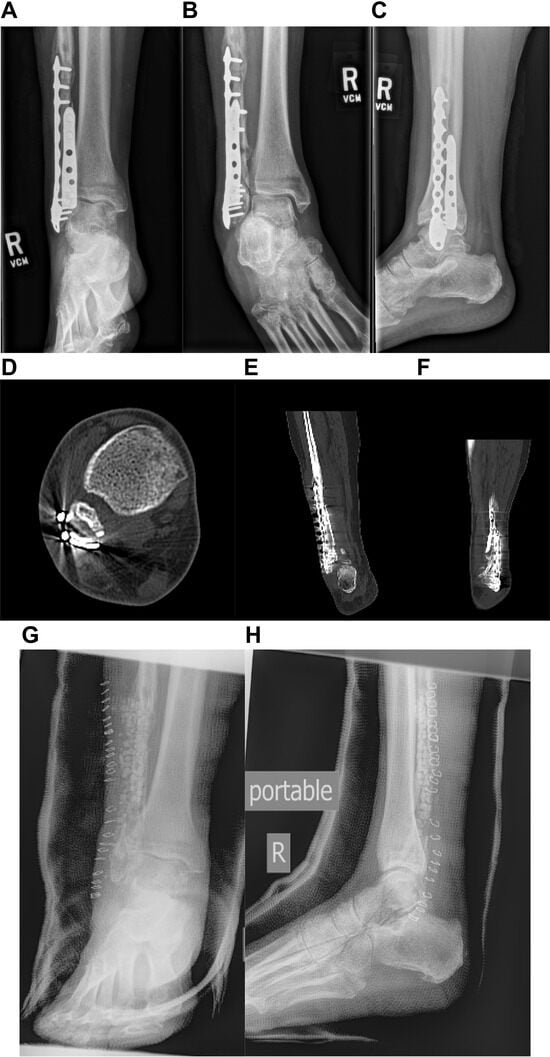
Figure 5.
Anteroposterior (A), mortise (B), and lateral (C) radiographs of septic nonunion of the distal fibular fracture 18 weeks following revisional surgery with a multiplanar plating. Axial (D), coronal (E), and sagittal (F) computed tomography images of septic nonunion before removal of infected hardware. Anteroposterior (G) and lateral (H) radiographs demonstrating removal of infected hardware with antibiotic beads inserted into the surgical site.
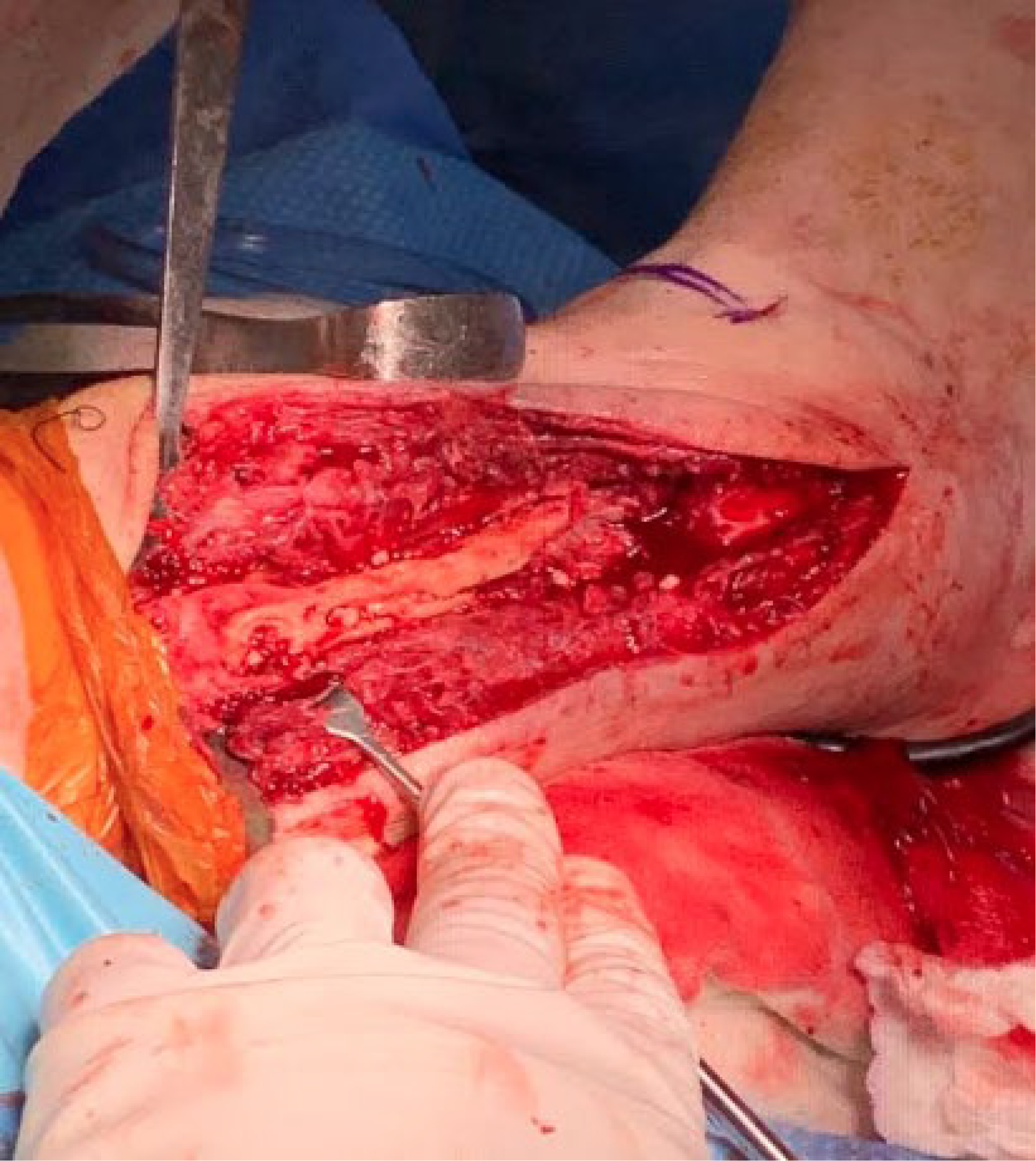
After removal of the hardware, the patient was referred to the senior author (M.S.) for surgical consultation. An operative ankle arthroscopy was performed to evaluate for any intra-articular extension to the ankle joint. Arthroscopic findings revealed significant acute hemorrhagic synovitis, along with a tear of the anterior talofibular ligament; however, no findings consistent with a septic joint were noted. Intraoperatively, the fibula was seen with a moth-eaten appearance, significant periosteal reaction and thickening, with fragmentation and fissuring noted up to the mid-diaphyseal region, thus necessitating excision of 12 cm of the mid- to distal fibula (Fig. 6).
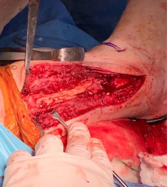
Figure 6.
Intraoperative view of infected fibular bone with moth-eaten appearance, significant periosteal reaction and thickening, with fragmentation and fissuring.
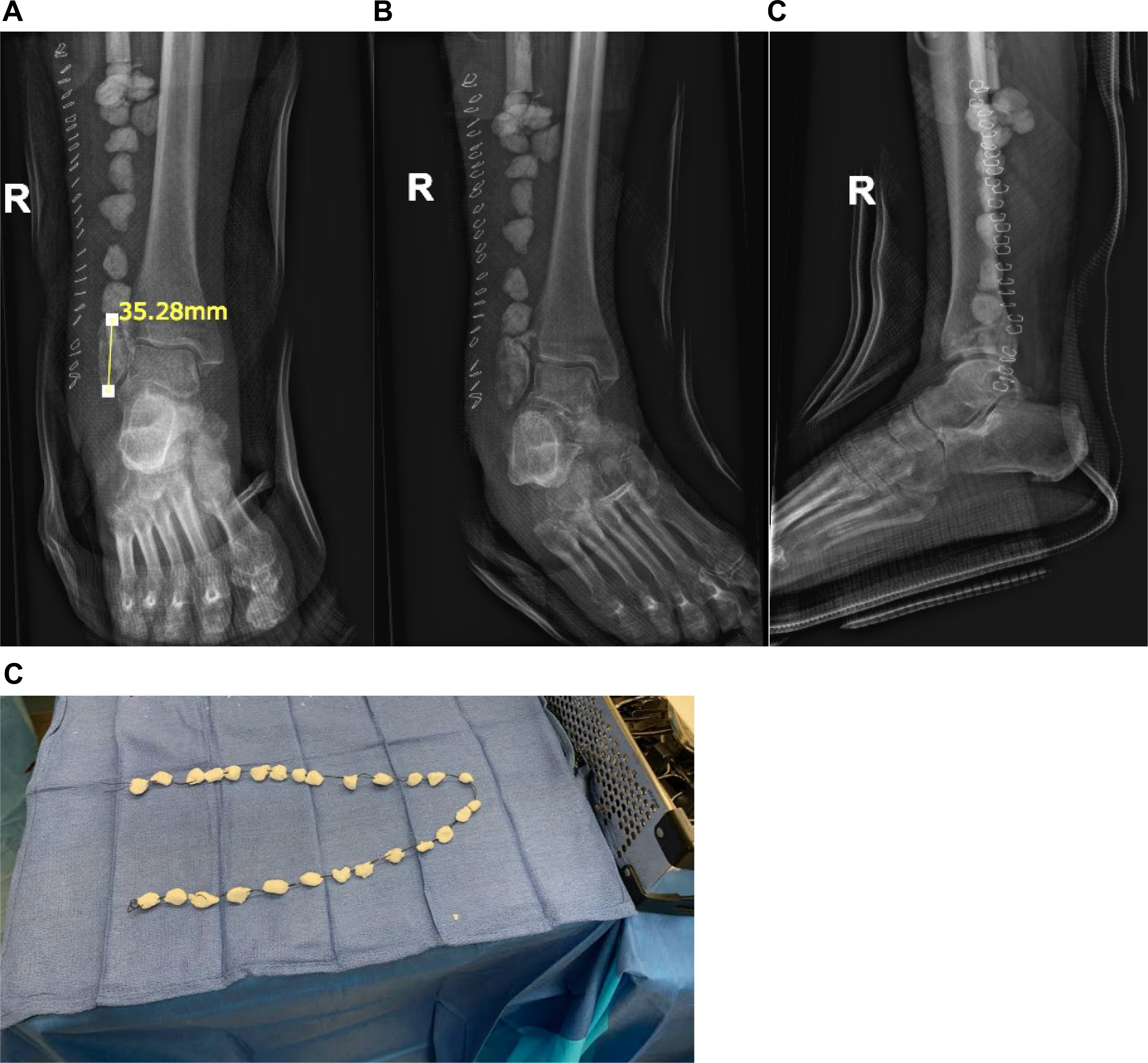
After the infected portion of the fibula was resected, 3.5 cm remained of the distal residual segment. The distal and proximal medullary canals were reamed, and polymethylmethacrylate (PMMA) cement impregnated with vancomycin and tobramycin was injected into the medullary canal. The PMMA cement contained 1 g of vancomycin and 160 mg tobramycin shaped into beads approximately 1–1.5 cm in diameter. These beads were then attached to a #2 nylon suture and secured onto the proximal and distal fibular segments (Fig. 7). Negative-pressure wound therapy was initiated over the incision site to assist in incision closure, and the right lower extremity was placed in a posterior splint with a stirrup. Intraoperative specimens were taken of the proximal and distal fibular bone margins as well as specimens from the tibia and submitted for microbiological and pathologic analysis. Final tibia and fibula bone cultures were negative, and histopathology reports showed no osteomyelitis of the tibia or proximal margin of the fibula. However, the residual distal fibula specimen was found to have areas of the medullary infiltrate of neutrophilic granulocytes, along with lymphocytes, plasma cells, and macrophages indicating acute and chronic osteomyelitis. On discharge, the patient was transitioned to a short leg cast and remained nonweightbearing to the right lower extremity with the continued administration of vancomycin and cefepime via PICC line for 2 weeks while she recovered in a rehabilitation facility.
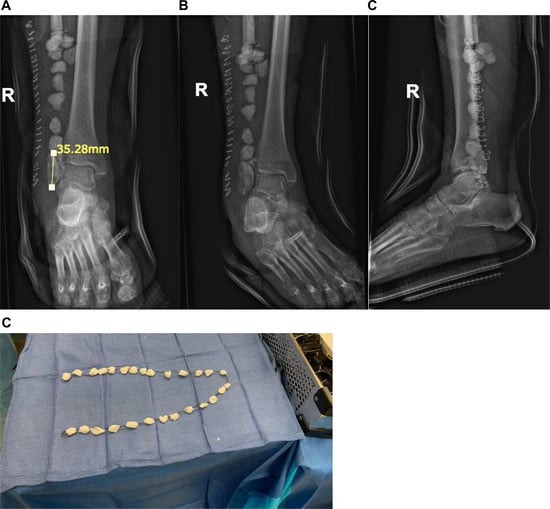
Figure 7.
A, Anteroposterior radiograph of fibular deficit replaced with antibiotic-impregnated polymethylmethacrylate beads and demonstrating a 3.5-cm distal fibular segment to maintain the ankle mortise. B, Mortise and C, lateral radiographs of fibular deficit replaced with antibiotic-impregnated polymethylmethacrylate beads. D, Intraoperative image of PMMA beads infused with 1 mg of vancomycin and 160 mg of tobramycin.
During the postoperative period, the patient was rehospitalized for a short course due to suicidal ideations. Once mentally stable, the patient returned for an arthroscopic debridement and irrigation of the right ankle, biopsy of the right fibula, and replacement of antibiotic beads. The arthroscopy revealed no evidence of infection within the ankle joint. The pseudomembrane that encapsulated the PMMA beads was preserved, with the cement excised. A new set of antibiotic PMMA cement consisting of 1 g of vancomycin and 160 mg of gentamicin shaped into 1–1.5 cm beads was placed on a #2 nylon and positioned inside of the pseudomembrane under fluoroscopic guidance. Specimens taken from the distal fibular segment were submitted for microbiological and pathologic analysis. Although cultures exhibited no growth, final histopathologic analysis revealed chronic inflammation suspicious of chronic osteomyelitis. The patient was placed in a short leg cast for continued nonweightbearing status and subsequently discharged back to the rehabilitation facility.
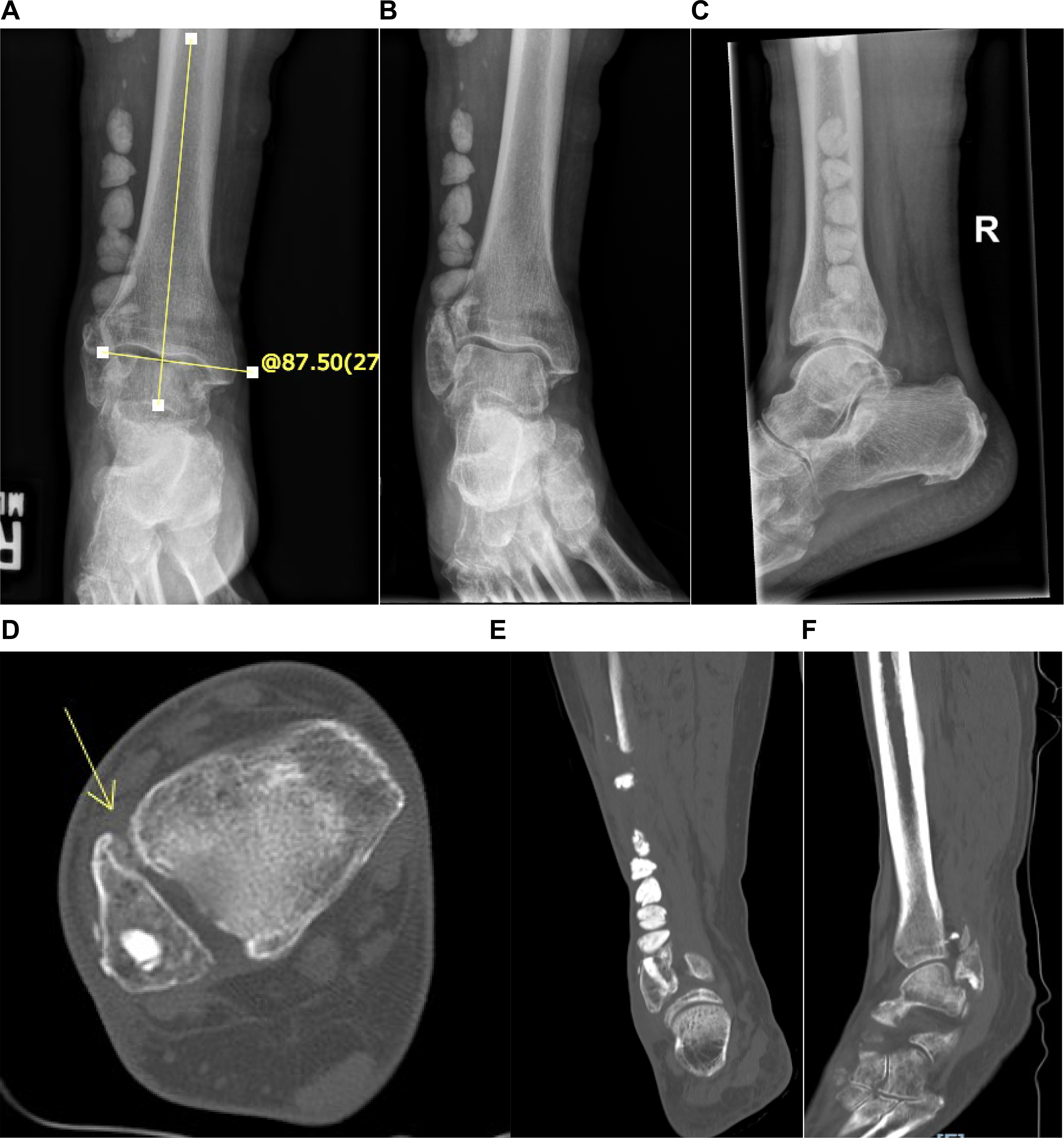
The postoperative course remained uneventful with a gradual transition to weightbearing in a CAM boot with an assisted device. Six months following the final procedure, the patient was able to walk in a CAM boot with no complaints and was instructed to transition to regular shoe gear with a gauntlet type ankle brace. The patient was also able to perform a gait analysis exam without any assistive devices. Final radiographs and computed tomography demonstrated a rectus ankle joint with mild degenerative changes to the articular surfaces, and no definitive lytic lesion or radiographic indications of acute osseous infection (Fig. 8).
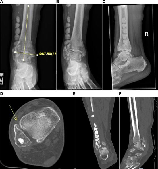
Figure 8.
A, Anteroposterior radiograph of the right ankle, 6 months following final surgery demonstrating a talotibial angle (TTA) of 87.5°, indicating a stable rectus ankle joint with mild degenerative changes and no definitive lytic lesions. Mortise (B) and lateral (C) radiographs of the right ankle, 6 months following final surgery. Axial (D), coronal (E), and sagittal (F) computed tomography views of the right lower extremity 6 months following final surgery with axial and coronal images displaying antibiotic bone cement well seated within the medullary canal of the fibula. Pseudoarthrosis noted to the axial view seen at the distal anterior syndesmosis contributing to ankle stability (arrow).
Discussion
A post-traumatic septic nonunion is a rare occurrence with incidence as low as 1% in closed low energy fractures with no data reported on the incidence of septic nonunion occurring in the distal fibula specifically.19 Treatment options are limited but include long-term suppressive antibiotics with immobilization and resection of the infected bone.19 Several techniques have been suggested in the cases where large deficits remain following debridement of infected bone including bone transport: the Masquelet technique with cement spacers and grafting, allograft or autograft, and vascularized free or pedicled grafts.10–18 To our knowledge, this is the first case describing a modified Masquelet technique in which a 2-staged antibiotic laden PMMA cement spacer was used as definitive treatment for a septic nonunion of the distal fibula to salvage the ankle joint.
Complications encountered postoperatively can be attributed to modifiable and nonmodifiable risk factors. The patient in this study was a smoker who was noncompliant with weightbearing protocols postoperatively, which increased the patient’s risk for developing complications during the postoperative period. Smokers have an increased prevalence of postoperative complications following ankle fracture surgery with a 3-times higher rate of wound complications as compared to nonsmokers.20,21 Smoking causes tissue hypoxia, dysfunction of both the innate and adaptive immune system, and increased level of oxidative stress products.22 Tobacco use also affects bone healing by decreasing bone density, via a decrease in periosteal gene expression for bone morphogenic proteins and decreased bone marrow progenitor cells.22
Additionally, according to Dodson et al,23 smokers require a longer period of immobilization to allow for proper bone healing. Krannitz et al24 reported that smoking increased radiographic healing time for ankle fractures by 20%, and noncompliance with a weightbearing regimen increased radiographic healing time by 23%.24 With continued smoking and noncompliance with nonweightbearing instructions, the patient developed a nonunion with a superficial wound that progressed to infection of the bone and hardware.
To compensate for the patient’s comorbidities and effectively treat the septic nonunion of the fibula, we elected to modify the classic Masquelet technique and used a cement spacer as definitive treatment. The basis for our technique was drawn from literature regarding treatment of infected joint replacements.14–18 The effect of the antibiotic spacer is two-fold. First, the antibiotics within the spacer allow for localized elution of antimicrobials. With the combination of parenteral therapy, the concentration of antibiotics to the infected region is increased without increasing the risk of systemic toxicity seen with IV treatment alone.15 Additionally, due to the slow elution from the antibiotic beads, the likelihood for chronic suppressive therapy is decreased, thus reducing the risk of adverse effects from prolonged administration of systemic antibiosis.15 Premade antibiotic beads are commercially available, with 7 mm being the standard bead size, impregnated with gentamicin.25 The antibiotic bead chain we fabricated consisted of beads ranging from 1 to 1.5 cm in diameter, which is almost twice the size of the standard beads. However, our beads were impregnated with both gentamicin and vancomycin. Thus, we elected to create larger size beads to accommodate for the increased amount of antibiotic used. With the use of two antibiotics together, specifically vancomycin and an aminoglycoside antibiotic, elution rates were higher than when used alone.26 Secondly, the spacer prevents fibrous invasion within the defect while inducing the formation of a pseudomembrane at the site of osseous defect27 This membrane is formed as a result of a foreign body reaction, containing a high concentration of vascular enothelial growth factor (VEGF) and transforming growth factor beta 1 (TGFß1). VEGF promotes angiogenesis while TGFß1 plays an important role in bone metabolism and extracellular matrix protein synthesis.27
The recommended length of distal fibula remaining to maintain stability of the ankle joint ranges from 6 to 10 cm.28–31 Based on a biomechanical study by Pacelli et al,32 instability of the ankle, which they defined as > 2 mm of translation between the tibia and fibula or catastrophic failure, was not observed until the length of the residual fibula was < 10% of the total fibular length. The fibular length of the patient in this study was approximately 36.5 cm. The length of the residual fibula was approximately 3.5 cm. Although the length of the residual fibula was slightly less than 10% of the fibular length, no instability at the ankle was noted, with the talotibial angle on an anteroposterior ankle view measuring at 87.5°, which falls within the normal range Najefi et al33 reported on, which was 88.5° ± 3.2.
Uchiyama et al31 recommends fixation of the syndesmosis or bracing in the presence of a large fibular defect to prevent ankle joint instability. In this case, a fusion of the syndesmosis was considered. However, there were concerns arthrodesis would promote contiguous spread of osteomyelitis and therefore was not performed. On final CT scans, the structures within the distal anterior region of the syndesmotic region were noted to be calcified providing a pseudoarthrosis. This calcified tissue within the syndesmosis provided further ankle stability. Clinical stability was evident via gait analysis performed on subsequent follow up with radiographic stability evident on postoperative films.
Several treatment modalities were considered; however, given the patient’s history of smoking and noncompliance with nonweightbearing instructions, options like the classic Masquelet technique, bone transport, allograft or autograft, and vascularized free fibula grafts were deemed nonviable. These techniques require nonweightbearing for extended periods of time as well as adequate potential for wound and bone healing, which is disrupted with tobacco use. The traditional Masquelet technique requires extrinsic stabilization beyond the margins of the graft with an external fixator.11 Considering the patient’s psychiatric history and history of noncompliance, we felt an external fixator would be contraindicated and likely result in further risk of infection and limb loss. Although bone transport allows for early weightbearing, patient compliance is imperative and the patient demonstrated a significant and recalcitrant history of noncompliance.12 Autograft requires quality bone stock from the patient while allograft integration can be very slow and would require even longer periods of nonweightbearing.12 Like autografts, free vascularized fibula grafts require quality bone stock as well as quality vascularization, which the patient lacked due an extensive history of tobacco use.13
We acknowledge several limitations within the present study. An inherent limitation is the retrospective nature of the case. Additionally, the authors acknowledge that the relatively short follow-up time, as well as the patient’s history of noncompliance, may result in a failure of treatment in the long term. Despite these limitations, the patient has demonstrated compliance with all postoperative instructions since the first surgery with the senior author.
In conclusion, we present a novel case of a septic nonunion of an isolated fibular fracture in the setting of tobacco use and weightbearing noncompliance with salvage of the ankle joint using an antibiotic spacer. At short-term follow-up, the patient is able to walk on the affected limb with no clinical instability or complications and with radiographs demonstrating adequate alignment of the ankle joint. Additional studies are necessary for long-term follow-up to fully assess the clinical and radiographic outcomes.
Financial Disclosure
None reported.
Conflict of Interest
None reported.
References
- Court-Brown CM, McBirnie J, Wilson G: Adult ankle fractures—an increasing problem? Acta Orthop Scand 69: 43, 1998.
- Elsoe R, Ostgaard SE, Larsen P: Population-based epidemiology of 9767 ankle fractures. Foot Ankle Surg 24: 34, 2018.
- Miller AG, Margules A, Raikin SM: Risk factors for wound complications after ankle fracture surgery. J Bone Joint Surg Am 94: 2047, 2012.
- Jordan RW, Chapman AWP, Buchanan D, et al: The role of intramedullary fixation in ankle fractures - A systematic review. Foot Ankle Surg 24: 1, 2018.
- Tas DB, Smeeing DPJ, Emmink BL, et al: Intramedullary fixation versus plate fixation of distal fibular fractures: a systematic review and meta-analysis of randomized controlled trials and observational studies. J Foot Ankle Surg 58: 119, 2019.
- Gross T, Kaim AH, Regazzoni P, et al: Current concepts in posttraumatic osteomyelitis: a diagnostic challenge with new imaging options. J Trauma 52: 1210, 2002.
- Kim DH, Kim HS, Nam KH, et al: Adverse drug reactions of long-term intravenous antibiotics in patients with pyogenic spondylitis. Korean J Spine 11: 113, 2014.
- Schrank GM, Wright SB, Branch-Elliman W, et al: A retrospective analysis of adverse events among patients receiving daptomycin versus vancomycin during outpatient parenteral antimicrobial therapy. Infect Control Hosp Epidemiol 39: 947, 2018.
- Lipsky BA, Berendt AR, Cornia PB, et al: 2012 Infectious Diseases Society of America clinical practice guideline for the diagnosis and treatment of diabetic foot infections. Clin Infect Dis: Off Publ Infect Dis Soc Am 54: e132, 2012.
- Spiegelberg B, Parratt T, Dheerendra SK, et al: Ilizarov principles of deformity correction. Ann R Coll Surg Engl 92: 101, 2010.
- Masquelet A, Kanakaris NK, Obert L, et al: Bone repair using the Masquelet technique. J Bone Joint Surg Am 101: 1024, 2019.
- Ashman O, Phillips AM: Treatment of non-unions with bone defects: which option and why? Injury 44(Suppl 1): S43, 2013.
- Shin EH, Shin AY: Vascularized bone grafts in orthopaedic surgery. J Bone Joint Surg Rev 5: e1, 2017.
- Durbhakula SM, Czajka J, Fuchs MD, et al: Spacer endoprosthesis for the treatment of infected total hip arthroplasty. J Arthroplast 19: 760, 2004.
- Ferrao P, Myerson MS, Schuberth JM, et al: Cement spacer as definitive management for postoperative ankle infection. Foot Ankle Int 33: 173, 2012.
- Proubasta IR, Itarte JP, Lamas CG, et al: Permanent articulated antibiotic-impregnated cement spacer in septic shoulder arthroplasty: a case report. J Orthop Trauma 19: 666, 2005.
- Regis D, Sandri A, Magnan B, et al: Six-year follow-up of a preformed spacer for the management of chronically infected total hip arthroplasty. Arch Orthop Trauma Surg 130: 1111, 2010.
- Themistocleous G, Zalavras C, Stine I, et al: Prolonged implantation of an antibiotic cement spacer for management of shoulder sepsis in compromised patients. J Should Elb Surg 16: 701, 2007.
- Metsemakers WJ, Kuehl R, Moriarty TF, et al: Infection after fracture fixation: current surgical and microbiological concepts. Injury 49: 511, 2018.
- Nåsell H, Ottosson C, Törnqvist H, et al: The impact of smoking on complications after operatively treated ankle fractures—a follow-up study of 906 patients. J Orthop Trauma 25: 748, 2011.
- Saleh H, Konda S, Driesman A, et al: Wound-healing issues following rotational ankle fracture surgery: predictors and local management options. Foot Ankle Spec 12: 409, 2018.
- Beahrs TR, Reagan J, Bettin CC, et al: Smoking effects in foot and ankle surgery: an evidence-based review. Foot Ankle Int 40: 1226, 2019.
- Dodson NB, Ross AJ, Mendicino RW, et al: Factors affecting healing of ankle fractures. J foot Ankle Surg 52: 2, 2013.
- Krannitz KW, Fallat LM, Schwartz SM: Radiographic healing of conservative versus operative management of supination-external rotation II fractures in a smoking and premature weight-bearing population. J Foot Ankle Surg 46: 218, 2007.
- Penner MJ, Masri BA, Duncan CP: Elution characteristics of vancomycin and tobramycin combined in acrylic bone-cement. J Arthroplast 11: 939, 1996.
- Klemm K: The use of antibiotic-containing bead chains in the treatment of chronic bone infections. Clin Microbiol Infect: Off Publ Eur Soc Clin Microbiol Infect Dis 7: 28, 2001.
- Pelissier P, Masquelet AC, Bareille R, et al: Induced membranes secrete growth factors including vascular and osteoinductive factors and could stimulate bone regeneration. J Orthop Res 22: 73, 2004.
- Babhulkar SS, Pande KC, Babhulkar S: Ankle instability after fibular resection. J Bone Joint Surgery Br 77: 258, 1995.
- Kanaya K, Wada T, Kura H, et al: Valgus deformity of the ankle following harvesting of a vascularized fibular graft in children. J Reconstr Microsurg 18: 91, 2002.
- Lang CJ, Frederick RW, Hutton WC: A biomechanical study of the ankle syndesmosis after fibular graft harvest. J Spinal Disord 11: 508, 1998.
- Uchiyama E, Suzuki D, Kura H, et al: Distal fibular length needed for ankle stability. Foot Ankle Int 27: 185, 2006.
- Pacelli LL, Gillard J, McLoughlin SW, et al: A biomechanical analysis of donor-site ankle instability following free fibular graft harvest. J Bone Joint Surg Am 5: 597, 2003.
- Najefi A-A, Buraimoh O, Goldberg A, et al: Should the tibiotalar angle be measured using an AP or mortise radiograph?: Does it matter? J Foot Ankle Surg 5: 930, 2019.
© 2024 American Podiatric Medical Association