


Abstract
Calcified tissue exposed in a leg ulcer can become infected and develop into a nidus of infection leading to sepsis. This case details a patient with a leg wound secondary to skin biopsy. This leg ulceration did not heal due to an underlying calcified mass and led to five hospital admissions for sepsis. She was diagnosed as having calcinosis cutis, which was suspected to be the source of her infections. The calcified mass was resected, and she healed uneventfully without further infections. Calcified soft-tissue masses should be considered in nonhealing leg ulcers and ulcers with multiple recurrent infections. Radiographs can be used to diagnose this condition, and surgical excision can be considered in cases of infection.
Calcinosis cutis is a rare disorder in which hydroxyapatite crystals or amorphous calcium phosphate is deposited in the skin and soft tissues [1]. Calcinosis cutis is divided into five forms: dystrophic, metastatic, idiopathic, iatrogenic, and calciphylaxis [2]. The dystrophic type is the most common form, in which there is associated normal serum laboratory values for calcium and phosphorus. With this form, there are underlying diseases such as systemic sclerosis, dermatomyositis, mixed connective tissue disease, and lupus. It is thought that this induces tissue damage and causes a nidus for infection [2]. The metastatic type occurs in undamaged tissues and is associated with abnormal levels of serum calcium and phosphorus depositions. These abnormal deposits are most caused by chronic kidney disease [1,2]. There is no tissue damage or abnormal values associated with the idiopathic type. Some associated causes include tumoral calcinosis, subepidermal calcified nodules, and scrotal calcinosis [2]. Some cases maybe induced iatrogenically by administration of calcium or phosphate causing deposits of calcium crystals [2]. Calciphylaxis is seen in chronic renal failure and dialysis. The disease is associated with calcification of small- and medium-sized vessels of fat and skin tissues [2].
Treatment for calcinosis cutis can be quite challenging and may differ based on the type. Although there is not a standard treatment for calcinosis cutis, many treatment options have been shown to be beneficial in case studies, including warfarin, bisphosphonates, minocycline, ceftriaxone, diltiazem, aluminum hydroxide, probenecid, intralesional corticosteroids, intravenous immunoglobulin, curettage, surgical excision, carbon dioxide laser, and extracorporeal shockwave lithotripsy (ESWL) [3].
Some novel techniques for calcinosis cutis, including minimally invasive and local treatments, include ESWL, laser therapy, and topical and intralesional sodium thiosulfate. The ESWL technique destroys calcifications by acoustic shockwaves [4]. It destroys the structure of calcifications without their mechanical extraction and is more effective for larger calcifications [5,6]. Satisfactory analgesic effects have been found with ESWL, and it could be considered as a pain relief and palliative treatment for calcinosis cutis [4,5]. Limitations of ESWL may include decreased accessibility to some lesions [4,5]. Laser therapy is a less invasive, faster, and convenient treatment that may require only a single session with complete removal of small calcifications (up to 2 cm long) [5,7]. Common adverse effects with laser therapy include skin discoloration, scarring, infection, and reccurances [5]. Sodium thiosulfate may act as a chelating agent that increases calcium solubility [5]. In addition, the substance may have anti-inflammatory, vasodilative, and antioxidative properties that may support tissue healing in calcinosis cutis [5]. Limitations with topical sodium thiosulfate treatment may include patient compliance, long-term adherence, pain, and infection [5].
Some reports have documented the benefits of surgical management and excision of calcinosis cutis [8]. Indications for surgical excision include presence of painful masses, recurrent infection, ulcerations, local functional impairment, and cosmetic motivation [9]. In addition, surgical management and removal of the calcium deposits is used for the treatment of infected deposits [10]. Physicians should be cautious in that excision or drainage of calcinosis cutis should be limited to selected sites where pain and disability outweigh the risks of recurrence and further calcifications caused by surgical trauma [11].
Surgical intervention has been effective in selective cases of calcinosis cutis. In 2008, Wu and Metz [11] described a case in which a 17-year-old presented with subcutaneous lesions of calcinosis cutis in the fingers. The patient underwent incision and drainage of the nodules, with significant improvement in symptoms and function. Minami et al [12] described two cases of widespread calcinosis cutis in patients with long-standing systemic lupus erythematosus. Calcifications around the forearm were surgically removed due to pain in the area. The nodules did not reappear in either of these patients.
Although calcinosis cutis is a rare disorder, there are even fewer cases reported highlighting calcinosis cutis in the lower extremity. There are cases involving congenital incidences of calcinosis cutis, but the present case study focuses on infected calcinosis cutis.
A literature search revealed only three reported cases of superinfected calcinosis cutis in the lower extremities. Cousins et al [13] reported calcinosis cutis in a 43-year-old woman with systemic lupus erythematosus who presented with a large, ulcerating, draining mass of the left thigh that measured 10×20×20 cm. The calcified mass interfered with movement at the waist and the hip. The lesion was effectively excised, with the patient being pain-free and devoid of gross calcifications; her range of motion at the hip was nearly normal, and her activity level was markedly increased. In 2020, Johnson et al [10] reported a case in which a 77-year-old woman with no significant medical history presented with multiple calcified nodules bilaterally. One of the nodules located at the lateral aspect of the left lower extremity led to cellulitis. The patient was then taken to the operating room twice for sharp excisional debridement with application of amniotic graft and acellular dermal matrix graft with placement of negative pressure wound therapy. The ulceration eventually healed and remained healed for 18 months postoperatively. Bernardo Cofiño et al [14] described a case in which an 83-year-old woman presented with rapidly progressive cellulitis in the bilateral lower extremities with multiple ulcerations and calcium deposits in the subcutaneous tissue. With parenteral antibiotic drug treatment, there was improvement of the ulcers and disappearance of the erythema and discharge.
Calcinosis cutis may mimic osteomyelitis, and the treatment may be confused. In this case report, infected calcified nodules were treated effectively with surgical excision. The diagnosis of calcinosis cutis was made based on radiographs and magnetic resonance imaging (MRI) findings. Osteoma cutis is a differential diagnosis based on the location of calcified nodules. This case report aims to raise awareness of the presentation and course of calcinosis cutis and osteoma cutis in lower-extremity ulcerations and infections.
Case Report
A 94-year-old patient with a history of hypertension, atrial fibrillation, squamous cell carcinoma of the right leg, and multiple admissions to another hospital for sepsis from right leg cellulitis presented to our facility. She was seen as an outpatient by another practitioner, who recommended that the patient visit the emergency department with the concerns of recurrent cellulitis and osteomyelitis. She reported that her wound has opened and closed multiple times, with four admissions to another hospital in the past 6 months. She noted pain, redness, and swelling of the right leg. She denied nausea, vomiting, chills, and fevers, as well as a history of smoking. She stated that she had been taking linezolid 1 day before admission. This was her fifth hospital admission for cellulitis of the right leg.
In the emergency department, laboratory tests were performed. The patient was found to have a white blood cell count of 16,800/mm3, an absolute neutrophil count of 14,600/mm3 and 87%, an erythrocyte sedimentation rate of 66 mm/h, a C-reactive protein level of 150 mg/L, and a lactic acid level of 4.1 mmol/L. Sepsis protocol was initiated. The patient was treated with aztreonam 2 g intravenously and vancomycin 750 mg intravenously initially. She was then transferred from the emergency department to the intensive care unit. Podiatric medicine, pulmonology, and infectious disease were all consulted.
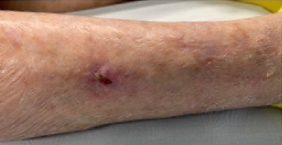
On physical examination, her light touch sensation, vibratory sensation, deep tendon reflexes, and protective sensation were all intact. Her dorsalis pedis and posterior tibial pulses were nonpalpable bilaterally. She had localized edema to the bilateral legs. The dermatologic examination showed a 1.0×1.0×0.5-cm ulceration with erythema and abnormal warmth present halfway between the ankle and knee of the right lower extremity. There was no drainage, no purulence, no malodor, no tunneling, and no undermining present.
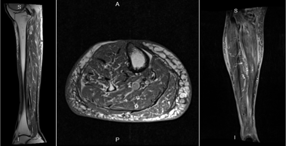
One day after admission she reached a maximum temperature of 101.2°F, a heart rate of 106/min, and a respiratory rate of 22/min. At the bedside, calcified lesions that were similar in appearance to bone fragments were removed from the ulceration. Plain film tibia/fibula radiographs were ordered and displayed clumped and sheetlike calcifications in the soft tissues of the right leg (Fig. 1). An MRI of the right lower extremity was ordered and showed no abscess and no evidence of osteomyelitis. The MRI displayed calcifications in the soft tissues of the right leg (Fig. 2). Per infectious disease recommendations, the patient was switched to cefepime 1 g intravenously every 8 hours and vancomycin 1 g intravenously daily. Blood cultures were negative.
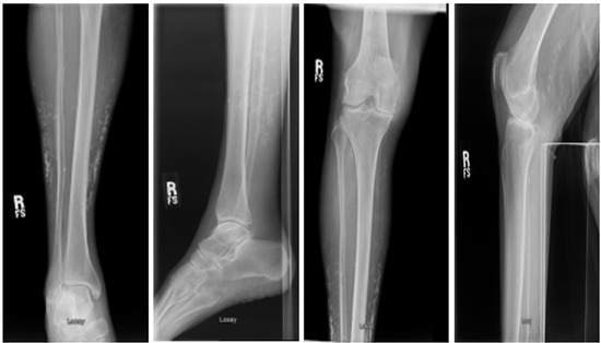
Figure 1.
Plain film radiographs of the right lower extremity.
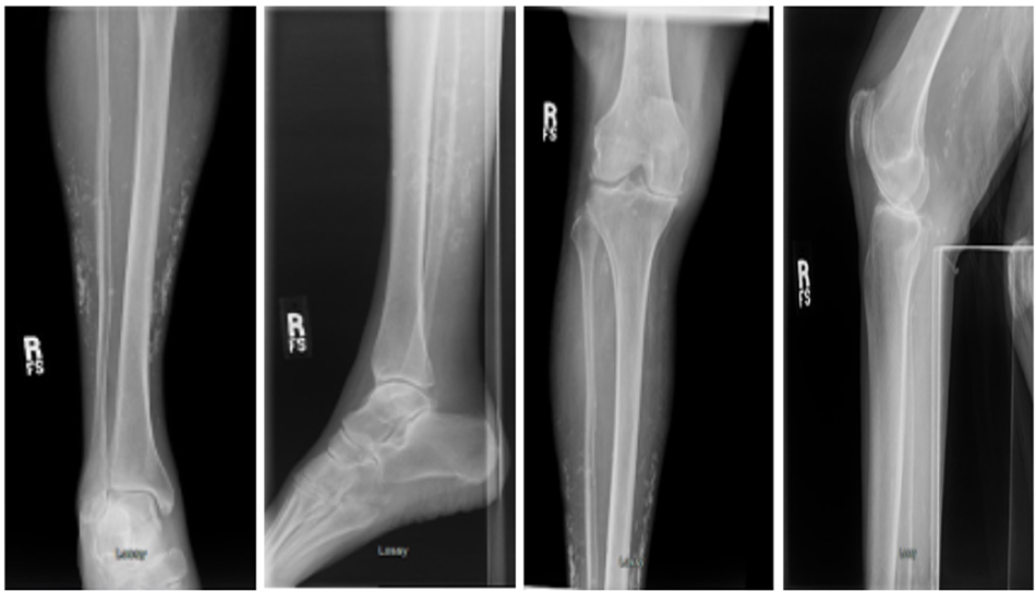
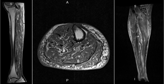
Figure 2.
Magnetic resonance image of the right lower extremity.
Although her laboratory values and vital signs were improving with continued intravenous antibiotic drug therapy, the wound and cellulitis did not improve clinically. Due to her recurrent episode of sepsis and cellulitis and with no improvement clinically, it was decided to take the patient to surgery for excision of the calcified mass and biopsy (Fig. 3).
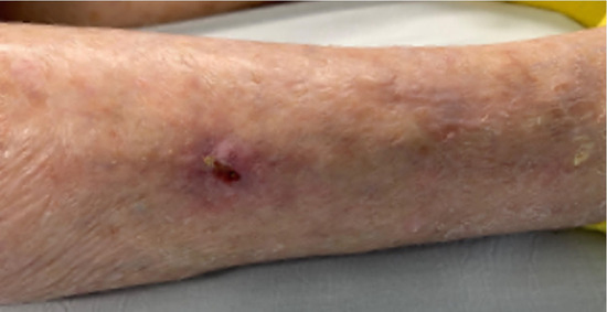
Figure 3.
Preoperative photograph of the right lower extremity.
Five days after admission to the hospital, she was taken to the operating room for excision of the ulceration and excision of the calcified nodules (Fig. 4). The surgical findings included an ulceration measuring 1.0×1.0×0.5 cm overlying the muscle belly of the anterior compartment and the wound with a hard calcified mass (Fig. 4). There was noted to be hard palpable calcified tissue extending approximately 5 cm distal to the ulceration (Fig. 4). There appeared to be discolored necrotic tissue in the base of the wound that was calcified as well, consistent with infection of the calcified mass with the appearance of osteomyelitis. An incision was placed over this calcified mass. On incision, the mass was hard and could not be incised using a No. 15 blade. This was well adhered to the deep layer of the skin. The skin was dissected from the mass. The mass appeared approximately 6 cm distal to the ulceration (Fig. 4).
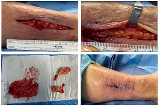
Figure 4.
Right leg excision of the ulceration and excision of the calcified mass and subcutaneous tissue in the operating room.
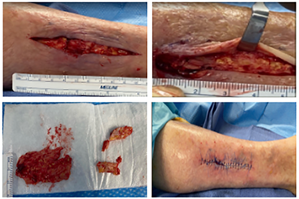
At this point, the anterior and posterior borders of the mass were identified, and the mass was reflected. After resection of the mass, the intermediate dorsal cutaneous nerve was identified and protected. The mass was resected and passed from the operative field (Fig. 4). The deep fascia was intact. The proximal wound was explored and noted to have further calcifications, which were excised using a No. 15 blade. A rongeur was used to resect any remaining fragments. We obtained one skin biopsy that included the deep calcified tissue for further diagnosis. The wound was closed primarily (Fig. 4). There was brisk capillary filling time to both sides of the incision after closure.
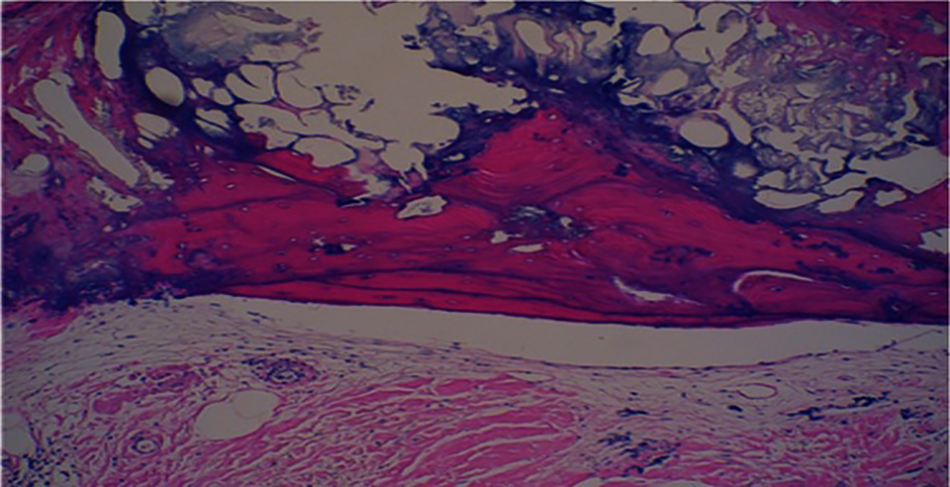
The tissue fragments were sent for histopathologic examination twice. The first specimen showed fragments of sclerotic fibroconnective tissue with calcification and fragments of viable bone. The second specimen was more suggestive of osteoma cutis. Grossly, the specimen was composed of multiple irregular tan-brown soft-firm tissue pieces. Microscopically, it showed multiple variable-sized bone formations in the dermis and subcutaneous tissue. The bone trabeculae were irregular, with associated osteophytes in the lacunae. Surrounding areas showed calcification. No cartilage formation was present in the tissues examined (Fig. 5). With these findings, clinical picture, and laboratory work, soft-tissue calcifications such as calcinosis cutis and osteoma cutis were diagnosed.
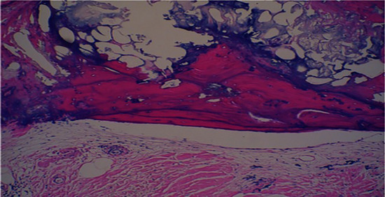
Figure 5.
Histopathologic photomicrograph displaying osteoma cutis: bone formation in the dermis and subcutaneous tissue without any intervening cartilage. Osteophytes can be seen in the lacunae in the bone trabeculae.
Postoperatively, vancomycin therapy was discontinued. Cefepime 1 g intravenously every 8 hours was continued per infectious disease recommendation. On discharge, the patient was scheduled to receive one dose of dalbavancin 1.5 g weekly for 2 weeks. The patient was discharged from the hospital 3 days postoperatively with improved erythema, edema, and pain, with no wound dehiscence.
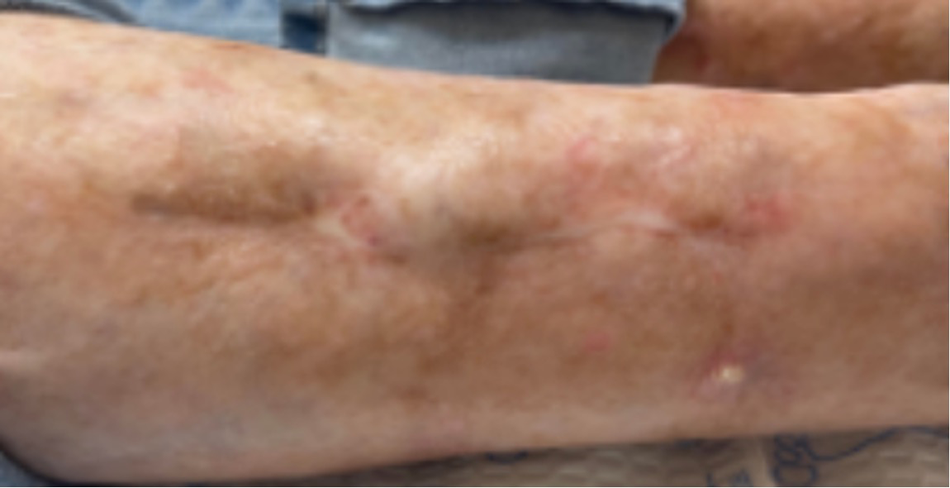
After surgical intervention, the patient underwent local wound care and was seen in the clinical setting weekly. Her wound dehisced and was treated with local debridement and a collagen dressing (Fig. 6). Eventually, the wound fully healed (Fig. 7). The wound has remained healed for 18 months and counting without complications (Fig. 8).
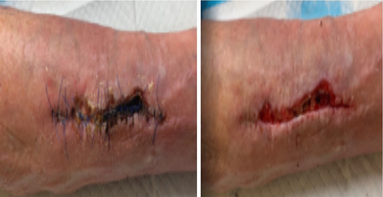
Figure 6.
Wound dehiscence.
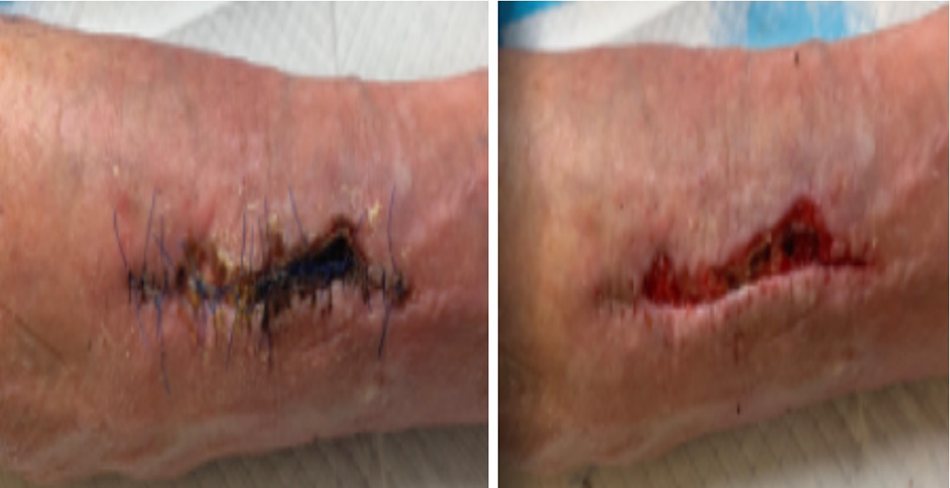
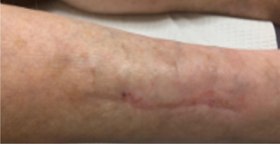
Figure 7.
Four-month follow-up visit poster surgical intervention showing full healing.
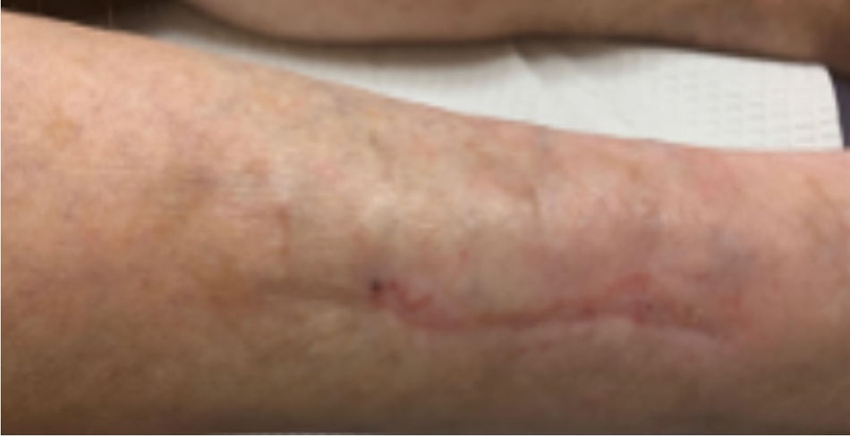
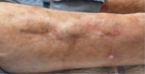
Figure 8.
Eighteen-month follow-up visit after the surgical intervention.
Discussion
Calcinosis cutis can severely affect a patient’s quality of life. Treatment for calcinosis cutis is challenging and depends on the cause. Recommendations are based mainly on case reports and case series. There is no standardized algorithm established for the treatment of calcinosis cutis. The present case showed promising results in a patient with recurrent cellulitis after surgical excision of an idiopathic calcinosis cutis mass with superimposed infection. In the present case, we believe that the subcutaneous calcium nodules became infected after biopsy to rule out skin cancer. This open portal over the calcium deposit allowed the calcium deposit to become infected. Once the subcutaneous calcium nodules at the site of infection were excised surgically, the cellulitis was resolved, and the patient did not have recurrence of cellulitis as she had four times before this admission.
We believe that the differential diagnosis of calcinosis cutis versus other soft-tissue calcifications should be considered early in the evaluation of a nonhealing leg ulcer. Conventional radiographs are generally sufficient for diagnosing calcinosis cutis and have been recommended for initial imaging [15]. In the present patient, radiographs displayed clumps and sheetlike calcifications at the area of ulceration. In the event of a leg ulcer with multiple infections and multiple hospital admissions for recurrent cellulitis, radiographs would be appropriate to evaluate for underlying calcifications. Histopathologic examination can then confirm the diagnosis.
Calcinosis cutis is characterized by fine granular deposition of calcium in the dermis and chunks of irregular calcium in the subcutaneous tissue. The common differential diagnoses include, but are not limited to, infectious etiologies such as cutaneous mycetoma, warts, milia, warts, molluscum contagiosum, osteoma cutis, and granulomatous reaction [2]. Among all of the differential diagnoses, osteoma cutis is interesting because there is heterotopic bone formation in the soft tissue. It usually starts from a focus of calcification [16]. There is membranous ossification, and, therefore, we will not see any cartilage formation [16]. Besides calcinosis cutis, some other histologic differential diagnoses are pilomatrixoma in its ossified form, osteochondroma, ossified hair follicle, and osteosarcoma in its extraskeletal form located in the dermis or in the subcutaneous tissue [16].
As mentioned previously herein, surgical intervention has been effective in selected cases. The studies by Wu and Metz11 and Minami et al [12] differ from the present study based on not only the location of the lesions but also the cause for surgical intervention. Both studies dictated the need for surgical excision because of pain, but our primary reason for surgical intervention was infection control. A literature search revealed only three reported cases of superinfected calcinosis cutis [10,13,14]. The present case report differed in that we reported a case with recurrent cellulitis, specifically four hospital admissions, that was resolved with surgical intervention of the superinfected calcinosis cutis.
We believe that further research is warranted to raise awareness of the presentation and course of calcinosis cutis and other forms of soft-tissue calcifications, such as osteoma cutis, in the lower extremities.
Conclusions
This case report highlights a presentation of soft-tissue calcifications with superimposed infection in a patient who was hospitalized five times for recurrent cellulitis. This case report aims to raise awareness of the presentation and course of calcinosis cutis as well as other soft-tissue calcifications, such as osteoma cutis, in the lower extremities.
Financial Disclosure: None reported.
Conflict of Interest: None reported.
References
- Larralde M, Giachetti A, Cáceres MR, et al: Calcinosis cutis following trauma. Pediatr Dermatol 22: 227, 2005.
- Le C, Bedocs PM: “Calcinosis Cutis,” in StatPearls [Internet], StatPearls Publishing, Treasure Island, FL, July 17, 2021.
- Reiter N, El-Shabrawi L, Leinweber B, et al: Calcinosis cutis: part II. Treatment options. J Am Acad Dermatol 65: 15, 2011.
- Sultan-Bichat N, Menard J, Perceau G, et al: Treatment of calcinosis cutis by extracorporeal shock-wave lithotripsy. J Am Acad Dermatol 66: 424, 2012.
- Nowaczyk J, Zawistowski M, Fiedor P: Local, non-systemic, and minimally invasive therapies for calcinosis cutis: a systematic review. Arch Dermatol Res 314: 515, 2022.
- Sparsa A, Lesaux N, Kessler E, et al: Treatment of cutaneous calcinosis in CREST syndrome by extracorporeal shock wave lithotripsy. J Am Acad Dermatol 53(suppl 1): S263, 2005.
- Joo YH, Kwon IH, Huh CH, et al: A case of persistent subepidermal calcified nodule in an adult treated with CO2 laser. J Dermatol 31: 480, 2004.
- Khudadah M, Jawad A, Pyne D: Calcinosis cutis universalis in a patient with systemic lupus erythematosus: a case report. Lupus 29: 1630, 2020.
- Lipskeir E, Weizenbluth M: Calcinosis circumscripta: indications for surgery. Bull Hosp Jt Dis Orthop Inst 49: 75, 1989.
- Johnson AR Jr, Mooshol D, Thakar V, et al: Surgical management of idiopathic ulcerative calcinosis cutis in the lower extremity: a case report. J Foot Ankle Surg 59: 603, 2020.
- Wu JJ, Metz BJ: Calcinosis cutis of juvenile dermatomyositis treated with incision and drainage. Dermatol Surg 34: 575, 2008.
- Minami A, Suda K, Kaneda K, et al: Extensive subcutaneous calcification of the forearm in systemic lupus erythematosus. J Hand Surg Br 19: 638, 1994.
- Cousins MA, Jones DB, Whyte MP, et al: Surgical management of calcinosis cutis universalis in systemic lupus erythematosus. Arthritis Rheum 40: 570, 1997.
- Bernardo Cofiño J, Trapiella Martínez L: Superinfected calcinosis cutis as a presentation of a limited form systemic sclerosis. Rheumatol Clin 10: 187, 2014.
- Shahi V, Wetter DA, Howe BM, et al: Plain radiography is effective for the detection of calcinosis cutis occurring in association with autoimmune connective tissue disease. Br J Dermatol 170: 1073, 2014.
- Limaiem F, Sergent SR: “Osteoma Cutis,” in StatPearls [Internet], StatPearls Publishing, Treasure Island, FL, July 22, 2021.
© 2025 American Podiatric Medical Association