


Abstract
Background: We aimed to find probable correlation between postoperative radiologic variables and clinical outcomes of surgically treated calcaneal fractures. Methods: In a retrospective study, 70 unilateral displaced intraarticular calcaneal fractures in adults with follow-up more than 1 year were asked to have a visit. Weightbearing radiographs of both ankles were taken and radiologic parameters, including the differences in values in Böhler and Gissane angles in comparison with the uninjured side, and calcaneocuboid and subtalar joint arthritis based on the Kellgren-Lawrence grading scale, were evaluated. They were considered to find any correlation with clinical outcomes assessed by American Orthopaedic Foot and Ankle Society Ankle-Hindfoot scale, visual analogue scale, Foot Function Index, and Tegner Activity Scale. Results: A total of 61 men (87.1%) and nine women (12.9%) with a mean age of 38.9 ± 12.7 years (range, 18–67 years) were included. Mean follow-up visit for the patients was 25.1 ± 12.7 months. Mean scores of American Orthopaedic Foot and Ankle Society Ankle-Hindfoot scale, visual analogue scale, Foot Function Index, and Tegner Activity Scale were 86.7 ± 12.9, 21.3 ± 22.2, 13.1 ± 15.4, and 5.2 ± 1.1, respectively. The mean Gissane angle and Böhler angle differences were –0.2 ± 8.6 and –3.7 ± 7.2, respectively. Regarding the calcaneocuboid arthritis, 50 (71.4%), 14 (20.0%), and six patients (8.6%) were categorized in grades 0, 1, and 2, respectively. Also, subtalar arthritis was seen in 15 (21.4%), 24 (34.3%), 20 (28.6%), and 11 patients (15.7%), categorized as grades 0, 1, 2, and 3, respectively. No statistical correlation was found between any of the radiologic variables and clinical scores. Conclusions: There was no significant correlation between Böhler and Gissane angles and the clinical outcomes in surgically treated calcaneal fractures. Also, functional outcomes do not change considerably among different grades of arthritis in calcaneocuboid and subtalar joints, at least during short- to mid-term follow-up periods. Radiologic findings after open reduction and internal fixation of calcaneal fractures are not predictors of function of the patients.
The most common fracture among the tarsal bones is calcaneal fracture, which accounts for approximately 60% of them. [1] This means that the annual incidence of calcaneal fractures has been reported to be approximately 10.5 per 100,000 in male and 3.8 per 100,000 in female populations. [2] They usually occur as a result of a high-energy trauma, such as a fall or a motor vehicle accident. Approximately 71% of these fractures are intraarticular, with the greater part of these injuries involving both the calcaneocuboid and subtalar joints. [3] Final functional outcomes are closely linked to a perfect anatomical reduction of the fractured fragments to reduce the likelihood of degenerative arthrosis, hindfoot deformities, limb shortening, neuritis, and tenosynovitis of surrounding tendons. [4] For displaced intraarticular calcaneal fractures, open reduction and internal fixation (ORIF) is the treatment of choice to restore calcaneal morphology and achieve anatomical reduction of the articular surfaces. [5] A long-term rehabilitation program, even for 1 year after surgery, with emphasis on balance and strength of all muscle groups crossing the ankle joint, is necessary to have a good outcome. [6]
Although several studies have described the functional results and/or radiologic outcomes following ORIF of calcaneal fractures, [7–10] a few studies have evaluated the correlation between the postoperative radiologic variables and their clinical outcomes. [11–16] After reviewing these studies, no consensus regarding this issue has yet been achieved. Also, no study in the literature has evaluated the correlation of subtalar and calcaneocuboid joint arthritis with postoperative functional and clinical outcomes. Our hypothesis for the lack of agreement was that most of these studies assessed a small number of cases, did not use appropriate outcome measures, and did not evaluate a specific type of fracture or operation technique. The purpose of this study was to discover any correlation between radiologic measurements such as Böhler angle, Gissane angle, and calcaneocuboid and subtalar arthritis grading and clinical and functional outcomes assessed by the American Orthopaedic Foot and Ankle Society (AOFAS) Ankle-Hindfoot scale, the Foot Function Index (FFI), the visual analogue scale (VAS), and the Tegner Activity Scale (TAS).
Methods
After approval of the study by the ethics committee and the institutional review board in accordance with the Declaration of Helsinki (Ethic number: IR.SUMS.MED.REC. 1400.098), this retrospective cross-sectional study was conducted in Chamran Hospital (the major orthopedic center) and Emtiaz Hospital (a level 1 trauma center) in Shiraz, Iran. From April of 2014 to March of 2019, all consecutive adult patients with Sanders type II and III intraarticular calcaneal fractures who underwent ORIF by means of the extensile lateral approach performed by the senior author (A.R.V.) were enrolled. Exclusion criteria included refusal to give informed consent to participate in the study, any associated fracture, bilateral fractures, associated peroneal tendon dislocation, open fractures, primary subtalar arthrodesis, Sanders type IV calcaneal fractures, postoperative wound breakdown and superficial or deep infection, diabetes mellitus, age younger than 18 years, postoperative follow-up of less than 1 year, and secondary subtalar arthrodesis after fixation of a calcaneal fracture.
Demographic characteristics and side and type of calcaneal fractures based on Sanders classification [17] were compiled from the files of the patients and operation notes. Patients were contacted to have a follow-up visit with new weightbearing radiographs.
Clinical Evaluation
Clinical outcomes in the follow-up visit were evaluated using the following tools:
- AOFAS Ankle-Hindfoot scale, evaluating pain, functional aspects, and alignment of the hindfoot by means of nine different items. [18]
- VAS, ranging from 0 (“no pain”) to 100 (“pain as bad as it could be”).
- The FFI was created to measure foot function in the subscales of disability, pain, and activity restrictions using 23 subjective questions. Based on percentile reports in our study, the scale ranges from 0 for the best functional foot condition, to 100 for the worst. [19]
- The TAS was developed to provide a standardized method for determining the level of activity before and following an event on a numeric scale, where 0 indicates disability and 10 indicates the highest activity possibility. [20]
Radiologic Evaluation
One of the other authors measured the Böhler and Gissane angles on the weightbearing plain radiographs of the patients based on defined methods of angular measurements. [21] To standardize the measurements, we calculated the difference in values in Böhler angles of the operated ankle and the uninjured ankle in standing radiographs. Also, we checked the difference in values in Gissane angles between both ankles. Negative values indicated a decrease in the Böhler angle and an increase in the Gissane angles of the injured side in comparison with the uninjured side. In addition, the Kellgren-Lawrence (KL) grading scale was used to grade arthritic changes in both subtalar and calcaneocuboid joints. [22]
Statistical Analysis
SPSS version 16.0 for Windows was used for analysis (SPSS Inc, Chicago, Illinois). Frequency, mean, and standard deviation were used to express descriptive data. Furthermore, regression analysis was used to calculate the correlation coefficient between clinical outcomes and radiologic examinations. The alpha value was set to 0.05.
Results
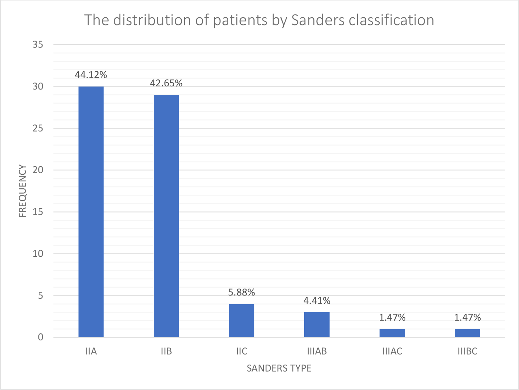
A total of 70 patients, 61 men (87.1%) and nine women (12.9%), with a mean age of 38.9 ± 12.7 years (range, 18–67 years), were included. Mean follow-up visit for the patients was 25.1 ± 12.7 months. Fracture of the right calcaneus was more common than fracture of the left calcaneus (57.1% versus 42.9%). As shown in Figure 1, most of the cases were categorized as Sanders type IIA and IIB.
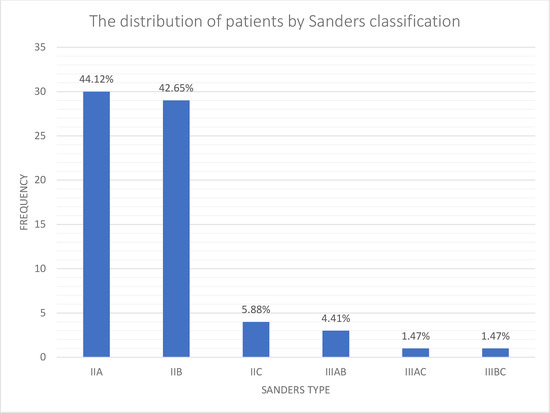
Figure 1.
The distribution of patients by the Sanders classification.
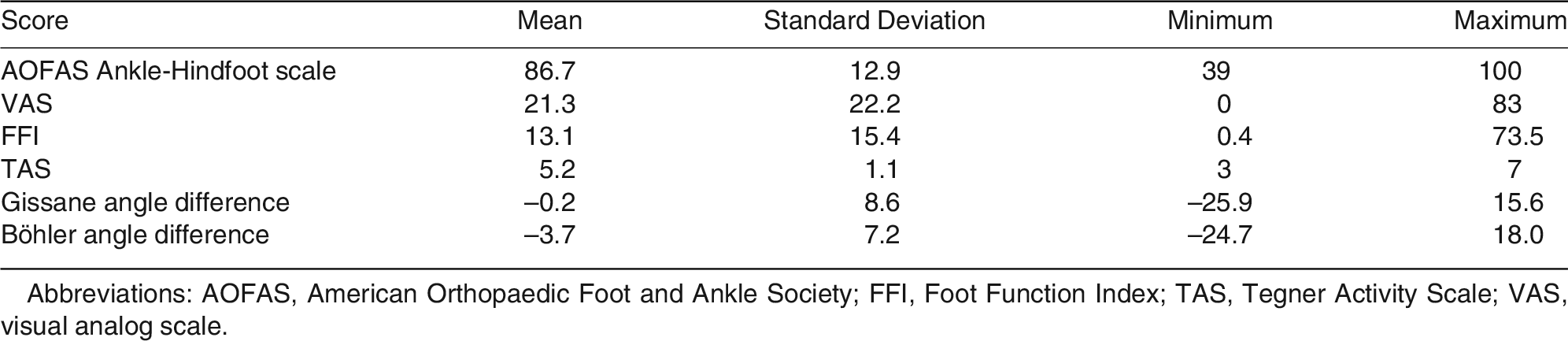
Functional and radiologic characteristics of the patients are summarized in Table 1. Regarding the calcaneocuboid joint arthritis based on the KL grading scale, 50 (71.4%), 14 (20.0%), and six patients (8.6%) were categorized as grades 0, 1, and 2, respectively. Also, subtalar arthritis based on the KL grading scale was seen in 15 (21.4%), 24 (34.3%), 20 (28.6%), and 11 patients (15.7%), categorized as grades 0, 1, 2, and 3, respectively.

Table 1.
Clinical and Radiologic Characteristics of the Patients
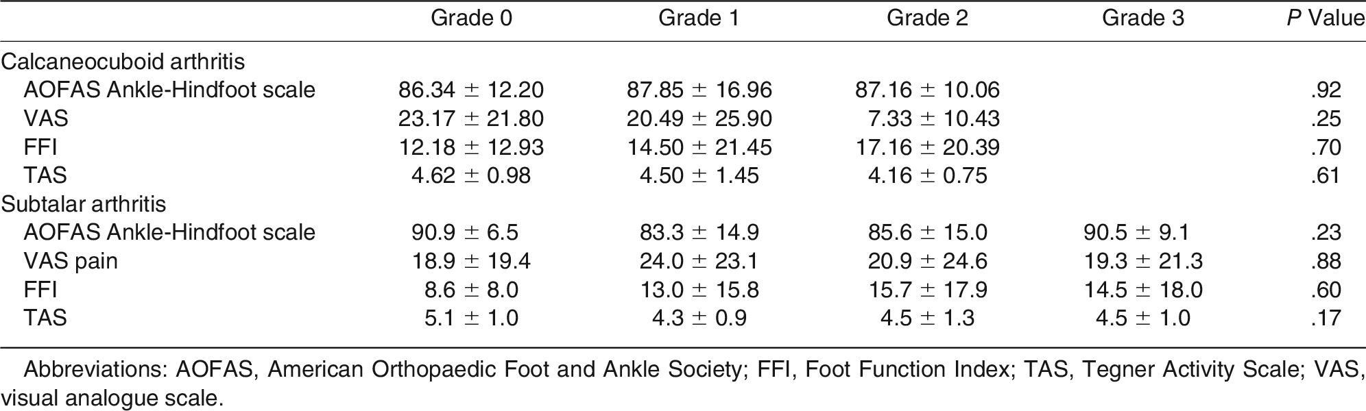
Regression analysis tests between clinical scores and Gissane and Böhler angle differences with the uninjured side, as the reference, did not show any significant correlation (Table 2). Scatterplot graphs are shown in Figures 2 and 3. Moreover, functional outcomes did not significantly change among different grades of arthritis in calcaneocuboid and subtalar joints (Table 3).
Table 2.
Correlation Coefficient and P Value Between Clinical Outcome Scores and Radiologic Variables
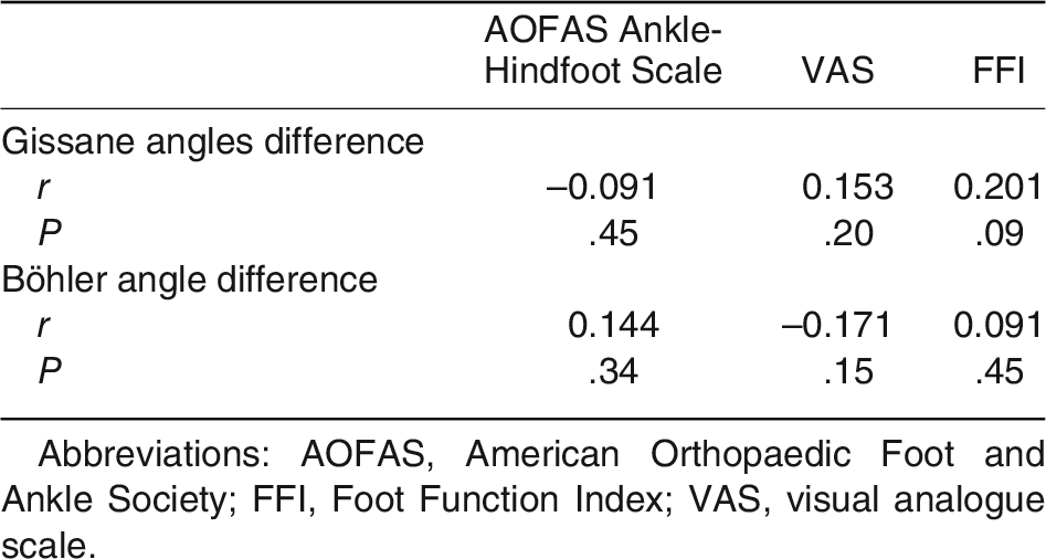
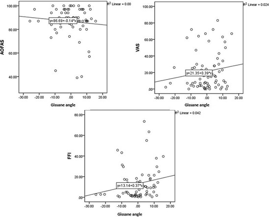
Figure 2.
Scatterplot graphs of clinical outcome scores based on Gissane angle difference.
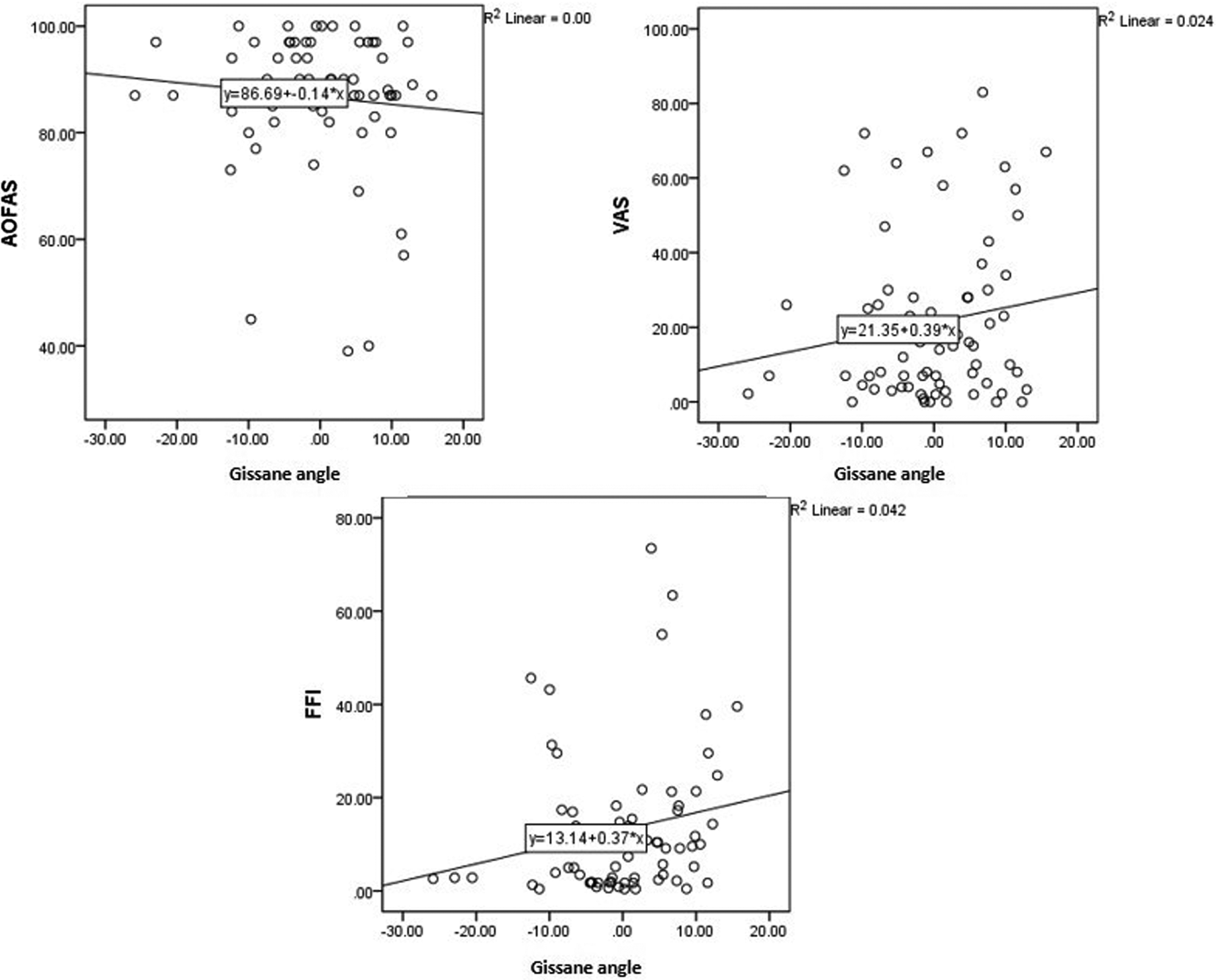
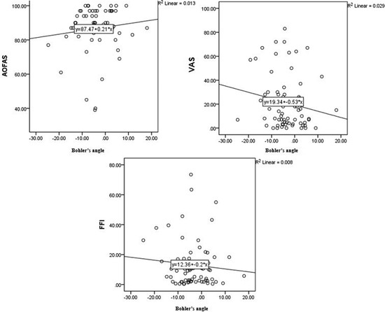
Figure 3.
Scatterplot graphs of clinical outcome scores based on Böhler angle difference.
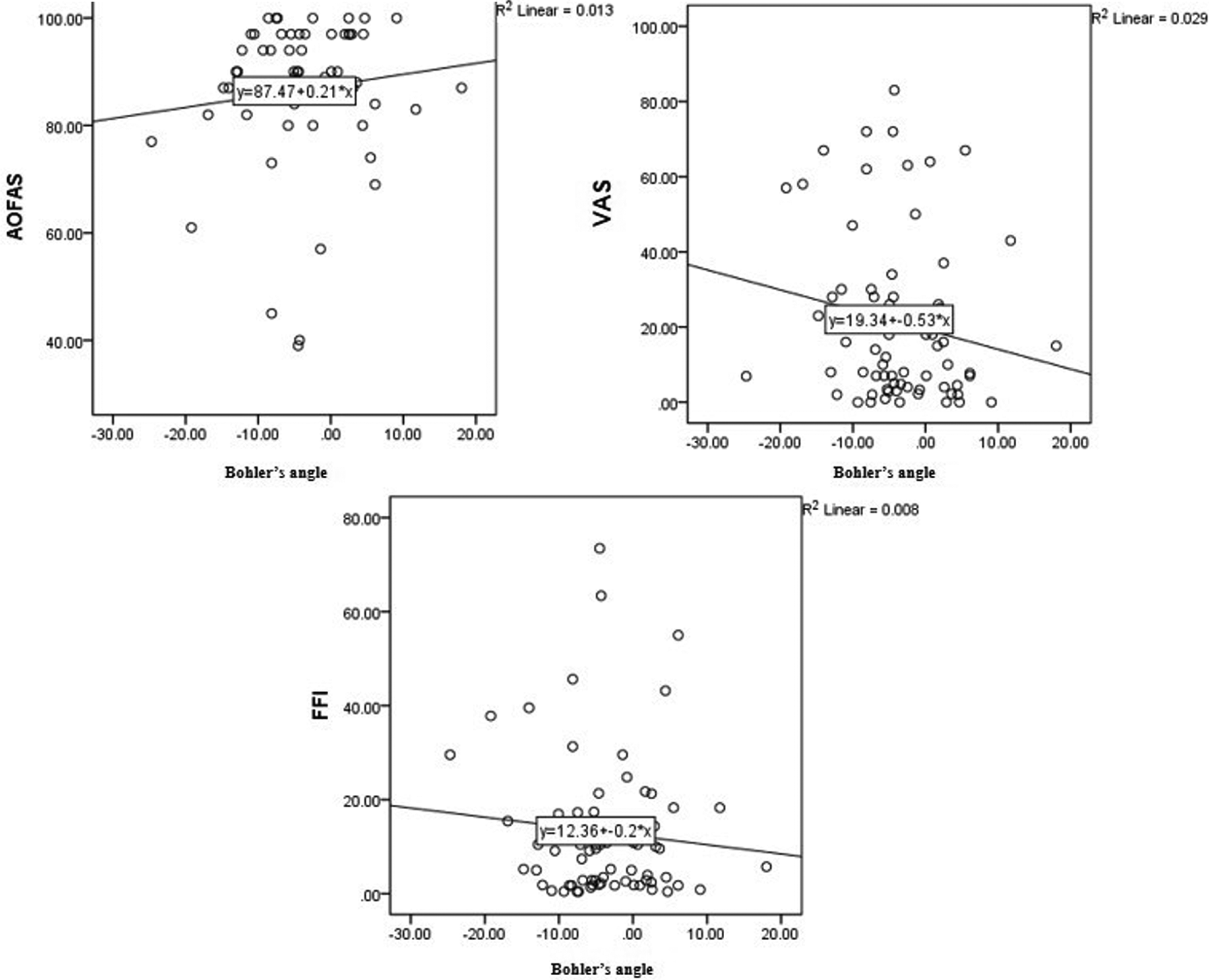
Table 3.
Comparison of Clinical Outcomes with Calcaneocuboid and Subtalar Arthritis Based on the Kellgren-Lawrence Grading Scale
Discussion
Radiologic evaluation is a useful modality for assessing foot and ankle problems. [23] The correlation between function and quality of life of the patients after the surgical procedures in the foot and ankle and radiologic measures are not always appreciated. [24] Likewise, our study found no correlation between postoperative clinical findings and radiologic variables in patients treated surgically for fracture of the calcaneus.
The mean value of the AOFAS Ankle-Hindfoot scale, in our study, was approximately 86.7, which is a little above what was mentioned in previous studies (range, 65 to 85). [8,9,11] The mean VAS score at the last follow-up visit was 2.1 of 10, similar to the study by Ortu et al, [8] whereas previous studies reported higher VAS scores, between 5 and 9. [11,15] The mean FFI in our patients (13.1) is comparable to the other studies evaluating FFI in surgically treated calcaneal fractures. [25,26] These differences may be the result of various fracture types, postoperative protocols, cooperation of the patients, and experience of the surgeons. Notably, one reason for the better outcomes in our series was the exclusion of complicated cases with open fractures, postoperative wound breakdowns, and infections.
Although several radiographic measurements such as the DeLangre angle, the facet inclination angle, the front angle, the calcaneal inclination angle, the talar declination angle, the posterior facet angle, the talo-tuber angle, the talocalcaneal angle, the tuber plantar angle, the anterior-front angle, and the calcaneal compression angle were introduced and evaluated for postoperative calcaneal fractures, [11,14] Böhler and Gissane angles are among the most commonly used parameters for evaluation of calcaneal fractures. [27] Measuring these angles is simple, and surgeons are familiar with their calculation. Despite comparing these angles with the reference values in the previous studies, [11] we calculated the difference in values of the aforementioned angles with regard to the uninjured side to standardize and increase the accuracy of measurements.
By reviewing the literature, the following studies could describe correlation between some radiologic variables and functional outcome in surgically treated calcaneal fractures. Magnan et al [12] could depict better clinical outcome, checked by the Maryland Foot Score, in cases with anatomical realignment of the calcaneus and restoration of the calcaneal height in 54 patients after ORIF of calcaneal fractures. Also, Qiang et al, [13] by evaluating computed tomographic images of 156 patients along with AOFAS and the 36-Item Short-Form Health Survey, found that restoration of Böhler and Gissane angles is associated with better AOFAS score but no correlation with the 36-Item Short-Form Health Survey. Furthermore, Su et al [16] discovered a moderate to low correlation between the change in Böhler angle, when compared to the normal side, and the AOFAS Ankle-Hindfoot scale in the Chinese population. They concluded that only postoperative Böhler angle has a substantial correlation with the functional recovery following ORIF of calcaneal fractures. Likewise, Ågren et al [15] showed a correlation between improved VAS pain and better Böhler angle restoration. They did not, however, examine the relationship between postoperative angles and clinical outcomes.
In contrast, Schepers et al, [11] after studying 33 patients at a mean follow-up of 29 months, stated that radiographic assessment had a poor correlation with the final outcome. They checked several angles and distances and could not find any correlation between clinical outcomes evaluated by the AOFAS, the VAS, and radiographic measurements. Moreover, Baptista et al [10] did not find important correlation between Böhler angle, Gissane angle, tibiotalar angle, talocalcaneal height, and clinical outcomes. They evaluated only postoperative skin complications, disabling residual pain, and need for secondary surgery. We think that these variables cannot exactly determine the functional results of the patients. In addition, Nooijen et al [14] found no apparent correlation between radiologic findings and clinical outcomes evaluated by the AOFAS and the FFI. They did not evaluate arthritic changes in subtalar and calcaneocuboid joints. These controversies could be attributed to the differences in sample sizes, fracture types, surgical techniques, surgeon experience, postoperative rehabilitation protocols, and follow-up periods.
Our findings revealed that the majority of the cases (71.4%) had no arthritic changes in the calcaneocuboid joints, but only 21.4% had normal subtalar joints without radiologic evidence of arthritis. This finding is consistent with the findings of Ibrahim et al, [28] who reported that the majority of cases had moderate to severe subtalar arthritis. Their study found no significant difference in the severity of subtalar arthritis between the operative and nonoperative groups with intraarticular calcaneal fractures, but they did not examine its probable relationship with clinical outcome. Although previous studies could not find any significant association between postoperative clinical outcomes with congruity of the joint, [12,13] they did not grade the severity of arthritic changes in the calcaneocuboid and subtalar joints. Our results showed that there is no relationship between the severity of the arthritis in either the calcaneocuboid joint or the subtalar joint and the postoperative functional outcomes.
One of the strengths of this study was that all operations were performed by a single orthopedic foot and ankle surgeon. This might prevent the common technical bias in studies by multiple surgeons. Another strength is calculating the Böhler and Gissane angle differences with regard to the uninjured calcaneus. Also, we evaluated the correlation of the grade of calcaneocuboid joint and subtalar joint arthritis with clinical outcomes, which is unique in this study. Moreover, we excluded associated injuries such as peroneal tendon instability accompanying calcaneal fractures [29] or cases with postoperative complications such as infection, [30] which can obviously modify the postoperative outcomes.
We did not assess the changes of immediate postoperative Böhler and Gissane angles with the final follow-up visit and compare them with clinical outcomes. If we had a longer postoperative follow-up, grading of arthritic changes and their effect on clinical outcomes might be changed; thus, further studies with long-term follow-up are required.
Conclusions
In conclusion, there is no significant correlation between Böhler and Gissane angles and the clinical outcomes in surgically treated calcaneal fractures. Also, functional outcomes do not change considerably among different grades of arthritis in calcaneocuboid and subtalar joints, at least at short- to mid-term follow-up. Radiologic findings after ORIF of calcaneal fractures are not predictors of function of the patients. Further studies with longer follow-up may be needed to exactly determine the correlation between arthritic changes in the subtalar and calcaneocuboid joints and the functional and clinical outcomes after ORIF of calcaneal fractures.
Financial Disclosure: None reported.
Conflict of Interest: None reported.
References
- Galluzzo M, Greco F, Pietragalla M, et al: Calcaneal fractures: radiological and CT evaluation and classification systems. Acta Biomed 89(suppl 1): 138, 2018.
- Humphrey J, Woods A, Robinson A: The epidemiology and trends in the surgical management of calcaneal fractures in England between 2000 and 2017. Bone Joint J 101: 140, 2019.
- Vosoughi AR, Borazjani R, Ghasemi N, et al: Different types and epidemiological patterns of calcaneal fractures based on reviewing CT images of 957 fractures. Foot Ankle Surg 28: 88, 2022.
- Kinner B, Schieder S, Müller F, et al: Calcaneocuboid joint involvement in calcaneal fractures. J Trauma Acute Care Surg 68: 1192, 2010.
- Popelka V, Zamborsky R: Operative treatment algorithm for intra-articular calcaneal fractures. Acta Orthop Belg 84: 469, 2018.
- Yoosefinejad AK, Khademi S, Vosoughi AR: Consequences of extensile lateral approach to the calcaneal fractures on balance and isokinetic strength of muscle groups crossing the ankle joint. Foot Ankle Surg 28: 732, 2022.
- Fourgeaux A, Estens J, Fabre T: Three-dimensional computed tomography analysis and functional results of calcaneal fractures treated by an intramedullary nail. Int Orthop 43: 2839, 2019.
- Ortu S, Brusoni M, Pisanu F: Functional and radiological outcomes in displaced heel fractures: open reduction and internal fixation versus external fixation. Orthop Rev 12: 8506, 2020.
- De Groot R, Frima A, Schepers T, et al: Complications following the extended lateral approach for calcaneal fractures do not influence mid- to long-term outcome. Injury 44: 1596, 2013.
- Baptista M, Pinto R, Torres J: Radiological predictive factors for the outcome of surgically treated calcaneus fractures. Acta Orthop Belg 81: 218, 2015.
- Schepers T, Ginai A, Mulder P, et al: Radiographic evaluation of calcaneal fractures: to measure or not to measure. Skeletal Radiol 36: 847, 2007.
- Magnan B, Samaila E, Regis D, et al: Association between CT imaging at follow-up and clinical outcomes in heel fractures. Musculoskelet Surg 94: 113, 2010.
- Qiang M, Chen Y, Jia X, et al: Post-operative radiological predictors of satisfying outcomes occurring after intra-articular calcaneal fractures: a three dimensional CT quantitative evaluation. Int Orthop 41: 1945, 2017.
- Nooijen LE, Spierings KE, Sanders FR, et al: Determining the correlation between lateral radiograph morphology and the outcome following surgically treated intra-articular calcaneal fractures. Foot Ankle Spec 14: 105, 2021.
- Ågren P-H, Mukka S, Tullberg T, et al: Factors affecting long-term treatment results of displaced intraarticular calcaneal fractures: a post hoc analysis of a prospective, randomized, controlled multicenter trial. J Orthop Trauma 28: 564, 2014.
- Su Y, Chen W, Zhang T, et al: Bohler’s angle’s role in assessing the injury severity and functional outcome of internal fixation for displaced intra-articular calcaneal fractures: a retrospective study. BMC Surg 13: 40, 2013.
- Vosoughi AR, Shayan Z, Salehi E, et al: Agreement between Sanders classification of intraarticular calcaneal fractures and assessment during the surgery. Foot Ankle Surg 26: 94, 2020.
- Vosoughi AR, Roustaei N, Mahdaviazad H: American Orthopaedic Foot and Ankle Society ankle-hindfoot scale: a cross-cultural adaptation and validation study from Iran. Foot Ankle Surg 24: 219, 2018.
- Budiman-Mak E, Conrad K, Stuck R, et al: Theoretical model and Rasch analysis to develop a revised Foot Function Index. Foot Ankle Int 27: 519, 2006.
- Tegner Y, Lysholm J: Rating systems in the evaluation of knee ligament injuries. Clin Orthop Relat Res 198: 43, 1985.
- Lamm BM, Stasko PA, Gesheff MG, et al: Normal foot and ankle radiographic angles, measurements, and reference points. J Foot Ankle Surg 55: 991, 2016.
- Kellgren JH, Lawrence J: Radiological assessment of osteo-arthrosis. Ann Rheum Dis 16: 494, 1957.
- DiGiovanni CW, Greisberg J: “Foot and Ankle Core Knowledge,” in Orthopaedics, Elsevier Health Sciences, Philadelphia, 2007.
- Hoveidaei AH, Roshanshad A, Vosoughi AR: Clinical and radiological outcomes after arthrodesis of the first metatarsophalangeal joint. Int Orthop 45: 711, 2021.
- Biz C, Barison E, Ruggieri P, et al: Radiographic and functional outcomes after displaced intra-articular calcaneal fractures: a comparative cohort study among the traditional open technique (ORIF) and percutaneous surgical procedures (PS). J Orthop Surg Res 11: 1, 2016.
- Backes M, Schep NW, Luitse JS, et al: The effect of postoperative wound infections on functional outcome following intra-articular calcaneal fractures. Arch Orthop Trauma Surg 135: 1045, 2015.
- Epstein N, Chandran S, Chou L: Current concepts review: intra-articular fractures of the calcaneus. Foot Ankle Int 33: 79, 2012.
- Ibrahim T, Rowsell M, Rennie W, et al: Displaced intra-articular calcaneal fractures: 15-year follow-up of a randomised controlled trial of conservative versus operative treatment. Injury 38: 848, 2007.
- Vosoughi AR, Afaridi E, Solooki S, et al: Prevalence and Predictors of Peroneal Tendon Instability Accompanying Calcaneal Fractures. Foot Ankle Int 44: 825, 2023.
- Vosoughi AR, Medhati P, Hosseini E, et al: Clinical outcomes following treatment of deep surgical site infection after fixation of calcaneal fractures: A retrospective case-control study. Foot Ankle Surg 29: 334, 2023.
© 2024 American Podiatric Medical Association