


Abstract
First metatarsophalangeal joint (MPJ) arthritis is a very common form of arthritis seen in the foot. Some signs and symptoms include pain, swelling, decreased passive and active range of motion, difficulty with shoe gear, and so forth. Surgically, options for alleviating symptomatic hallux limitus and arthritis fall into two broad categories: joint sparing and joint sacrificing. In this case study, we present a patient with a bilateral failed total silastic implant of the first MPJ and our proposed revision using an osteochondral bone allograft to fill the deficit left behind from silastic implant removal. The ability for immediate weightbearing and to perform this procedure bilaterally is an advantage to this surgical treatment option compared with other described revision techniques. Postoperatively, the patient has adequate range of motion and no pain when ambulatory. We believe this osteochondral allograft implant may be a viable option for revision first MPJ arthroplasty in select patient populations.
First metatarsophalangeal joint (MPJ) arthritis is the most common form of arthritis in the foot [1,2]. Some signs and symptoms include pain, swelling, decreased passive and active range of motion, difficulty with shoe gear, and so forth. The first MPJ, like other joints, can become arthritic for many reasons, but the result of denuding or loss of articular cartilage leads to pain and deformity. Articular cartilage is an avascular structure that does not regenerate once denuded or damaged. Because of cartilage loss and the inability to restore native cartilage, treatment options revolve around alleviating the symptoms. Conservative treatment options include carbon fiber inserts with Morton extension, physical therapy, rocker bottom shoe gear, nonsteroidal anti-inflammatory drugs, and steroid injections into the joint.
Surgically, options for alleviating the symptoms of hallux limitus or arthritis of the first MPJ fall into two broad categories; joint sparing and joint sacrificing. Joint-sparing procedures allow for the maintenance of the articulation and aim to decompress the joint space with procedures such as a Moberg osteotomy and chilectomy. Joint-sacrificing procedures can either maintain or sacrifice range of motion. Motion-sacrificing procedures include arthrodesis which alleviates symptoms by stopping motion over denuded cartilage and limits retrograde dorsal jamming that tends to cause pain and irritation. Some motion-sparing procedures include arthroplasty with the implementation of implants and interposition grafts. Arthroplasties have been reported in the literature to maintain first MPJ range of motion and alleviate pain [3,4].
There has been a plethora of implants and advancements when it comes to joint replacement in the foot and ankle and the first MPJ has not been neglected. Hemi-implants and total implants have come and gone with debated success. Early first MPJ joint replacement operations initially demonstrated pain relief with perceived maintenance of motion. However, long-term follow-up revealed instability, malalignment, bone erosion, and decreased range of motion [1]. Furthermore, long-term studies show significant reoperation and failure rates of implants in the first MPJ, leading to recorded revision rates of 25% [6]. Salvage arthrodesis has become the main operative tool for failed arthroplasties for foot and ankle surgeons and has been noted to have favorable outcomes; however, it is not free of complications. Salvage arthrodesis of the 1st MPJ unite slower, have higher reoperation rates, and have lower American Orthopaedic Foot and Ankle Society scores when compared to those treated with primary arthrodesis [5].
In this case study, we present a patient with bilateral failed total silastic implant of the first MPJ and our proposed revision using a proximal and distal cancellous bone plug with nonviable cartilage. This unique case demonstrates complications of silastic implant and a proposed intervention that allows for quick recovery and maintained motion and integrity of the first MPJ. This technique entails removal of the implant with subsequent reaming of the first metatarsal deficit and proximal phalanx deficit, with an osteochondral allograft replacing the site of silastic implant stems at both the first metatarsal head and the base of the proximal phalanx. Use of the osteochondral allograft allowed the patient to bear weight on the bilateral foot immediately postoperatively in surgical shoes. This permitted a single operation on bilateral first MPJs.
Multiple case studies have been presented on autografts and allografts of first MPJ osteochondral defects; however, the author does not know of any case studies that describe their use in failed first MPJ implants [7]. This technique can add additional options for patients who require revision surgery because of failed implants of the first MPJ, which can offer a challenge for patient and surgeon alike. With notable revision rates in first MPJ implants and the complications associated with salvage arthrodesis, we see the osteochondral allograft implant as a viable option for revision first MPJ arthroplasty in a select patient population.
Case Report
History of Present Illness
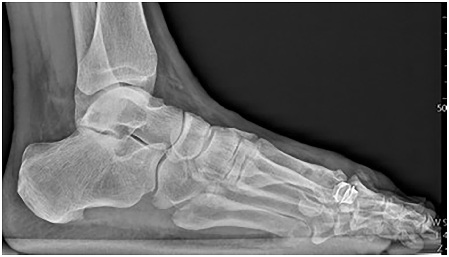
A 58-year-old man presented with bilateral first MPJ pain. He related a history of bilateral total first MPJ silastic implant approximately 13 years ago performed by another community physician. The patient’s primary complaint of pain occurs when ambulating in work boots, and he further related that working a full day is difficult because of the pain. The patient stated that he had difficulty putting the toe through the full range of motion, which had become most pronounced over the past 2 years. Radiographs of the bilateral foot showed implant failure at the hinged silastic implant and grommet displacement (Fig. 1–2). The patient did not want to undergo arthrodesis and would like bilateral extremity procedures performed at the same time because of work commitments. A lengthy discussion was held with the patient regarding multiple treatment options and the risks and benefits of each. The patient elected for surgical implant removal with osteochondral allograft implantation of bilateral first MPJs.
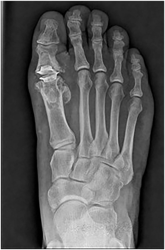
Figure 1.
Preoperative image of failed Swanson silastic implant. Note displacement of grommet.
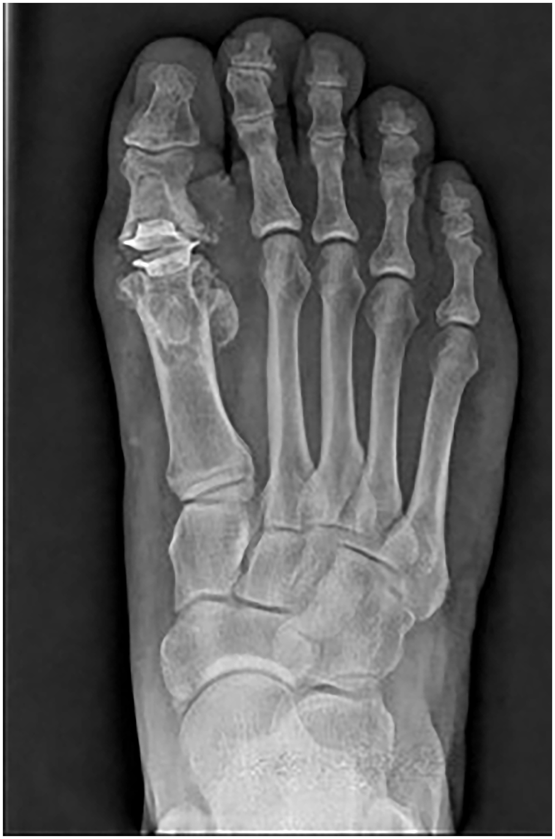
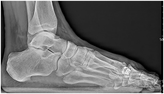
Figure 2.
Preoperative image of failed Swanson silastic implant. Note displacement of grommet.
Surgical Technique
Incision was made at dorsal medial aspect of right first MPJ where two semielliptical incisions were made spanning the first MPJ joint and the previous surgical incision-site cicatrix. This allowed for excision of scar from previous surgery. Dissection continued to the extensor hallucis longus tendon, which was exposed and retracted laterally; all vital structures were visualized and retracted as necessary. Once to the level of the first MPJ capsule, there was noted to be significant discoloration and foreign body granulomas appreciated to the first MPJ, as shown in Figure 3. With the capsule dissected and the first MPJ exposed, the implant was fractured at the hinge of the implant, with grommet displacement as noted in Figures 1, 2 and 4. The silicone implant was removed in its entirety, and there was notable bony destruction and cystic changes to the first MPJ. Extensive debridement was necessary to remove all foreign body granulomas and devitalized/cystic bone. After debridement, copious amounts of normal saline was used to rinse out the surgical site. Resection of the implant is depicted in Figure 5.
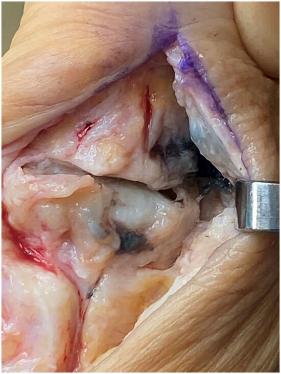
Figure 3.
Intraoperative image of failed Swanson silastic implant showing significant foreign body reaction and joint destruction.
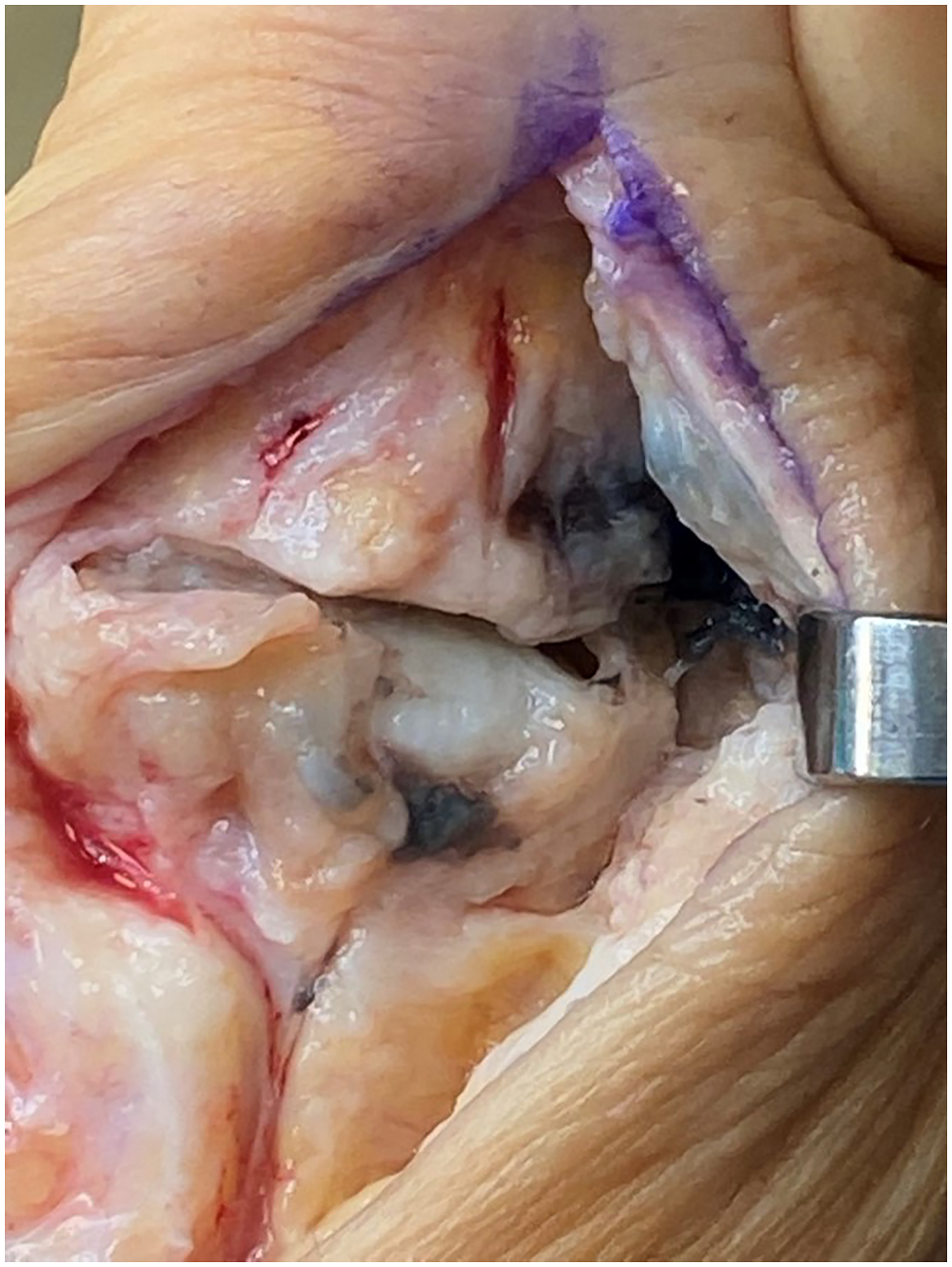
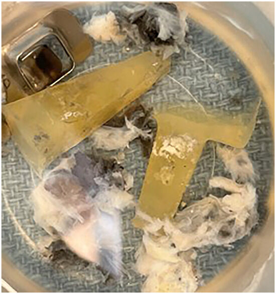
Figure 4.
Failed implant with metal grommets and silastic implant.
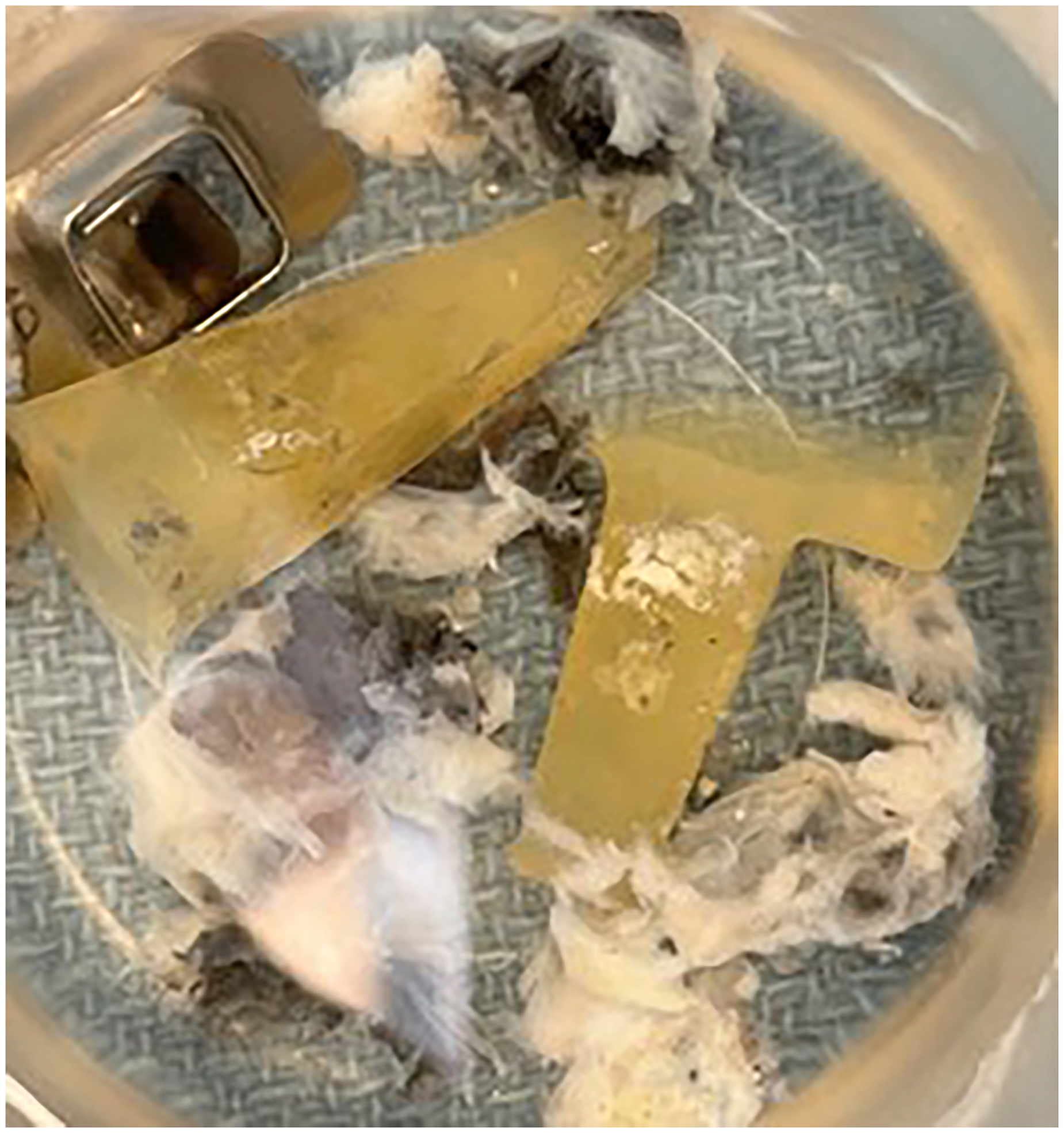
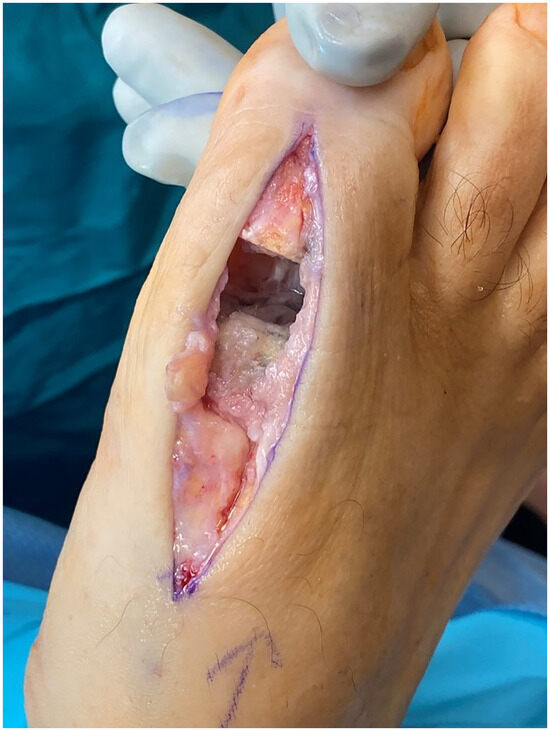
Figure 5.
After removal of implant and resection of bone at the distal first metatarsal and proximal phalanx base.
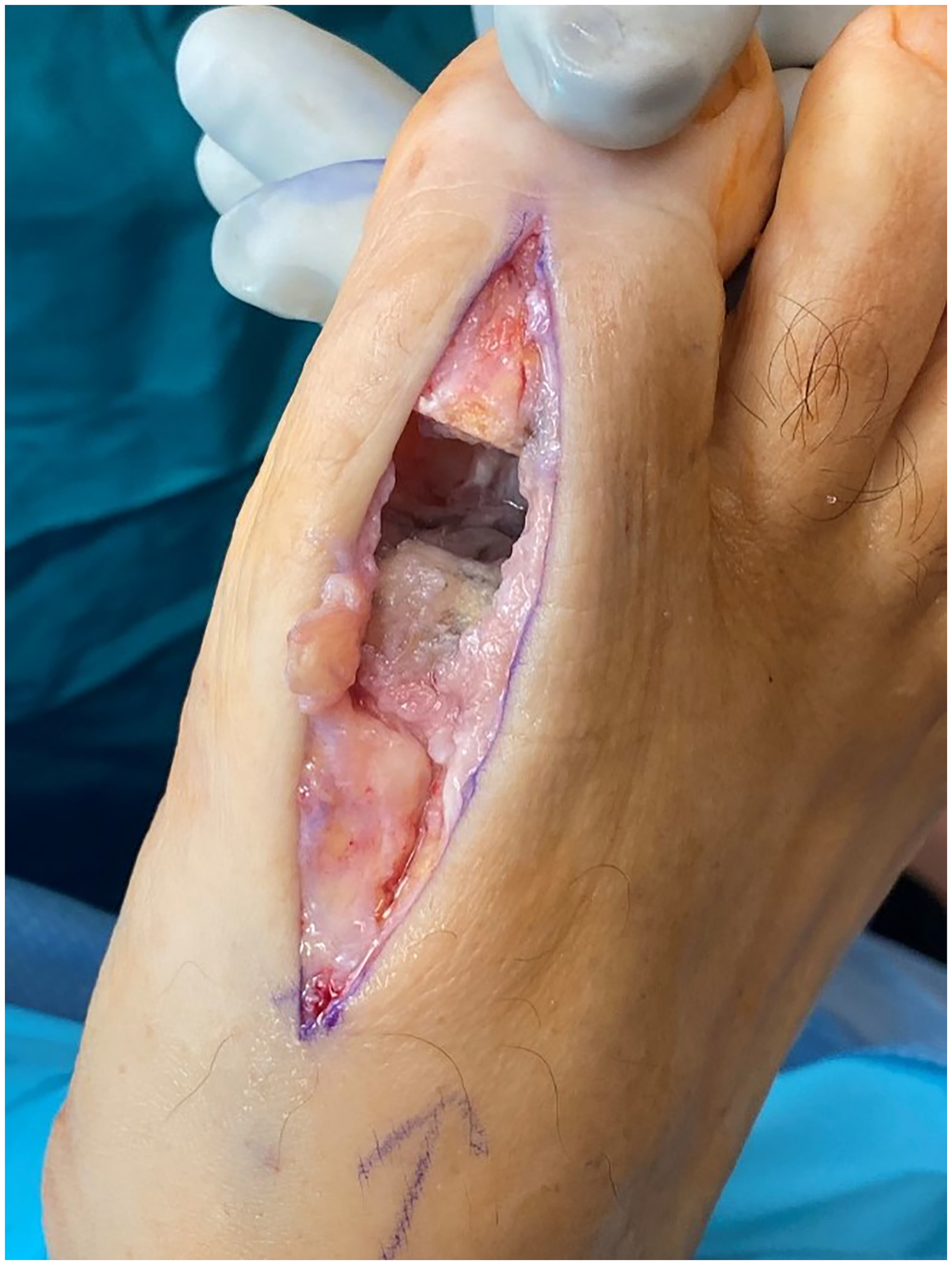
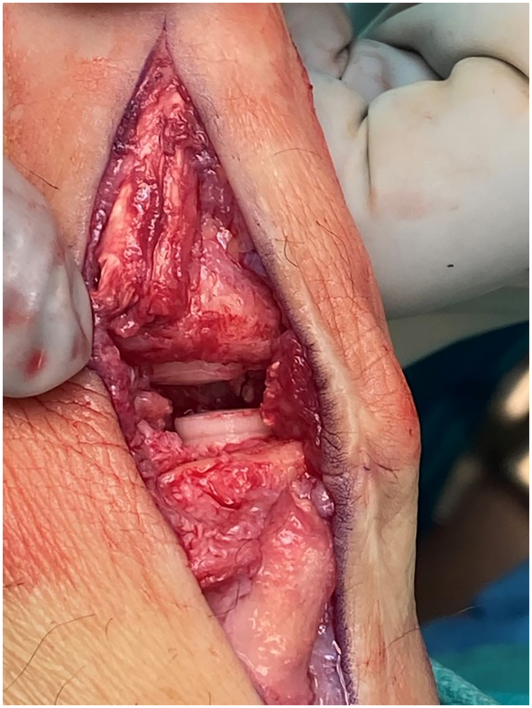
The first metatarsal head was inspected and noted to have a large cartilaginous and bony deficit from the implant that measured approximately 9 mm in diameter. A guide pin was then inserted centrally into the deficit and reamed with a 10-mm-diameter reamer to a depth of 14 mm. A measuring device was inserted into the reamed metatarsal to accurately determine the length of the allograft to be transplanted as noted in Figure 6. The osteochondral allograft was then placed in room-temperature saline for 5 minutes and then measured to ensure 1 to 2 mm of cartilage would be proud at the first metatarsal head. The allograft was submerged in blood drawn from the patient for approximately 3 minutes. The cancellous end of the graft was then beveled with a rongeur, and the graft was then implanted by gently pressing the graft into the reamed bone. Figure 6 shows the press-fit allograft with 1 to 2 mm of proud cartilage at both the metatarsal head and the base of the proximal phalanx.
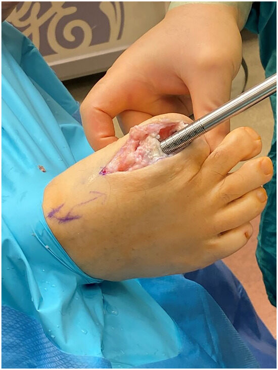
Figure 6.
Measurement of needed allograft bone implant.
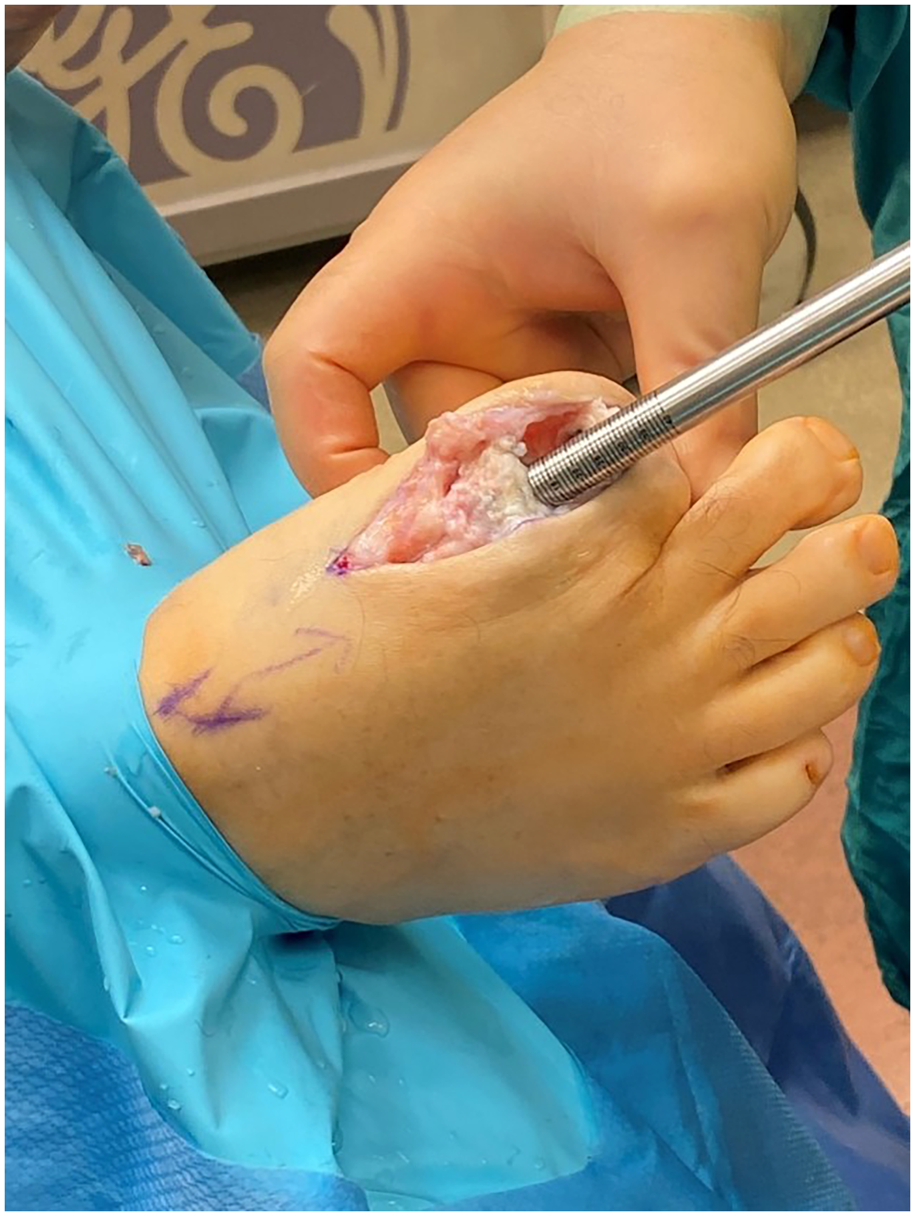
Attention was directed to the base of the proximal phalanx, where osteophyte formation was removed using a rongeur and sagittal saw. The base of the proximal phalanx was noted to have an 8-mm-diameter defect left by removal of the silastic implant. A guide pin was inserted centrally to the deficit reamed with an 8-mm-diameter reamer to 10 mm in depth. Next, the measuring device was inserted into the base of proximal phalanx to accurately determine the length of osteochondral allograft to be transplanted. The allograft was then placed in room-temperature saline for 5 minutes and then measured to ensure 1 to 2 mm of the cartilage cap would be proud. A bone saw was used to reduce excess length of the allograft. The allograft was placed in blood drawn from the patient for approximately 3 minutes. The cancellous end of the graft was then beveled, and graft was implanted by gently pressing it into the base of proximal phalanx (Fig. 7).
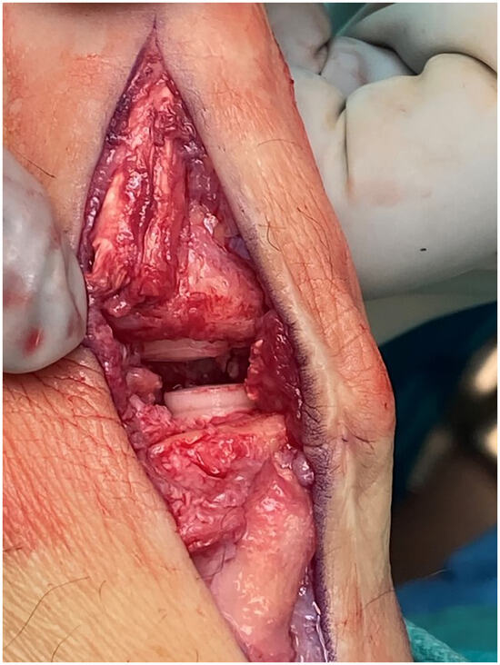
Figure 7.
Intraoperative photograph showing allograft placement at both distal first metatarsal and proximal phalanx base articulations.
Inspection of the first MPJ revealed plantar laxity of the flexor hallucis longus tendon (FHL). This brought concerns for possible floating toe; therefore, FHL transfer was performed. Flexor hallucis longus tendon was transferred to the base of the proximal phalanx through a drill hole in the base of the proximal phalanx passing the suture through this hole and into the FHL tendon under appropriate plantarflexion.
The surgical site was then rinsed with copious amounts of saline. The capsule was reapproximated using 2-0 Vicryl (Ethicon Inc, Somerville, New Jersey). Subcutaneous closure was reapproximated using 3-0 Vicryl, and the skin was reapproximated using 4-0 Monocryl (Ethicon) in a running subcuticular fashion. Next, a dry sterile dressing consisting of Steri-Strips (3M, St. Paul, Minnesota), Xeroform (McKesson, Irving, Texas) 4 × 4, and Kling (Johnson & Johnson, New Brunswick, New Jersey) was applied to the right foot. The ankle tourniquet was then deflated, and prompt vascular return was noted to the right foot. The same surgical procedures and technique was performed on the left first MPJ.
Discussion
Patient selection and patient expectations can be difficult to manage in forefoot surgery. In this case, we believe we were able to provide our patient with an acceptable surgical outcome that matched the expectations. This patient presented with the goals of getting back to work and walking pain-free as quickly as possible. He expressed the desire to have both feet operated on at the same operative day to shorten his time off work and recovery time. These expectations and requests by the patient made removal of implant with arthrodesis a nonviable option at this time. Arthrodesis would entail a period of nonweightbearing and thus would not be performed bilaterally by the operating surgeon. Furthermore, arthrodesis would likely require autogenous graft or allograft to maintain the length of the first ray, which has been shown to have higher complication rates as compared to primary arthrodesis. Revision first MPJ arthrodesis has shown to be more difficult, with high reoperation rates, nonunion, and donor-site morbidity, as autogenous bone grafting may be required [5]. The patient did not elect for arthrodesis for these reasons.
After discussing the many risks and benefits of his surgical options, the patient elected to undergo osteochondral allograft implantation of the first MPJ bilaterally. As described in the technique above, the first metatarsal head and base of the proximal phalanx were reamed based on the intraoperative deficit. This deficits were noted to be 8 mm and 10 mm, and the respective reamers were used to remove all affected bone after removal of silastic implant. This allowed for removal of foreign body debris, which may have been present secondary to the silastic implant and grommet. With regard to depth of reaming, it is important to accurately measure using a depth gauge (Fig. 6) to allow for 1 to 2 mm of allograft kept proud at both the metatarsal and proximal phalanx end of first MPJ. We believe by leaving this allograft 1 to 2 mm proud on both the metatarsal and proximal phalanx sides of the first MPJ, we allow for decompression of the joint and a smooth range of motion. Because of variability of deficit, we recommend having several sizes of allograft plug available, keeping in mind the length of plug can be cut shorter and beveled for press fitting.
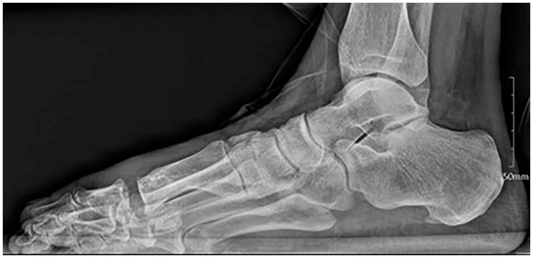
Postoperatively, the patient was placed in bilateral surgical shoes and discharged on the day of surgery with crutches and orders for weightbearing as tolerated. The patient has since followed up at 2 weeks postoperatively for suture removal and was without wound complications. After suture removal, the patient transitioned to normal shoe gear and was back to work at week 6. Radiographs were then taken at 6-month follow-up that show acceptable alignment with maintained first-ray length (Fig. 8, Fig. 9, Fig. 10, Fig. 11). The patient reported overall improved pain and satisfaction.
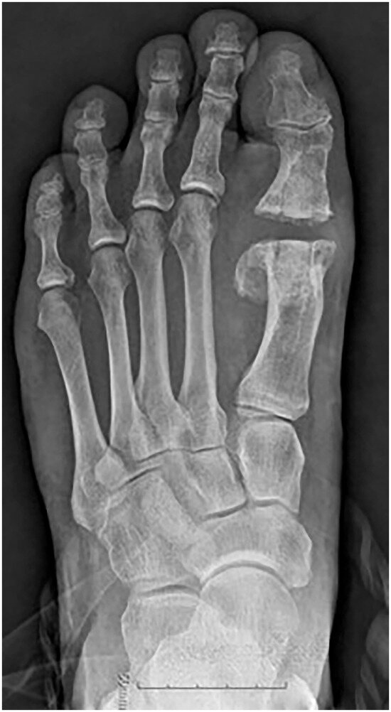
Figure 8.
Postoperative anteroposterior image of the left foot.
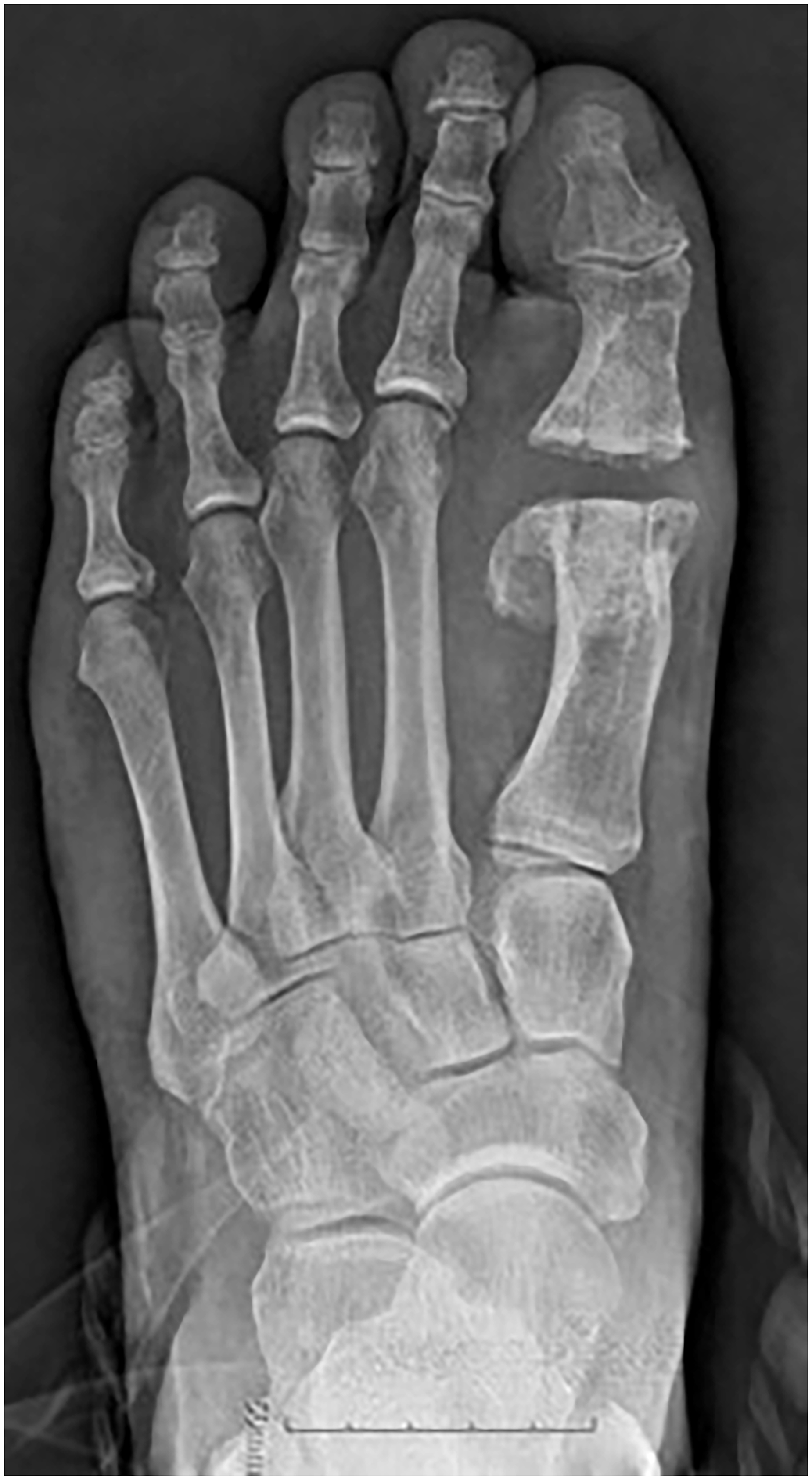
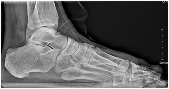
Figure 9.
Postoperative lateral image of the left foot.
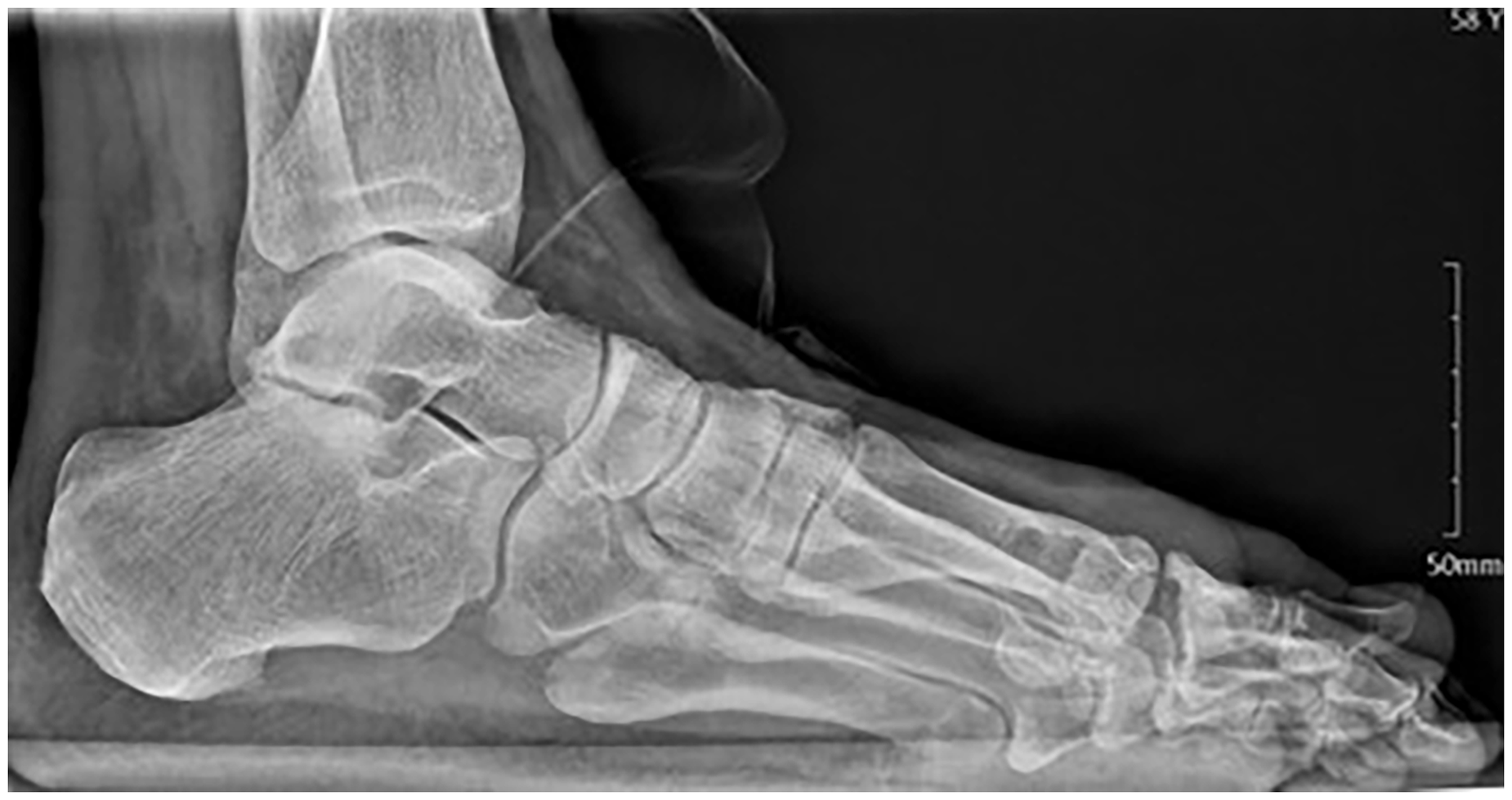
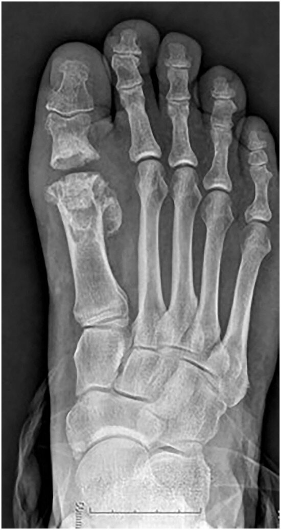
Figure 10.
Postoperative anteroposterior image of the right first metatarsophalangeal joint shows minimal shortening and adequate alignment/joint space.
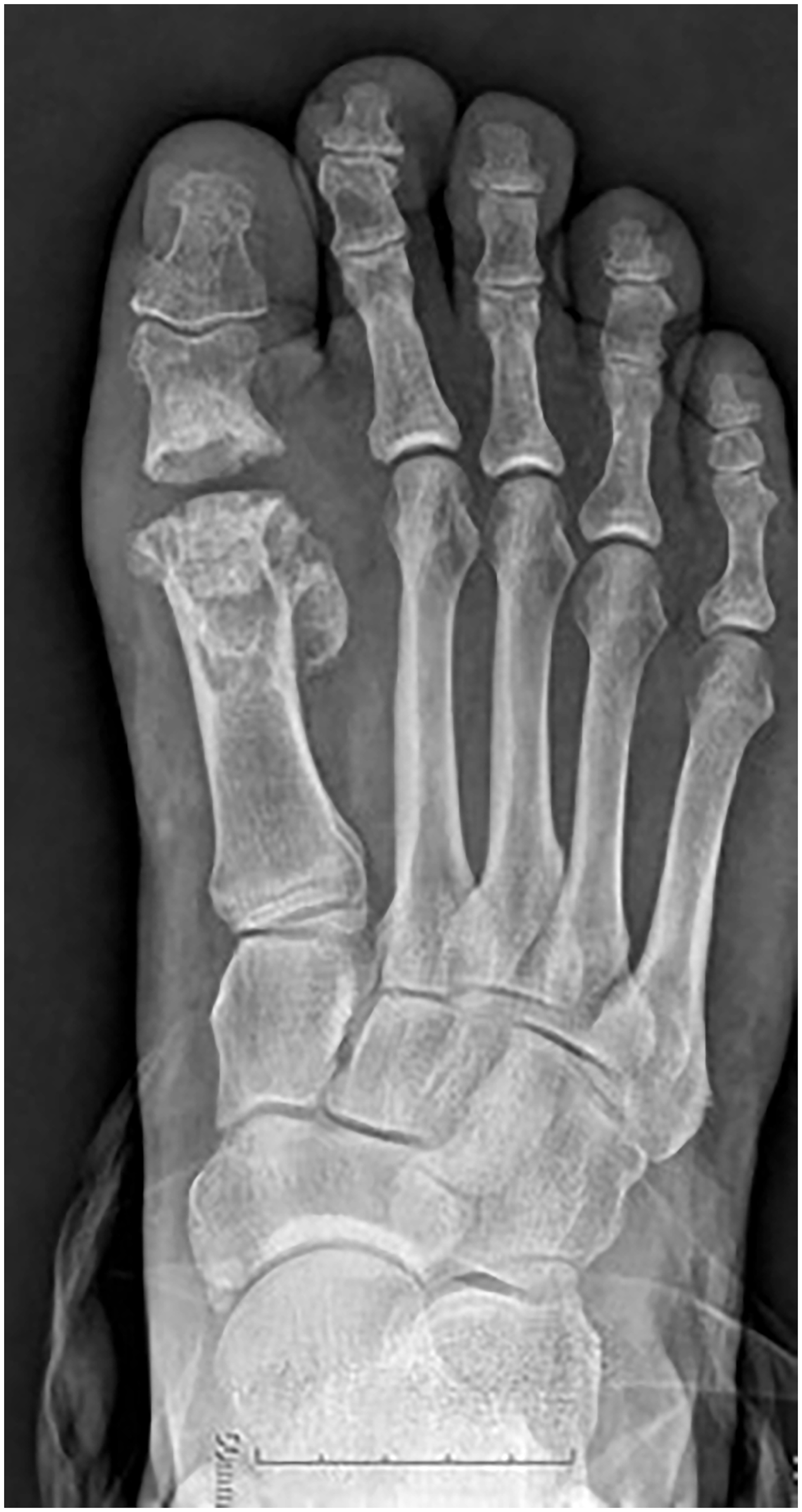
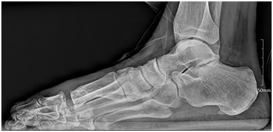
Figure 11.
Postoperative lateral image of right first metatarsophalangeal joint shows minimal shortening and adequate alignment/joint space.
The option for osteochondral allograft implantation was made by the patient and surgeon, as it did not impede future revision surgical intervention should this become necessary. Implantation of the osteochondral allografts into the deficits left behind from the removal of the silastic implants allow for maintained structural integrity of the first metatarsal and proximal phalanx. The restored cartilaginous surface from the allograft allows for reduced pain and increased motion. Finally, to implant the osteochondral allograft, intramedullary reaming was necessary, which eliminated the residual silicone particulate left in the medullary canal of the metatarsal and proximal phalanx. By removing silicone particulate and foreign body, we hoped to reduce any further degeneration and foreign body reaction, which was noted intraoperatively.
Postoperative radiographs revealed minimal shortening and, clinically, the patient is with adequate range of motion and minimal to no pain with ambulation. The patient was able to return to work and met his goal of returning to work with minimal time off. We believe osteochondral allograft bone plug with nonviable cartilage can be a viable option for revision first MPJ arthroplasty in a select patient population. This procedure will also allow for the option to further perform arthrodesis in the future if pain or debility arise.
Conclusions
Osteochondral allograft bone plug implantation with nonviable cartilage can be a viable alternative when removing first MPJ implants of any type. In our case, bilateral silastic first MPJ implants were removed because of implant failure and secondary foreign body granuloma formation from silicone breakdown. The ability for immediate weightbearing and to perform this procedure bilaterally is an advantage to this surgical treatment option as compared to other described revision techniques. At this time, revision arthrodesis is the standard treatment; however, this is often associated with delayed union, nonunion, and need for autogenous or allograft for maintenance of first-ray length. Arthrodesis also limits future surgical options if this becomes necessary. With patients living longer and maintaining activity at greater ages, there is a potential of increased need for revision surgery in patients who have had first MPJ implant arthroplasty. This technique adds to options for patients and surgeons alike when dealing with these complicated revision cases. The procedure also permits for the option to further perform arthrodesis in the future if pain or debility arise as length and integrity of bone are maintained.
Financial Disclosure
None reported.
Conflict of Interest:
None reported.
References
- Anderson MR, Ho BS, Baumhauer JF: Current concepts review: hallux rigidus. Foot Ankle Orthop 3: 247301141876446, 2018.
- Ashford RL, Tollafield D, Vogiatzoglou F, et al: A retrospective analysis of Swanson Silastic double-stemmed great toe implants with titanium grommets following podiatric surgery for arthritic joint disease. Foot 10: 69, 2000.
- Barouk LS, Barouk P: Joint-preserving surgery in rheumatoid forefoot: preliminary study with more-than-two-year follow-up. Foot Ankle Clin 12: 435, 2007.
- Berlet GC, Hyer CF, Lee TH, et al: Interpositional arthroplasty of the first MTP joint using a regenerative tissue matrix for the treatment of advanced hallux rigidus. Foot Ankle Int 29: 10, 2008.
- Gross CE, Hsu AR, Lin J, et al: Revision MTP arthrodesis for failed MTP arthroplasty. Foot Ankle Spec 6: 471, 2013.
- Stone OD, Ray B, Thomsen CE, et al: Long-term follow-up of arthrodesis vs total joint arthroplasty for hallux rigidus. Foot Ankle Int 38: 375, 2016.
- Zelent ME, Neese DJ: Osteochondral autograft transfer of the 1st metatarsal head: a case report. J Foot Ankle Surg 44: 406, 2006.
© 2023 American Podiatric Medical Association