


Abstract
Isolated congenital pseudarthrosis of the fibula is a rare entity with a limited number of cases reported in the literature. Treatment is challenging because of recalcitrant nonunion and because no consensus about the best treatment plan exists. We report a case of isolated congenital fibular pseudarthrosis with valgus deformity of the ankle. The patient had a history of two failed operations. We used a novel surgical plan that combined tibiofibular synostosis with fibular segment transfer through a unilateral external fixator. The patient showed good early results with fibular union. We advocate the combination of tibiofibular synostosis and fibular segment transfer to restore the integrity and stability of the ankle in recalcitrant isolated congenital fibular pseudarthrosis cases with a history of failed surgery.
Isolated congenital pseudarthrosis of the fibula (ICPF) is an uncommon condition that is associated with neurofibromatosis in approximately 40% of patients. [1] Although fibular bowing or prepseudoarthrotic changes are present from birth, the diagnosis is often delayed until the activities of childhood cause the condition to become symptomatic. [2]
Dooley et al [3] described a four-part classification of congenital pseudarthrosis of the fibula; however, only types 2 and 3 are ICPF: part 1, fibular bowing without fibular pseudarthrosis; part 2, fibular pseudarthrosis without ankle deformity; part 3, fibular pseudarthrosis with ankle deformity but without the late development of tibial pseudarthrosis; and part 4, fibular pseudarthrosis with the late development of tibial pseudarthrosis. Nonunion usually occurs at the distal third of the fibula in ICPF; thus, the fibula becomes ineffective to provide lateral support to the talus through the lateral malleolus. [4] Lack of lateral support produces lateral instability of the ankle joint, resulting in severe valgus. Patients also may present with an abnormal gait or a prominent fibula. [5]
Because of bad functional results and the scarcity of this disease, different operative techniques are proposed and various therapeutic methods can be combined to improve results. Congenital pseudarthrosis of the fibula is frequently associated with a pseudarthrosis of the tibia, but its isolated form is uncommon. Isolated congenital pseudarthrosis of the fibula may be relatively asymptomatic, as the fibula bears only 7% to 15% of weight across the ankle. [6] However, lack of lateral thrust produced by the fibula and fibular shortening because of established pseudarthrosis predispose to the development of valgus instability. [7] A close relation between the fibula shortening and progressive valgus deformity has been well documented by Dias. [7] As the valgus deformity progresses, patients may have limping and pain on walking and radiologic changes in the tibia; even pathologic fracture may be seen. [3]
Because of the rarity of the condition, limited information, and uncertain results, the treatment of ICPF is also difficult. Various therapeutic methods have been proposed and comprehensive approaches are often required. When prepseudarthrosis is defined, observation and close monitoring are recommended. Prophylactic splinting or bracing is also an option to prevent the development of the deformity, [8] although its efficacy is controversial. [4]
After pseudarthrosis has developed, in skeletally immature patients with neutral ankle alignment, fibular osteosynthesis is indicated when the distal fibular fragment is large and the gap left by the excised lesion can be filled with a bone graft. [4]
In case of deformity of the ankle (Dooley type 3), distal tibiofibular synostosis is recommended, which was first advocated by Langenskiöld, [9] to stabilize the ankle and prevent the progressive valgus by fusing the metaphyses of the distal tibia and fibula. [4] However, the tibial and fibular physes have a unique pattern of growth, and a slight discrepancy of growth ratio is present between the distal tibia and fibula. [5] Therefore, the loss of the normal mechanics of the distal tibiofibular-talar articulation may lead to gradual valgus deformity in a growing child, although synostoses may prevent severe ankle valgus. [5,10]
Given the above findings, some authors recommend tibiofibular synostosis only for failed or nonfeasible osteosynthesis. [5] If tibiofibular synostosis is performed before the age of 10 years, medial hemiepiphysiodesis of the distal tibia should be considered to correct the valgus deformity. [4]
In skeletally mature patients, the Ilizarov technique can be used to restore fibular length by transferring the bone after supramalleolar osteotomy. [4] Bone grafting and a distal pedicled returned periosteal flap method can also be used as fibular continuity restitution procedures. However, these operative techniques also have a high risk of failure because of resorption of the graft and may require reoperations. [11,12] Here, we reported the treatment of a recalcitrant ICPF case by combining two different techniques: fibular segment transfer and tibiofibular synostosis with a unilateral external fixator.
Case Report
A 11-year-old girl presented with pain and deformation of the left ankle. Examination showed an incision scar at the distal lateral part of the fibula, and a valgus deformity of the ankle was present on standing. Her main complaint was ankle instability during weightbearing, disrupting her walking ability. Spine examination was normal, no café au lait spots were observed, and there was no family history of neurofibromatosis. The patient had already undergone two failed plate-screw fibular osteosynthesis with autograft application operations for the treatment of ICPF in different hospitals. Plain radiographs and computed tomography of the ankle revealed pseudarthrosis at the distal third of the fibula with a normal tibia (Dooley ICPF type 3) (Fig. 1).
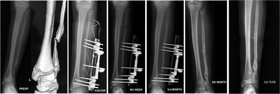
Figure 1.
Preoperative, early postoperative, and postoperative (week-6, month-3, month-6, and year-1) radiographs of the patient.
The patient was prepared for surgery. The surgical aim was to restore fibular length and obtain a stable tibiofibular relationship to prevent valgus instability of the ankle that was caused by fibular nonunion and defect.
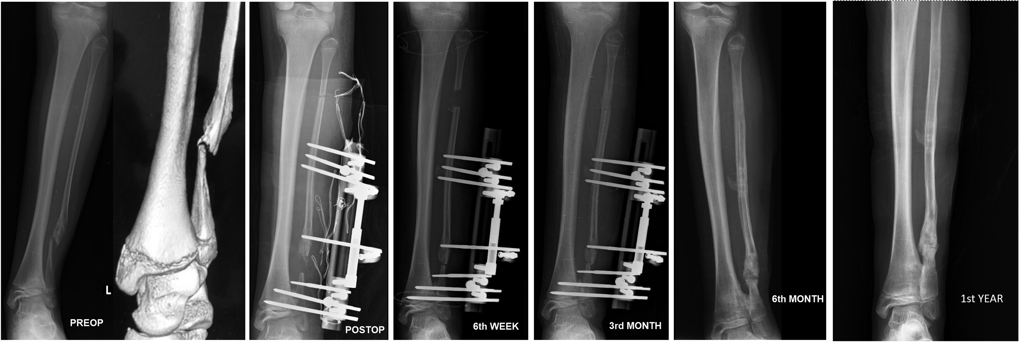
The patient underwent proximal fibular osteotomy after excision of the pseudarthrosis. Schanz screws were then inserted to transfer the fibular segment through the unilateral external fixator. Distal Schanz screws of the fixator were attached to the tibiofibular joint and tibiofibular fixation was achieved (Fig. 2). Caution was taken to place distal Schanz screws proximal and parallel to growth plates, to prevent any physeal damage that can cause a deformity during growth.
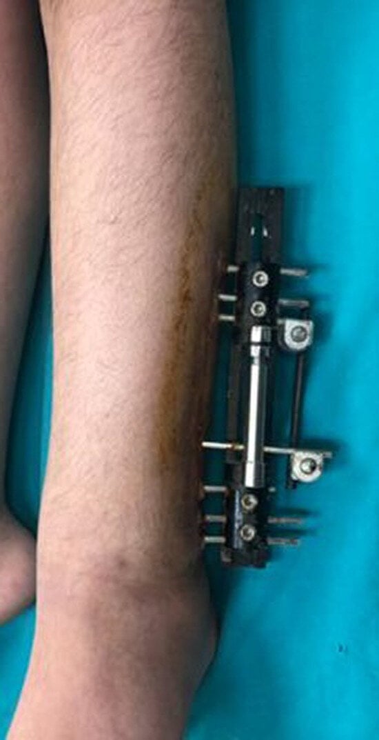
Figure 2.
Application of the unilateral external fixator.
Six weeks postoperatively, the proximal fibular defect had closed and consolidation occurred in the third month. External fixator was removed and weightbearing was allowed at the end of the third month. Her latest follow-up radiographs at postoperative month 6 showed fusion across the tibiofibular syndesmosis and union at the ICPF area (Fig. 1). After removal of the brace, the outcome was favorable, with the disappearance of clinical symptoms. The patient was mobilized without pain or any assisting device, and there was no valgus instability in the ankle. Her foot was plantigrade, she could walk freely without any sign of ankle instability, and no sequela was detected at first-year follow-up. The patient will be monitored closely to reveal whether any deformity requiring hemiepiphysiodesis or supramalleolar osteotomy procedures will develop.
Conclusions
Knowing the failure risks and unsuccessful fibular osteosyntheses history of our patient, we composed a new treatment modality. Excision of the nonunion part of the fibula and a successful transfer of fibular segment with a unilateral external fixator restored the integrity of the fibula, and attaching the distal Schanz screws of the fixator to the tibiofibular region at the same time managed the synostosis. Tibiofibular synostosis is highly important when fibular union cannot be restored. This method provides the combination of two different techniques, and thus can increase the success rate and provide stability even if one of the two components of the treatment fails. However, the patients should be monitored regarding development of any deformity, which might require further interventions such as hemiepiphysiodesis or supramalleolar osteotomy.
Financial Disclosure
None reported.
Conflict of Interest
None reported.
References
- DiGiovanni CW, Ehrlich MG: Treatment of congenital pseudarthrosis of the fibula with interposition allograft. Orthopedics 21: 1225, 1998.
- Eisenberg KA, Vuillermin CB: Management of congenital pseudoarthrosis of the tibia and fibula. Curr Rev Musculoskelet Med 12: 356, 2019.
- Dooley BJ, Menelaus MB, Paterson DC: Congenital presudoarthrosis and bowing of the fibula. J Bone Joint Surg Br 56: 739, 1974.
- Cho T-J, Choi IH, Chung CY, et al: Isolated congenital pseudarthrosis of the fibula. J Pediatr Orthop 26: 449, 2006.
- Martus JE, Johnston CE: Isolated congenital pseudoarthrosis of the fibula. J Pediatr Orthop 28: 825, 2008.
- Goh JC, Mech AM, Lee EH, et al: Biomechanical study on the load-bearing characteristics of the fibula and the effects of fibular resection. Clin Orthop Relat Res 279: 223, 1992.
- Dias LS: Valgus deformity of the ankle joint: pathogenesis of fibular shortening. J Pediatr Orthop 5: 176, 1985.
- Narasimhan RR, Banta JV: Congenital pseudarthrosis of the fibula. Orthopedics 24: 499, 2001.
- Langenskiöld A: Pseudarthrosis of the fibula and progressive valgus deformity of the ankle in children. J Bone Joint Surg Am 49: 463, 1967.
- Ng BKW, Saleh M: Fibula pseudarthrosis revisited treatment with Ilizarov apparatus: case report and review of the literature. J Pediatr Orthop B 10: 234, 2001.
- Cherrad T, Zejjari H, Bousbaä H, et al: Isolated congenital pseudarthrosis of the fibula: a report of two cases and review of the literature. J Clin Orthop Trauma 10: 797, 2019.
- Trigui M, de Billy B, Metaizeau J-P, et al: Treatment of congenital pseudarthrosis of the fibula by periosteal flap. J Pediatr Orthop B 19: 473, 2010.
© 2023 American Podiatric Medical Association