


Abstract
Background: With the advent of percutaneous plating techniques and anatomical locking plates, open plating combined with percutaneous plating may be a feasible option to reduce pilon fracture soft-tissue complications. The purpose of this study was to evaluate the outcomes of a combined open and percutaneous plating approach for the treatment of pilon fracture. Methods: Forty-two consecutive patients treated with a combined open and percutaneous plating approach between March of 2010 and February of 2020 for pilon fracture were reviewed retrospectively. The study population consisted of four female patients and 38 male patients with an average age of 47.5 years (range, 15–71 years). The mean follow-up duration was 25.7 months (range, 12–48 months). The combination of a small anterolateral approach and a small anteromedial approach (or a small direct medial approach) was used in all cases. A small posterolateral approach or a small posteromedial approach was added as necessary. Results: The average ranges of ankle sagittal motion and hindfoot coronal motion at 1 year postoperatively were 43.3° (range, 30°–60°) and 47.7° (range, 40°–55°), respectively. The mean 1-year postoperative visual analogue scale score and American Orthopaedic Foot and Ankle Society score were 0.90 (range, 0–4.0) and 94.5 (range, 78–100), respectively. All patients except one achieved bony union. The mean time to union (except in the one case of nonunion) was 4.5 months (range, 3–8 months). Minor wound breakdown occurred in five cases using combined approaches, but these eventually healed with local wound care. There were no major soft-tissue complications and no instances of deep infection. Conclusions: A combined open and percutaneous plating approach is a feasible option for the treatment of pilon fracture. This combined plating technique involving a combination of a small anterolateral approach and a small anteromedial approach (or a small direct medial approach) yielded satisfactory outcomes without major soft-tissue complications.
Pilon fracture of the distal tibia accounts for 10% of cases of lower limb fracture and usually results from high-impact injuries [1]. The treatment of pilon fracture remains challenging, particularly because of the potential for soft-tissue complications. Even with the many approaches that are available for pilon fracture, such as the anterolateral [2,3], anteromedial 4], modified anteromedial [5] straight anterior [6], posterolateral [7], posteromedial [7], and straight lateral approaches [8], major soft-tissue necrosis is still the most devastating complication [1,9].
With advances in the use of locking anatomical plate and minimally invasive plate osteosynthesis (MIPO) techniques, open plating combined with percutaneous plating can be a feasible option to reduce soft-tissue complications associated with pilon fracture. We used a combination of small anterolateral, anteromedial, direct medial, posteromedial, and posterolateral incisions, with the exception of an anterior midline approach, for the management of pilon fracture. The combination of a small anterolateral approach and a small anteromedial approach (or a small direct medial approach) was used in all cases. A small posterolateral approach or a small posteromedial approach was added as necessary. This method allows for the use of open plating for treating the intra-articular fracture portion of the pilon fracture and of percutaneous plating for treating the extra-articular fracture portion of the pilon fracture without compromising the anterior compartment of the ankle.
The purpose of this study was to evaluate the functional outcomes, bony outcomes, and complications of a combined open and percutaneous plating technique with a combination of small approaches for pilon fracture.
Methods
We retrospectively reviewed 42 consecutive patients who were treated for pilon fracture. All patients were treated using a combination of small anterolateral, anteromedial, direct medial, posteromedial, and posterolateral incisions, with the exception of an anterior midline approach, for pilon fracture between March of 2010 and February of 2020. There were four female patients and 38 male patients, with an average age of 47.5 years (range, 15–71 years) included in this study. At minimum, 1 year of follow-up data were evaluated for each patient.
This study was approved by the institutional review board. This article does not contain any studies with human participants or animals performed by any of the authors. Informed consent was not needed from all patients included in this study. Even though this study was a retrospective case series, clinical and radiographic data were prospectively collected at the beginning of the study. Clinical charts were reviewed and the following data were recorded: time from injury to surgery, functional outcomes (ie, arc range of motion, visual analog scale [VAS] score, and American Orthopedic Foot and Ankle Society [AOFAS] ankle–hindfoot score), and any complications. The radiographic data included fracture pattern and time to fracture union. Preoperative radiographs of both the ankle and tibia were performed. Postoperative radiographs of the injured ankle and tibia were obtained in all cases. Preoperative and postoperative computed tomographic (CT) scans with two- and three-dimensional reconstructions were also obtained in all cases. The Orthopedic Trauma Association classification was used to assess the pilon fractures.
Contraindications to open reduction and internal fixation included uncontrolled diabetes mellitus, spontaneous soft-tissue necrosis around the ankle during time to surgery, and noncompliance. Older age was not a contraindication to surgery. Cases of open pilon fracture and bilateral pilon fracture were excluded to reduce selection bias. All operations were performed by the corresponding author (J.P.).
Soft-tissue complications were divided into the categories of major and minor soft-tissue complications. Major soft-tissue complications were defined as soft-tissue problems that required subsequent surgical procedures to achieve soft-tissue coverage. Minor soft-tissue complications were defined as soft-tissue problems that were able to heal with dressing changes without a need for subsequent surgical procedures (eg, superficial wound edge necrosis, superficial wound dehiscence).
Surgical technique
Each pilon fracture was initially fixed using a temporary unilateral external fixator for soft-tissue care (first stage of the operation), according to a staged treatment protocol [1,10]. An associated fibular fracture was simultaneously fixed with either open plating through the long posterolateral approach of the distal fibula or percutaneous plating using the MIPO technique through a small posterolateral approach of the distal fibula with a separate proximal skin incision.
After the swelling was reduced at approximately 1 or 2 weeks after the injury, a definitive internal fixation was planned. The operation was performed with the patient in the supine position on an ordinary radiolucent table under fluoroscopic guidance. Among the many confirmed approaches for the treatment of pilon fracture, we selected a combination of small anterolateral, anteromedial, direct medial, posteromedial, and posterolateral incisions, with the exception of an anterior midline approach.
A combination of a small anterolateral approach and a small anteromedial approach [11] (or a small direct medial approach) was used in all cases. A small posterolateral approach or a small posteromedial approach was added as needed.
This method allows for open plating to be used for the intra-articular fracture portion of the pilon fracture and for percutaneous plating to be used for the extra-articular fracture portion of the pilon fracture without compromising the anterior compartment of the ankle [12–14].
First, a small anterolateral approach (Fig. 1) was used. In this, an anterolateral skin incision approximately 5 cm in length was made along the lateral border of the extensor digitorum longus and peroneus tertius around the ankle joint. Deep dissection was also performed along the lateral border of the extensor digitorum longus and peroneus tertius.
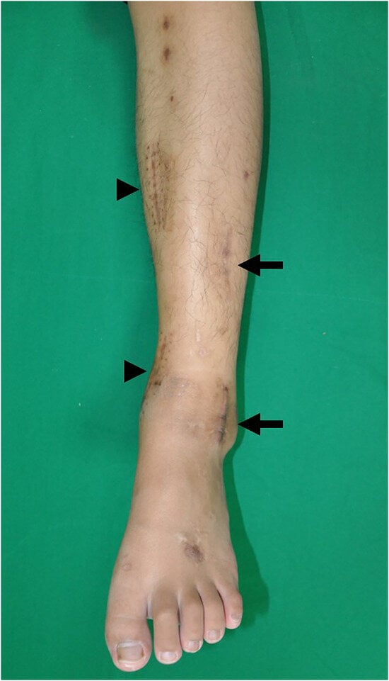
Figure 1.
An anterolateral distal tibia plate was slid submuscularly through an anterolateral skin incision and a separate proximal skin incision (arrows) and a medial distal tibia plate was inserted subcutaneously through an anteromedial skin incision and another proximal skin incision (arrowheads).
We accessed the lateral aspect of the pilon fracture using a small anterolateral approach of the ankle. The intra-articular fracture of the pilon fracture was directly reduced with or without manual traction of the foot for better exposure of the tibial plafond.
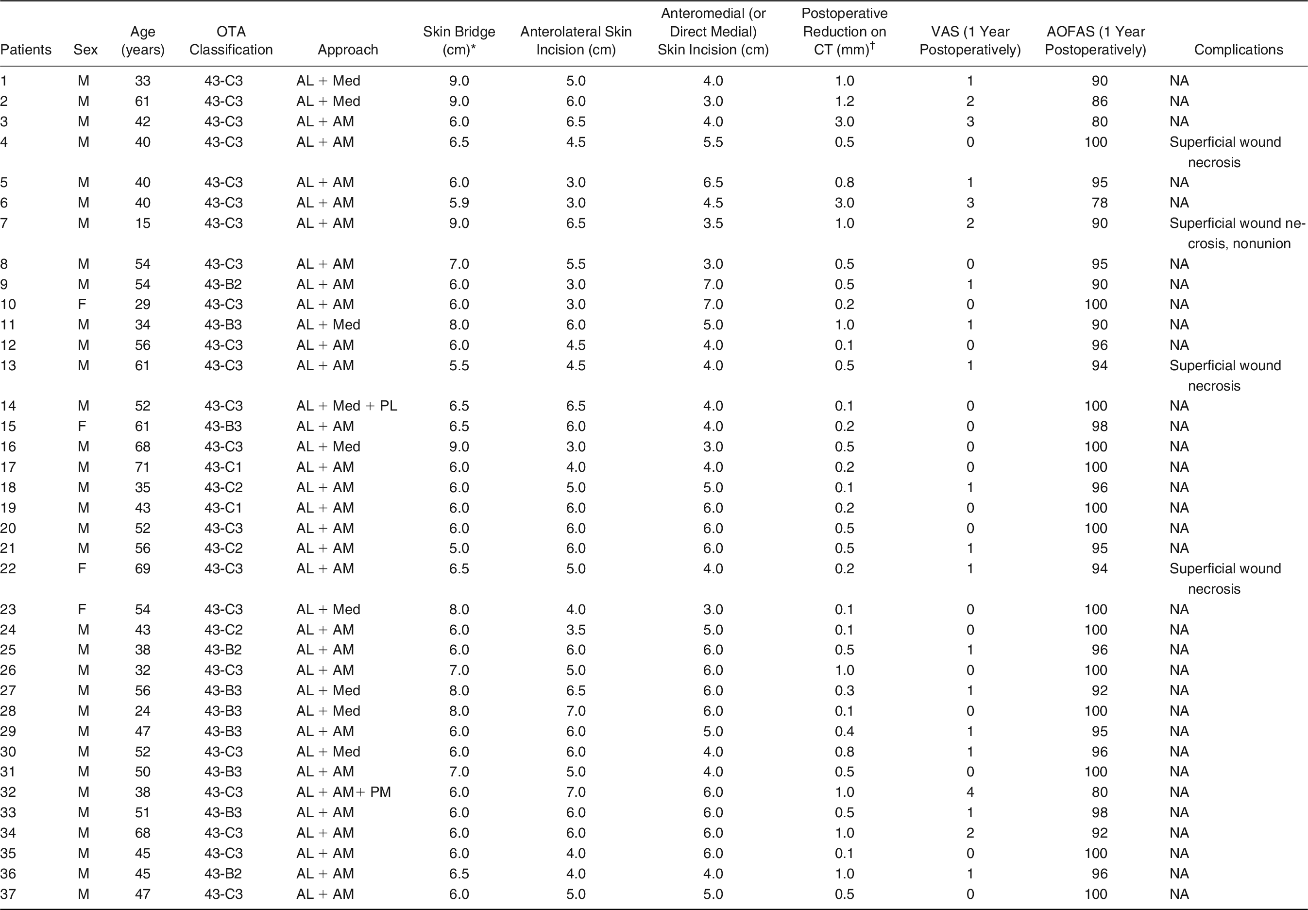
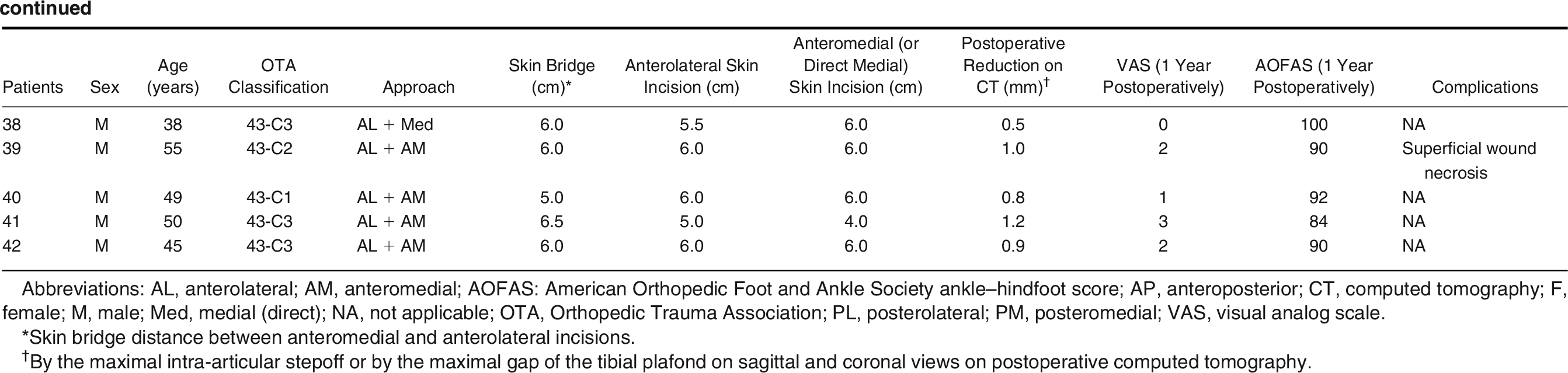
If open reduction through a small anterolateral approach only was sufficient, a small direct medial approach was added to increase the skin bridge between these two small approaches. Conversely, if open reduction through a small anterolateral approach only was insufficient, a small anteromedial approach (Fig. 1) was added instead of a small direct medial approach to attain better exposure of the ankle joint to facilitate open reduction of the tibial plafond. According to fracture pattern, a small posterolateral or a small posteromedial approach was added (Table 1).

Table 1.
Patient Demographic Data (n = 42)
The anteromedial skin incision was approximately 5 cm in length and was made at the level of the ankle joint, directly along the medial border of the anterior tibial tendon. Skin incisions could be slightly extended if needed, on a case-by-case basis.
The skin bridge between the two approaches was approximately 6 cm. Deep dissection was also performed along the medial border of the anterior tibial tendon. Both the small anterolateral and small anteromedial approaches involved skin incisions made in line with the deep dissection plane, with establishment of a full-thickness flap to make the anterior compartment of the ankle less injured. Therefore, this combined approach minimized damage to the soft tissue that may already have been jeopardized by the initial injury. This flap could also reduce damage to the contents of the anterior compartment including the tibialis anterior, extensor hallucis longus, extensor digitorum longus, peroneus tertius muscles, and neurovascular structures. Temporary fixation using Kirschner wire or screws for the intra-articular fracture portion of the pilon fracture was performed.
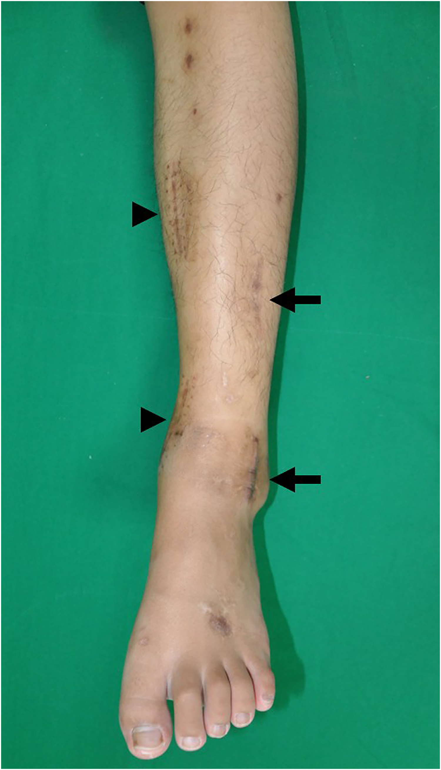
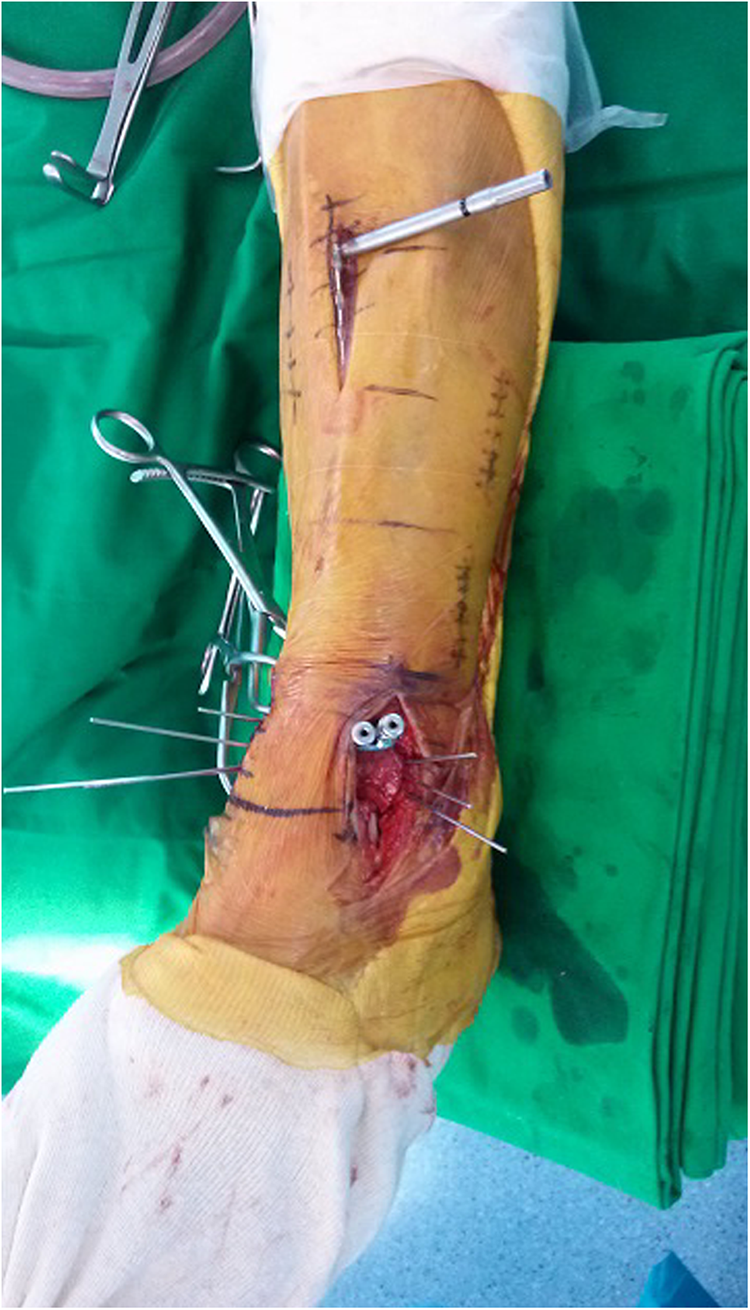
Next, the extra-articular metaphyseal aspect of the pilon fracture was indirectly reduced. Finally, an anterolateral distal tibia plate (Zimmer, Warsaw, Indiana) was slid and fixed submuscularly through a small anterolateral approach and a separate proximal skin incision (Fig. 2). A medial distal tibia plate (Zimmer) was also inserted and fixed using the subcutaneous plating technique, by means of a small anteromedial (or a small direct medial) approach and another proximal skin incision. The locking plate was secured to both the distal and proximal fragments with the fracture reduced and maintained in the desired position using either a joystick technique, Kirschner wire, or femoral distractor under fluoroscopic guidance. At least four screws were placed at both the proximal and distal fragments. The number of locking screws was individualized according to the fracture pattern and stability. In some cases, cannulated screwing was performed instead of medial distal tibia plating. Bone grafting was not performed in all cases. The wound was then closed layer by layer with drain insertion. An external fixator was not applied after a definitive internal fixation (second stage of the operation).
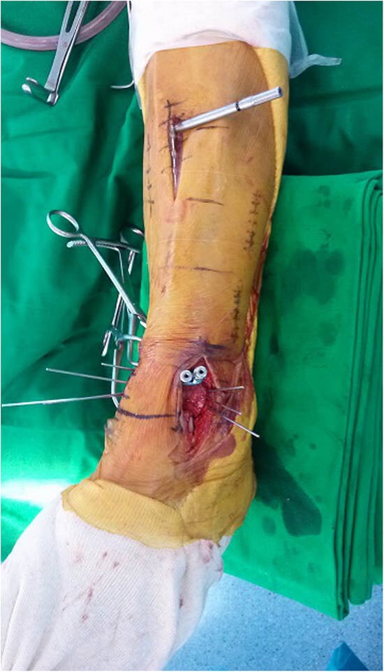
Figure 2.
Intraoperative photograph showing that an anterolateral distal tibia plate was slid submuscularly through an anterolateral skin incision and a separate proximal skin incision.
Radiographic Evaluation
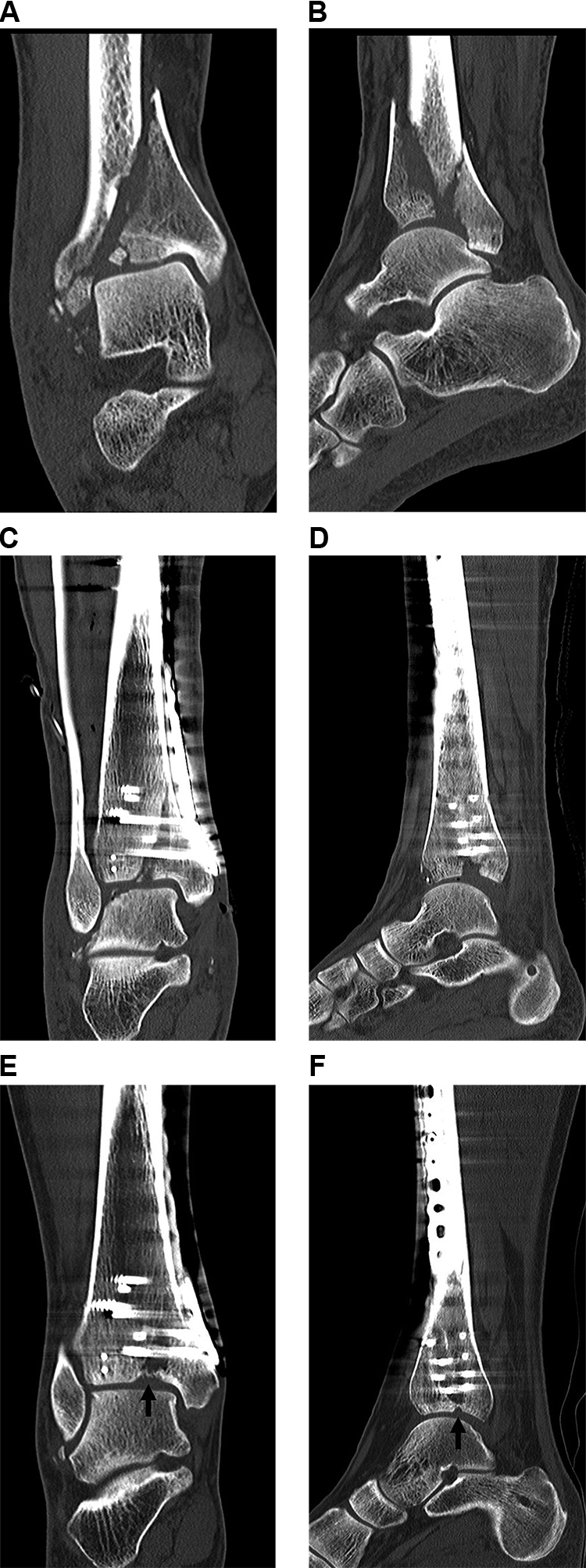
The tibial alignment angles in the anteroposterior and lateral views were measured between the longitudinal axis of the tibial shaft and that of the ankle joint. Postoperative reduction quality of the tibial plafond was measured by the maximal intra-articular stepoff or by the maximal gap of the tibial plafond on sagittal views and coronal views on postoperative CT scans (Fig. 3). A central defect (Fig. 4) of the tibial plafond after reduction that resulted from very severe comminution of the distal tibia articular surface was not considered as the maximal intra-articular stepoff or the maximal gap of the tibial plafond.
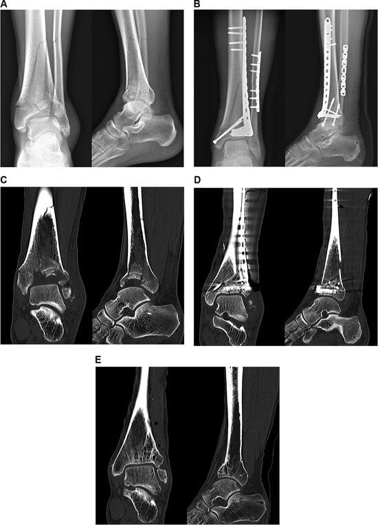
Figure 3.
Preoperative anteroposterior and lateral (A) radiographs showing a comminuted pilon fracture combined with a simple distal one-third fibula fracture. Postoperative anteroposterior and lateral (B) radiographs demonstrating good fixation through open and percutaneous plating. Serial follow-up of coronal and sagittal views on preoperative computed tomographic (CT) scan (C), immediate postoperative CT scans (D), and CT scan at 2 years after the index surgery (E) showed good results with minimal posttraumatic osteoarthritis.
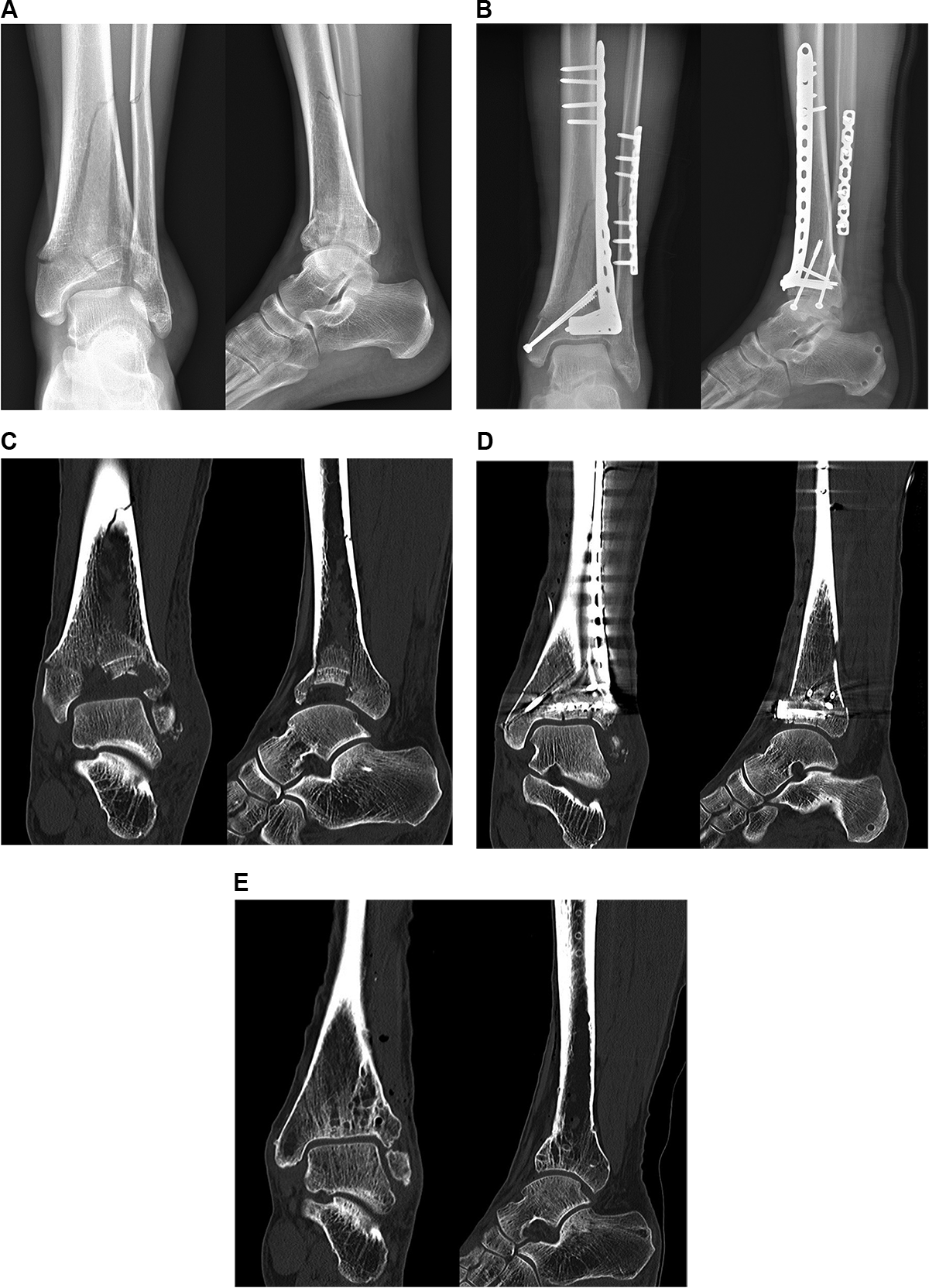
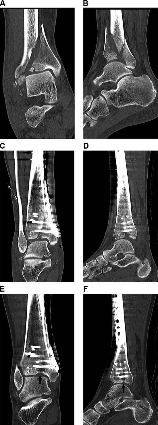
Figure 4.
Coronal (A) and sagittal (B) preoperative computed tomographic (CT) scans demonstrate severe comminution including a very small intra-articular fragment. Central defect of the tibial plafond after reduction was found in the immediate postoperative coronal (C) and sagittal (D) CT scans. Coronal (E) and sagittal (F) CT scans at 2 years after the index surgery showed a bony bridge (arrows) of subchondral bone with minimal posttraumatic osteoarthritis.
Bony union was radiographically defined by the disappearance of fracture lines on radiographs, including the anteroposterior and lateral views. Clinical bony union was defined by the ability to stand without fracture-site pain. A case of nonunion was defined by the radiographic persistence of a fracture line without progression and pain at the fracture site with walking. This line was required to be present on three sequential monthly radiographs that were taken at least 4 months after surgery. Malalignment was defined as a greater than 5° angulation, a greater than 15° malrotation, and a greater than 2-cm discrepancy in length.
All angles were measured in the same period by two independent observers (with >10 years of orthopedic experience), both of whom were blinded to each other’s findings. Four weeks later, the measurements were repeated in the same fashion to determine intraobserver reliability.
Postoperative Treatment
A short leg splint was applied for 2 weeks until soft-tissue swelling reduction was achieved. If possible, patients began isometric exercises on the first postoperative day. Sutures were removed 2 weeks postoperatively. In most cases, range-of-motion exercises of the ankle joint began approximately 2 weeks postoperatively, as the wound healed. After 6 weeks, patients were allowed to bear partial weight on their ankle with a boot, as tolerated. Patients were then advanced to full-weightbearing status (using an ankle boot) at a point approximately 8 weeks postoperatively. This progression was deemed appropriate by clinical and radiographic evidence of healing. Patients were advised to refrain from smoking and using nonsteroidal anti-inflammatory drugs in the postoperative period to prevent potential interference with bone healing.
Follow-Up
Radiographic union was assessed at each follow-up visit. Patients were evaluated at 1, 2, 3, 4, 6, 9, and 12 months postoperatively. A CT scan at 1 year after surgery was obtained in all cases to evaluate posttraumatic arthritis. After 1 year, patients were followed subsequently on an annual basis. If patients were free of progression at 4 months, they returned monthly for radiographic confirmation of union. The mean follow-up duration was 25.7 months (range, 12–48 months).
Statistical Analysis
The intraclass correlation coefficient (ICC) (2,1) with 95% confidence interval (CI) was used to analyze interobserver and intraobserver agreement regarding measurements. Student t tests were performed to compare ankle motion between the normal opposite ankle and the injured ankle at 1 year postoperatively. Pearson correlation test was performed between the following: VAS score, AOFAS score, postoperative reduction quality of the tibial plafond fracture, postoperative tibial alignment in anteroposterior and lateral views, and both ankle sagittal motion and hindfoot coronal motion at 1 year postoperatively. Values of P < .05 were considered to be statistically significant; SPSS (version 24; IBM Corp, Armonk, New York) was used for all statistical analyses.
Results
All fractures in this study resulted from falls from heights. There were three cases of 43-B2, seven cases of 43-B3, four cases of 43-C1, four cases of 43-C2, and 24 cases of 43-C3 fracture according to the Orthopaedic Trauma Association classification (Table 1). There were 28 cases of associated distal fibular fractures, which were fixed with either open plating or MIPO plating. The average time from injury to surgery (or delay to operation) was 10.2 days (range, 6–15 days).
The ICCs of interobserver agreement for postoperative reduction quality of the tibial plafond on postoperative CT and postoperative tibial alignment in the anteroposterior and lateral views were 0.85 (95% confidence interval [CI], 0.80–0.90), 0.88 (95% CI, 0.81–0.91), and 0.78 (95% CI, 0.75–0.82), respectively. The ICCs of intraobserver agreement for postoperative reduction quality of the tibial plafond on postoperative CT scans and postoperative tibial alignment in the anteroposterior and lateral views were 0.86 (95% CI, 0.81–0.91), 0.88 (95% CI, 0.80–0.92), and 0.80 (95% CI, 0.77–0.82), respectively.
The mean ranges of ankle sagittal motion (flexion plus extension) and hindfoot coronal motion (inversion plus eversion) of the normal opposite ankle were 57.5° (range, 40°–65°) and 49.0° (range, 40°–55°), respectively. The mean ranges of the ankle sagittal motion and hindfoot coronal motion at 1 year postoperatively were 43.3° (range, 30°–60°) and 47.7° (range, 40°–55°), respectively.
At 1 year after surgery, there was a significant statistical difference in the ankle sagittal motion (P < .001) between the normal opposite ankle and the injured ankle. In contrast, there was no significant difference in the coronal hindfoot motion (P = .172) between the normal opposite ankle and the injured ankle at 1 year after surgery.
Table 1 summarizes the characteristics of the tibial plafond fractures included in this study. A combination of a small anterolateral approach and a small anteromedial (or a small direct medial approach) was used in all cases. A small posterolateral or a small posteromedial approach was added as necessary in two cases (Table 1).
The average transverse skin bridge between a small anterolateral approach and a small anteromedial (or a small direct medial approach) of the tibia was 6.6 cm (range, 5.0–9.0 cm). The average transverse skin bridge between a small anterolateral approach of the tibia and a posterolateral approach of the distal fibula was 5.0 cm (range, 4.1–6.2 cm).
The average longitudinal length of skin incision in the small anterolateral approach was 5.2 cm (range, 3.0–7.0 cm). The average longitudinal length of skin incision of either the small anteromedial or small direct medial approach was 5.0 cm (range, 3.0–7.0 cm). The average postoperative tibial alignment angles in the anteroposterior and lateral views were 1.0° (range, 0°–5°) and 1.2° (range, 0°–5°), respectively.
The mean postoperative reduction quality of the tibial plafond on postoperative CT scan was 0.67 mm (range, 0.1–3.0 mm). In addition, the mean 1-year postoperative VAS and AOFAS scores were 0.90 (range, 0–4.0) and 94.5 (range, 78–100), respectively.
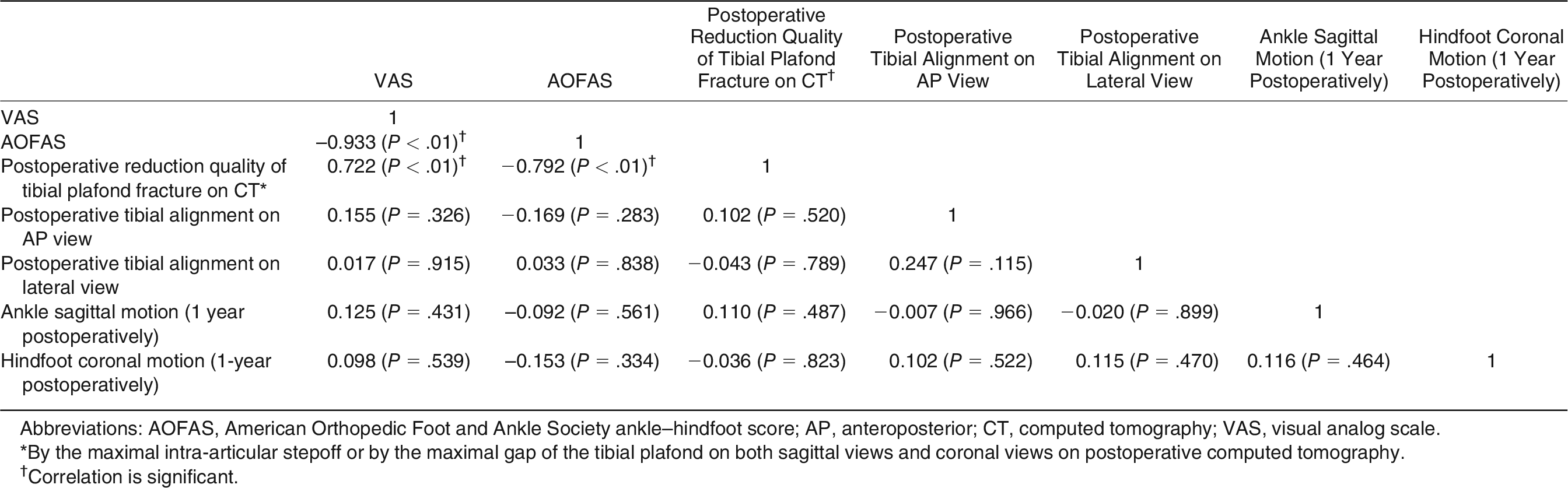
Table 2 summarizes the correlation outcomes in this study. The VAS score had a strong, negative correlation with AOFAS score (P < .01; Pearson correlation coefficient, 0.933) and a strong positive correlation with the postoperative reduction quality of the tibial plafond (P < .01; Pearson correlation coefficient, 0.722). In contrast, the VAS score was not correlated with the postoperative tibial alignment angle in the anteroposterior view (P = .326) or lateral view (P = .915), with ankle sagittal motion (P = .431) after 1 year, or with hindfoot coronal motion (P = .539) after 1 year.
Table 2.
Correlation Outcomes
Bony union was achieved in all cases except one. This single case of nonunion (Table 1) was revised at 6 months after the index surgery through the previous small anteromedial approach using an autogenous iliac bone graft and medial plate change. Radiographic union was subsequently achieved at 3 months after the plate revision.
The mean union time except in the one case of nonunion was 4.5 months (range, 3–8 months). There were no cases of ankle fusion because of posttraumatic arthritis during follow-up. There were also no major soft-tissue complications. Five cases of minor soft-tissue complications resolved with no secondary procedures (Table 1).
We do not routinely perform hardware removal. However, hardware removal is performed at a patient’s request. In 15 cases, the hardware was removed at approximately 1 year after bony union. There were no complications related to hardware removal.
Discussion
The treatment of pilon fracture remains a challenge for orthopedic surgeons, particularly because of the potential for soft-tissue complications [1,9]. Approaches that are available for pilon fracture include anterolateral [2], anteromedial [4], modified anteromedial [5], straight anterior [6], posterolateral [7], posteromedial [7], and straight lateral approaches [8]. However, major soft-tissue necrosis is still the most catastrophic complication that may present [1,9,15,16].
With the advances made in lock-in anatomical plate and MIPO techniques, a combined open and percutaneous plating approach can be a feasible option for reducing the soft-tissue complications of pilon fracture. We herein selected a combination of a small anterolateral approach and a small anteromedial (or a small direct medial approach) in all cases. A small posterolateral or small posteromedial approach was added as necessary.
Our method allows for open plating to be performed for the intra-articular fracture portion of the pilon fracture and for percutaneous plating to be used for the extra-articular fracture portion of the pilon fracture without compromising the anterior compartment of the ankle.
The anterolateral plate of the distal tibia was inserted subcutaneously through the anterolateral incision and another proximal skin incision. The medial plate of the distal tibia was inserted subcutaneously through the anteromedial or direct medial incision and another proximal skin incision (Figs. 1 and 2) [17,18].
Historically a 7-cm skin bridge has generally been recommended [19–21]. One study reported minimal soft-tissue complications with the use of a skin bridge between 5 and 6 cm [9]. The minimum skin bridge in our approach was 5 cm. We believe that a 5-cm skin bridge can be safe because soft-tissue dissection through our approach was less than that performed through conventional open plating. This may be because the MIPO technique that was used for the extra-articular fracture portion of the pilon fracture can protect the anterior compartment of the ankle.
There were no cases of major soft-tissue complications and five cases of minor soft-tissue complications that were healed with continuous wound care. These outcomes may suggest that our approach could be a feasible option for reducing soft-tissue complications during the treatment of pilon fracture.
The fibular incision was made at the posterolateral aspect of the distal fibula as distant from the anterolateral incision of the tibia as possible to minimize the possible skin necrosis between the fibular incision and the anterolateral incision of the tibia. There were no cases of skin necrosis at this area.
The essential principles for successful open plating for pilon fracture consist of the restoration of anatomical fibular length, anatomical restoration of the distal tibial articular surface, bone grafting of metaphyseal defects, and stable fixation of the fracture with medial buttress plating [22,23]. The original four principles still continue to guide current treatment decisions for pilon fracture. However, from among these four principles, we did not perform bone grafting of metaphyseal defects in all cases because the MIPO technique was applied for the metaphyseal portion of the pilon fractures.
There were no cases of collapse of the tibial plafond. One case of nonunion occurred that was revised with bone grafting and plate change and that healed 3 months after revision.
Although posttraumatic osteoarthritis of the tibiotalar joint may develop with time, there were no instances of tibiotalar fusion because of posttraumatic osteoarthritis in all cases. In two cases, well-reduced pilon fracture even with central defect of the tibial plafond that resulted from very severe comminution showed very mild posttraumatic osteoarthritis at 2 years after surgery (Fig. 4).
In this study, the VAS and AOFAS scores were strongly correlated with the postoperative reduction quality of the tibial plafond. The VAS and AOFAS scores had no correlations with the postoperative tibial alignment angles. This study may suggest that the postoperative reduction quality of the articular surface of the distal tibia (tibial plafond) is one of the important factors in the outcome of pilon fracture as long as the tibial alignment is well corrected within the acceptable range (Table 2).
The range of hindfoot coronal motion at 1 year after surgery was approximately equal to that of the normal opposite foot. In contrast, the range of ankle sagittal motion at 1 year postoperatively was reduced compared to that of the normal opposite foot. However, the ankle motion was not correlated with the VAS or AOFAS scores.
This study has several limitations, including its retrospective design, the minimum follow-up of 1 year, and an absence of a comparative case series. However, to the best of our knowledge, there have been no reports on a combined open and percutaneous plating technique using a combination of small approaches for the treatment of pilon fracture. In addition, our study correlated the functional outcomes and the reduction quality of the tibial plafond of pilon fracture using postoperative CT. In the future, a prospective comparative study would provide more information on the clinical outcomes of pilon fracture.
Conclusions
In conclusion, a combined open and percutaneous plating is a feasible option for the treatment of pilon fracture. This hybrid plating technique with a combination of a small anterolateral approach and a small anteromedial approach (or a small direct medial approach) does not compromise the anterior compartment of the ankle and showed promising outcomes without major soft-tissue complications.
Financial Disclosure
None reported.
Conflicts of Interest
None reported.
Contributions
The first two authors contributed equally to this work.
References
- Sirkin M, Sanders R, DiPasquale T, et al: A staged protocol for soft tissue management in the treatment of complex pilon fractures. J Orthop Trauma 18 (suppl): S32, 2004.
- Herscovici D Jr, Sanders RW, Infante A, et al: Bohler incision: an extensile anterolateral approach to the foot and ankle. J Orthop Trauma 14: 429, 2000.
- Kim GB, Shon OJ, Park CH: Treatment of AO/OTA type C pilon fractures through the anterolateral approach combined with the medial MIPO technique. Foot Ankle Int 39: 426, 2018.
- Borrelli J Jr, Catalano L: Open reduction and internal fixation of pilon fractures. J Orthop Trauma 13: 573, 1990.
- Assal M, Ray A, Stern R: The extensile approach for the operative treatment of high-energy pilon fractures: surgical technique and soft-tissue healing. J Orthop Trauma 21: 198, 2007.
- Sanders RW, Walling AK. “Pilon Fractures,” in Surgery of the Foot and Ankle, 8th Ed, edited by MJ Coughlin, RA Mann, CL Saltzman, p 1941, Mosby, Philadelphia, 2007.
- Dunbar RP, Barei DP, Kubiak EN, et al: Early limited internal fixation of diaphyseal extensions in select pilon fractures: upgrading AO/OTA type C fractures to AO/OTA type B. J Orthop Trauma 22: 426, 2008.
- Grose A, Gardner MJ, Hettrich C, et al: Open reduction and internal fixation of tibial pilon fractures using a lateral approach. J Orthop Trauma 21: 530, 2007.
- Howard JL, Agel J, Barei DP, et al: A prospective study evaluating incision placement and wound healing for tibial plafond fractures. J Orthop Trauma 22: 299, 2008.
- Blauth M, Bastian L, Krettek C, et al: Surgical options for the treatment of severe tibial pilon fractures: a study of three techniques. J Orthop Trauma 15: 153, 2001.
- Park J, Lee HB, Kim GL, et al: Surgical treatment approach for pilon and talar dome fracture. JAPMA 110: Article_14, 2020.
- Taylor GI, Palmer JH: The vascular territories (angiosomes) of the body: experimental study and clinical applications. Br J Plast Surg 40: 113, 1987.
- Taylor GI: The angiosomes of the body and their supply to perforator flaps. Clin Plast Surg 30: 331, 2003.
- Taylor GI, Pan WR: Angiosomes of the leg: anatomic study and clinical implications. Plast Reconstr Surg 102: 599, 1998.
- Kottmeier SA, Madison RD, Divaris N: Pilon fracture: preventing complications. J Am Acad Orthop Surg 26: 640, 2018.
- Trumble TE, Benirschke SK, Vedder NB: Use of radial forearm flaps to treat complications of closed pilon fractures. J Orthop Trauma 6: 358, 1992.
- Helfet DL, Shonnard PY, Levine D, et al: Minimally invasive plate osteosynthesis of distal fractures of the tibia. Injury 28 (suppl 1): A42, 1997.
- Krettek C, Muller M, Miclau T: Evolution of minimally invasive plate osteosynthesis (MIPO) in the femur. Injury 32 (suppl 3): SC14, 2001.
- Mast JW, Spiegel PG, Pappas JN: Fractures of the tibial pilon. Clin Orthop Relat Res 230: 68, 1988.
- Muller M, Allgower M, Schneider R: Manual of Internal Fixation: Techniques Recommended by the AO-ASIF Group, 3rd Ed, Springer-Verlag, New York, 1991.
- Ruedi TP, Murphy WM: “Tibia Distal [Pilon],” in AO Principles of Fracture Management, p 539, Thieme, Stuttgart, 2000.
- Ruedi TP, Allgower M: The operative treatment of intra-articular fractures of the lower end of the tibia. Clin Orthop Relat Res 138: 105, 1979.
- Babis GC, Vayanos ED, Papaioannou N, et al: Results of surgical treatment of tibial plafond fractures. Clin Orthop Relat Res 341: 99, 1997.
© 2023 American Podiatric Medical Association