


Abstract
Background: As common as plantar fasciitis is, there’s a lack of evidence regarding the true pathophysiologic process causing plantar fasciitis and plantar heel pain in general. This may partially explain the high variability and outcomes with current treatment of recalcitrant plantar fasciitis. Although Lemont reported myxoid degeneration of plantar fascia with histologic analysis of patients with fasciitis, muscle biopsy results were not reported. So far it appears we have not focused on the muscular component that may be present with plantar heel pain in general and in patients we diagnose with plantar fasciitis in particular. Methods: In this article we performed a retrospective analysis of biopsy results from five patients with the diagnosis of recalcitrant plantar fasciitis to determine whether this diagnosis was correct or whether other component pathologies contribute to the chronicity of symptoms or to the failure of treatment. Results: Three of the five pathology reports included specific mention of inflammation, degeneration and atrophy of the intrinsic musculature consistent with myositis. Two of these showed lymphocytic infiltration in the muscle consistent with inflammation, with no signs of inflammation in the fascia. One showed inflammation of the fascia without signs of inflammation of the muscle. Conclusions: This small study introduces the idea that intrinsic myositis may contribute to, or be responsible for some cases of plantar heel pain and plantar fasciitis. This may be important in changing the way we deal with plantar heel pain in the future.
Plantar fasciitis is one of the most common diagnoses of infracalcaneal heel pain. The incidence of plantar fasciitis is reported to be 10% of the general population [1]. The etiology is multifactorial, with various intrinsic and extrinsic factors. A few of these factors include pes planus, rearfoot valgus, obesity, and equinus [2,3].
Anatomy
The plantar fascia consists of three distinct fibrous bands that originate from the medial, central, and lateral aspects of the calcaneal tuberosity [4]. The central band originates from the medial tubercle of the calcaneal tuberosity, with attachments extending over the calcaneus [5]. This extension is continuous with the paratenon of the Achilles tendon [5]. The three fibrous bands of the plantar fascia are separated by intermuscular septa and extend distally to attach to the plantar plates and metatarsophalangeal joints [4].
Treatment
There are various conservative and nonconservative treatment options for plantar fasciitis. Conservative treatment options include corticosteroid injections, physical therapy, orthotic devices, extracorporeal shockwave therapy (ESWT), and intracorporeal pulse wave therapy. Some of the surgical options include fasciotomy, os calcis drilling, neurolysis, and radiofrequency coblation.
Controversy?
With high recurrence and complication rates associated with surgical intervention, conservative treatment continues to be the mainstay in plantar fasciitis treatment [6]. There is a paucity of evidence regarding the true pathophysiologic process of plantar fasciitis. This could explain the high recurrence and complication rates in surgical intervention, as well as the high variability in outcomes with conservative treatment of recalcitrant plantar fasciitis [7]. In 2003, Lemont et al [8] introduced the idea of plantar fasciosis using histologic analysis of patients with fasciitis. Although myxoid degeneration was reported, muscle biopsy results were not reported [8]. The purpose of this study was to present a case series to help better understand the underlying pathophysiology of plantar fasciitis and to introduce the topic of plantar myositis.
Materials and Methods
A retrospective evaluation was performed using data from January 2010 to May 2016. Eleven patients were identified using Current Procedural Terminology codes 28119 and 28008 for plantar fascia release and ostectomy of the calcaneus, respectively. Of these patients, six had biopsy of the resected fascia/bone spur. All of the patients were evaluated and treated at Foot and Ankle Associates (Oak Lawn, Illinois) by four surgeons. Five of the six patients had specimen analysis of the resected heel spurs/fascia/muscle by a hospital pathologist where the surgery was performed. One patient did not have a conclusive pathology result due to inadequate quantity of the specimen for analysis. Herein, we report on the five patients who underwent surgical intervention for chronic plantar fasciitis with positive pathologic findings.
Results and Summary
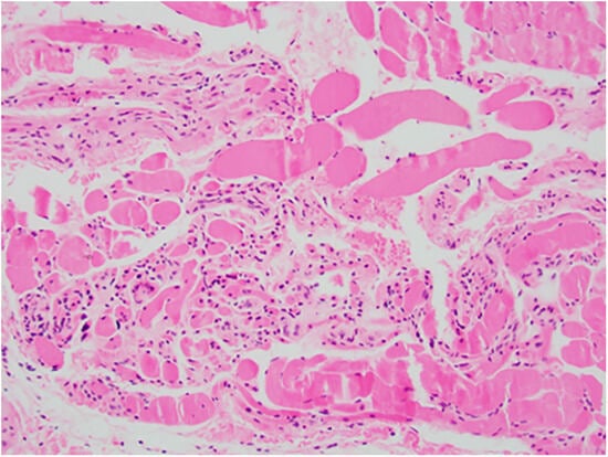
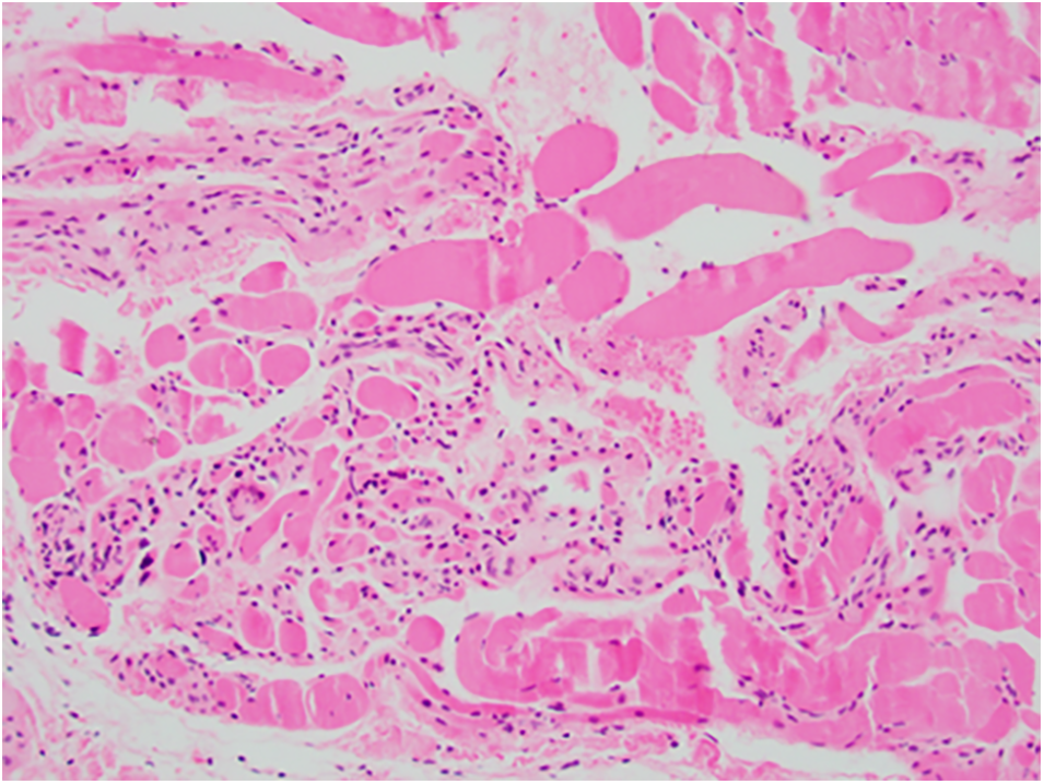
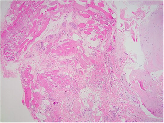
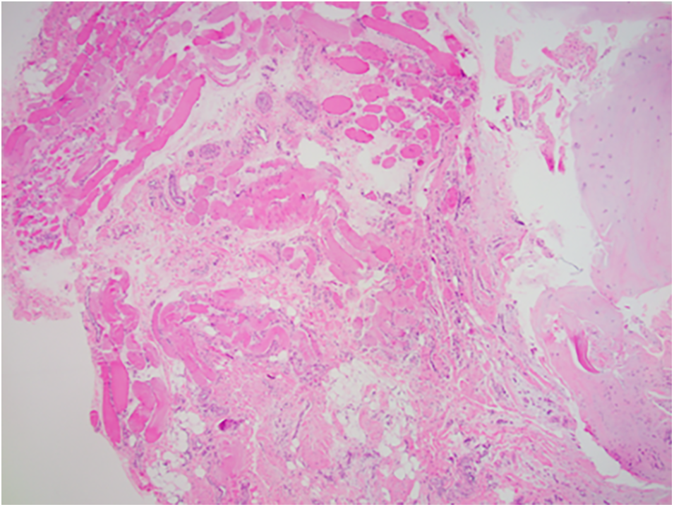
The average patient age was 47.4 years. The average body mass index (the weight in kilograms divided by the square of the height in meters) of the five patients was 34.27. All five of the specimens were positive for degenerative changes and inflammation of fascia and musculature. There were no osseous or soft-tissue malignancies noted in any of the five specimens. Three of the five pathology reports included specific mention of inflammation, degeneration, and atrophy of the intrinsic musculature, consistent with myositis. Two of these showed lymphocytic infiltration in the muscle consistent with inflammation with no signs of inflammation in the fascia. Two of the five showed inflammation of the fascia and the muscle and one showed inflammation of only the fascia. In Fig. 1, atrophic muscle with lymphocytic infiltration consistent with myositis is seen in a patient younger than 50 years. All five of the patients, who had recalcitrant plantar fasciitis, had positive intraoperative findings of inflammation in the plantar intrinsic musculature, fascia, bone or a combination of these. In Fig. 2, lymphocytic infiltration is seen in muscle, and the fascia does not show signs of inflammation.
Figure 1.
Photomicrograph showing atrophic muscle with lymphocytic infiltration consistent with myositis. (H&E, x100).
Figure 2.
Photomicrograph showing lymphocytic infiltration in muscle with no sign of inflammation in fascia. (H&E, x100).
Discussion
This article describes a small sample of patients who underwent surgical intervention for recalcitrant plantar fasciitis and gives pathologic evidence of a myositis component to plantar fasciitis. Patients with shorter duration (<4 months) of symptoms are routinely treated with conservative options. Current conservative treatment options include foot orthoses, corticosteroid injections, ESWT, stretching, physical therapy, prolotherapy, botulinum toxin type A injection, amniotic injection, and platelet-rich plasma injection. Note that there are currently no prospective randomized controlled trials or level I evidence for conservative treatment options. Corticosteroid injection studies have shown that a series of dexamethasone injections provided significant relief for patients for up to 3 months [9]. Recently, more studies have shown the high efficacy of ESWT in treating patients with plantar fasciitis [10]. A noticeable deficit in the literature concerning conservative treatment of plantar fasciitis is the role of intrinsic muscle strengthening in the treatment algorithm. An absence of research into the role of intrinsic strengthening in the plantar fasciitis algorithm could be a missing step leading to increased recurrence.
Very few studies exist describing the true pathophysiologic or histologic findings of the primary source of the subcalcaneal heel pain in recalcitrant plantar fasciitis. Ahmad et al [11] showed that the plantar heel spur size and shape was not directly correlated to plantar dysfunction and/or patient symptoms. The association between plantar calcaneal bone spurs and plantar fasciitis is controversial. Several studies have been conducted regarding the association between heel spurs and plantar fasciitis, but there is an inconsistency in the results. Osborne et al [12] and Johal and Milner [13] showed that patients with plantar fasciitis are more likely to have bone spurs, but they concluded that the patient symptoms are more directly associated with fat pad atrophy and plantar fascial thickness. Some studies have shown the efficacy of inferior calcaneal nerve (the first branch of the lateral plantar nerve) radiofrequency ablation to be effective in treating patients with recalcitrant plantar fasciitis. However, no studies have been performed implicating a specific pathophysiologic process in the inferior calcaneal nerve.
The true pathophysiologic process of chronic plantar fasciitis is poorly understood. The technical definition of inflammation involves a prostaglandin-mediated process. However, in recalcitrant plantar fasciitis there is thickening of the plantar fascia and proliferation of fibroblasts, and non–prostaglandin-mediated pathways to increase the production of excitatory neurotransmitters, which corticosteroids have been shown to inhibit. This means that plantar fasciitis could actually be a misnomer. The thickening of the plantar fascia, referred to as fasciosis, can explain a potential natural adaption by the human body in response to chronic degeneration, inflammation, and atrophy of intrinsic plantar musculature.
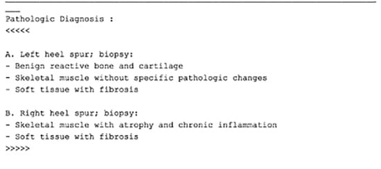
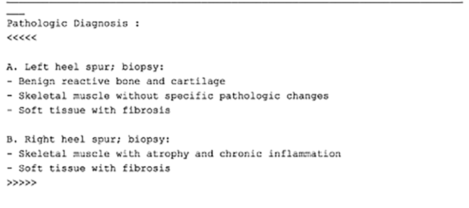
In this case series, the pathologic report shows that intrinsic plantar myositis, specifically flexor digitorum brevis myositis, can be concomitant with plantar fasciitis (Fig. 3). Intrinsic plantar myositis is described as degeneration, atrophy, and chronic inflammation of the muscle attachment to the plantar medial calcaneal tubercle. The intimate contact of plantar fascia to the intrinsic plantar musculature (flexor digitorum brevis) could explain the high degree of success with conservative treatment options such as corticosteroid injections, foot orthoses, nonsteroidal anti-inflammatory drugs, stretching, and other physical therapy modalities. Similarly, the high degree of recurrence of plantar fasciitis could also be attributed to inflammation of deeper structures involving bone and muscle.
Figure 3.
Pathology report confirming findings.
Two of the five patients’ pathology specimen analyses did not have specific mention of the condition of the muscle, which could be attributed to inadequate specimen for analysis or the differences in opinion among the pathologists between hospitals where surgeries were performed. The involvement of deeper structures, in this case the flexor digitorum longus muscle, attaching to the plantar medial calcaneal tubercle also explains a potential reason for poor long-term results with plantar fasciotomy. With plantar fasciotomy alone, the true underlying pathology would not be addressed.
There are several limitations to this study. First, this is a small sample size of a specific patient population. Plantar fasciotomy is a surgical procedure usually reserved for the worst cases of recalcitrant plantar fasciitis. Second, this was a retrospective case series, which inherently has selection bias limitations. Third, there is variability among pathologists and surgeons reporting the results and intraoperative findings.
Conclusions
This study provides pathologic evidence that there is a myositis component to the plantar fasciitis. This insight into the idea of “plantar myositis” can allow for clinicians to appropriately address this condition in both conservative and surgical management. This study also provides evidence for future higher-level studies to examine the true pathophysiologic process in plantar fasciitis and plantar flexor digitorum brevis myositis.
Financial Disclosure
None reported.
Conflicts of Interest
None reported.
References
- Irving, DB; Cook, JL; Menz, HB. Factors associated with chronic plantar heel pain: a systematic review. J Sci Med Sport 2006, 9, 11. [Google Scholar]
- Neufeld, SK; Cerrato, R. Plantar fasciitis: evaluation and treatment. J Am Acad Orthop Surg 2008, 16, 338. [Google Scholar]
- Wearing, SC; Smeathers, JE; Urry, SR; et al. The pathomechanics of plantar fasciitis. Sports Med 2006, 36, 585. [Google Scholar]
- Sarrafian, SK. “Functional Anatomy of the Foot and Ankle,”. In Sarrafian’s Anatomy of the Foot and Ankle: Descriptive, Topographic, Functional, 3rd Ed; Kelikian, AS, Ed.; Lippincott Williams & Wilkins: Philadelphia, 2011; p. 507. [Google Scholar]
- Stecco, C; Corradin, M; Macchi, V; et al. Plantar fascia and its relationship with Achilles and paratenon. J Anat 2013, 223, 665. [Google Scholar]
- Thomas, JL; Christensen, JC; Kravitz, SR; et al. The diagnosis and treatment of heel pain: a clinical practice guideline revision 2010. J Foot Ankle Surg 2010, 49, S1. [Google Scholar]
- Davies, MS; Weiss, GA; Saxby, TA. Plantar fasciitis: how successful is surgical intervention? Foot Ankle Int 1999, 20, 803. [Google Scholar]
- Lemont, H; Ammirati, KM; Usen, N. Plantar fasciitis: a degenerative process (fasciosis) without inflammation. JAPMA 2003, 93, 234. [Google Scholar]
- McMillan, MA; Landorf, KB; Gilheany, MF; et al. Ultrasound guided corticosteroid injection for plantar fasciitis: randomised controlled trial. BMJ 2012, 344, e3260. [Google Scholar]
- Aqil, A; Siddiqui, MR; Solan, M; et al. Extracorporeal shock wave therapy is effective in treating chronic plantar fasciitis: a meta-analysis of RCTs. Clin Orthop Relat Res 2013, 471, 3645. [Google Scholar]
- Ahmad, J; Karim, A; Daniel, JN. Relationship and classification of plantar heel spurs in patients with plantar fasciitis. Foot Ankle Int 2016, 37, 994. [Google Scholar]
- Osborne, HR; Breidahl, WH; Allison, GT. Critical differences in lateral X-rays with and without a diagnosis of plantar fasciitis. J Sci Med Sport 2006, 9, 231. [Google Scholar]
- Johal, KS; Milner, SA. Plantar fasciitis and the calcaneal spur: fact or fiction? Foot Ankle Surg 2012, 18, 39. [Google Scholar]
© 2023 American Podiatric Medical Association