Idiopathic toe-walking (ITW) is a condition in which children walk with a toe-toe gait pattern in the absence of any known cause [
1,
2]. In the stance phase of gait the heel is in contact with the ground at three of five subphases (initial contact, loading response, and midstance), and most of the gait pathologies are observed in these subphases [
3]. Toe-walking is the inability to generate a heel strike at initial contact and the absence of full foot contact during the stance phase; it is a common pattern observed in healthy, developing children before age 3 years [
4]. Toe-walking may gradually lead to ankle plantarflexion contracture over time [
5]. Furthermore, the natural history of persistent toe-walking places children at risk for structural and functional abnormalities such as falling [
6], permanent outward tibial torsion [
7], and limitation of ankle mobility [
8].
Gait assessment of children with ITW consists of determining the severity (how high the heels are from the ground) and extent (frequency) of toe-walking. Severity is usually evaluated using computerized or observational gait analysis with additional physical measurements, including ankle flexibility and manual muscle tests [
9]. Although goniometric assessment of ankle range and computerized gait analysis are the most commonly reported outcome measures in studies of children with ITW [
10], observational gait assessment is a quick, inexpensive method, which is efficiently performed by health professionals, to assess patients’ gait patterns and create intervention plans to address the impairments and gait deviations [
11]. However, different than in daily activities, children with ITW tend to modify their gait when they realize that they are being observed, which changes and usually delays the beginning of the treatments. In addition, the frequency and severity of toe-walking may also vary depending on speed [
12].
Treatment of ITW includes active and passive stretching of the calf muscles [
13], botulinum toxin A (BoNT-A) injections [
14], serial casting, [
15] surgery [
16], and orthoses [
17,
18], the most common assistive treatment tool. However, the benefit of these treatments is still questionable [
19], and a standard treatment protocol has not yet been established in clinics [
17]. In a recent review, which discussed the effects of BoNT-A injections, footwear, exercises, and different types of orthoses in ITW, the authors concluded that the outcome data in the related studies were too limited to assess their effects. Besides, the review revealed that it is not known whether treatment with BoNT-A injections and serial casting together is more effective than serial casting alone in children with ITW [
20]. Functional bandaging is an intervention tool used by health professionals to address impairments and activity limitations in people with musculoskeletal disorders to improve muscle strength, postural stability, and proprioception [
21]. To our knowledge, no studies have been published assessing gait with dorsiflexion-assist bandage application to improve heel contact during gait in children with ITW. An alternative treatment intervention of adhesive bandaging may help increase heel contact during stance, facilitate appropriate proprioception through the heel and hindfoot, preserve ankle range of motion, and, therefore, reduce the incidence of surgery to reduce a fixed contracture [
22].
By comparing the gait of typically developing children, the aim was to evaluate the acute effects of dorsiflexion-assisted functional bandaging applied in children with ITW to achieve heel strike at initial contact, gain heel contact at loading response, prevent premature heel-off at midstance, and investigate whether this application helps to improve gait quality, providing an alternative conservative intervention for children with ITW.
Methods
This is a basic longitudinal study that tested the children at three time points: before bandaging (T0), immediately after initial bandaging (T1), and 1 week after initial bandaging (T2).
Participants
Participants included 19 children diagnosed as having ITW (age range, 4–16 years; mean ± SD age, 7.36 ± 3.16 years) who were followed up by the Child Neurology and Orthopedics and Traumatology Divisions of Istanbul University, Istanbul Faculty of Medicine (Istanbul, Turkey), and ten typically developing paired children (mean ± SD age, 7.30 ± 2 years). All of the participants with ITW were recruited to the study from the Istanbul Medicine Faculty Gait Analysis Laboratory (Istanbul, Turkey) and were diagnosed as having ITW by neurologists in the same university.
The inclusion criteria included no heel strike at initial contact and observed premature heel-off or no heel contact at the midstance phase bilaterally; no neurologic, physiologic, or orthopedic problems; and no previous interventions with an exercise program, BoNT-A injection, or orthopedic surgery.
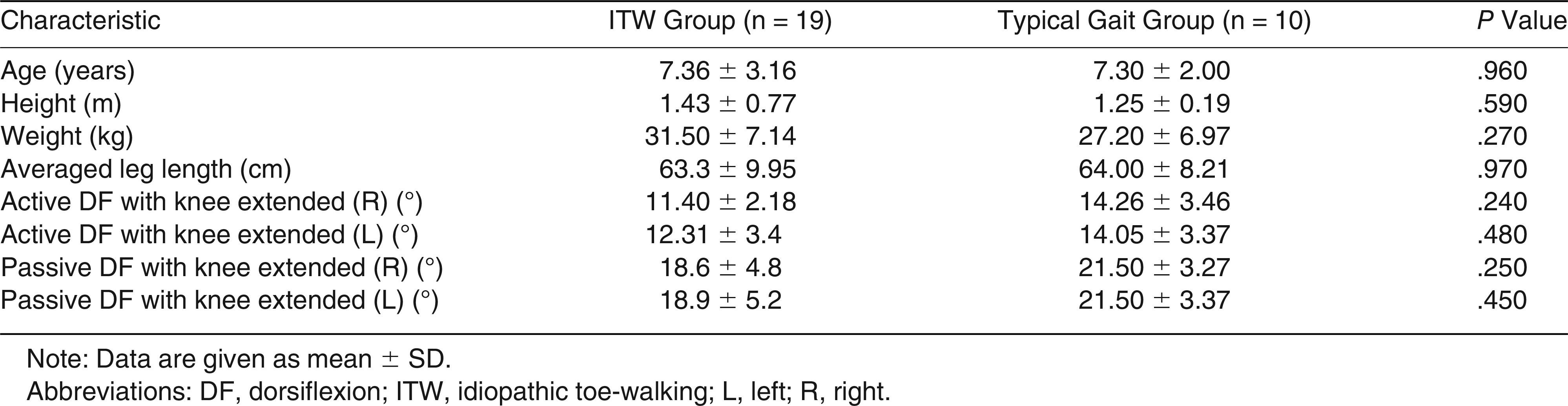
Table 1 gives the participant characteristics, including age, height, weight, averaged bilateral leg length, and goniometric active and passive dorsiflexion range of motion with the knee extended. All of the ankle dorsiflexion range of motion measurements were performed three times, and the mean values were used in the data analysis for each side. All of the participants were evaluated with the Silfverskiold test, which evaluates ankle dorsiflexion in knee flexion and extension for detecting the isolated gastrocnemius tightness. For each participant, the same examiner (D.T.) performed the physical examination (goniometric joint ROM of ankle and leg-length measurements), videotaping and applying the bandages. Another physical therapist (N.E.A.) analyzed the videos using tools, so the data analysis was performed by someone blinded to the clinical data intervention.
The Toe Walking Tool (TWT) was used as a screening tool for children with ITW. To diagnose toe-walking that was not caused by a medical condition and ensure the diagnosis of ITW, all of the participants were evaluated by the TWT, which is a valid and reliable assessment tool. The TWT includes four sections: demographics, indicators of trauma, indicators of neuromuscular influence, and indicators of neurogenic influence [
23]. According to the TWT questionnaire, children with ITW in the present study have no neurogenic, neuromuscular, or traumatic indicators. Although ankle flexibility is tested during the lunge test in the TWT, ankle dorsiflexion range was measured by goniometer in a nonweightbearing position in this study (
Table 2).
Information about the study was provided to all of the participants and their families through written informed consent and written permission obtained for images provided by parents for the study to be published. The protocol of this study was in accordance with the guidelines of the Declaration of Helsinki and was approved by the ethical committee of Istanbul University.
Measures
Video-Based Observational Gait Analysis.
Gait deviations of participants, while wearing only their underclothes, were analyzed by video-based observational gait analysis (VBOGA). The presence or absence of heel contact was evaluated by using a pressure sensor and VBOGA without shoes. The children with ITW were evaluated before (T0) and immediately after (T1) bandaging and 1 week after wearing the same bandage (T2). The primary outcome measure in this study was VBOGA. The physical therapist (D.T.) who videotaped the videos had worked more than 5 years as a physical therapist and had at least 5 years of experience in treating and analyzing gait deviations by means of observational gait analysis.
Edinburgh Visual Gait Score.
The Edinburgh Visual Gait Score (EVGS) has been proved to be valid and reliable and helps to gain quantitative data on top of the typical qualitative data when observing gait [
24,
25]. Quality of gait for all of the participants was also analyzed by the EVGS, which scores sagittal and coronal plane gait quality in 17 variables. In this study, all of the participants were evaluated with 12 sagittal plane variables of EVGS. Each variable can be scored on a 3-point scale: 0 (normal), 1 (slight deviation), or 2 (severe deviation) for each joint and each side [
24]. The EVGS has been developed primarily for children with cerebral palsy, and its reliability in children with ITW has not been studied [
25]. The assessor (N.E.A.) was well trained and experienced in the evaluation and application of the EVGS as a senior physical therapist who had worked for more than 10 years in a motion laboratory and had experience in VBOGA and three-dimensional gait analysis in children.
Toe-Walking Frequency
Children with ITW, who have no fixed contracture in their plantar flexors, can often choose to walk with a heel-toe gait on request. The visual analog scale (VAS) was used to assess the frequency of toe-walking observed by the family recently. This frequency is a 10-cm-long percentage frequency (0%, never walking; 100%, walking continuously). For subjective comments, we asked the parents the frequency of toe-walking in a community setting where the child is not aware of being observed, and it is asked in T0 and T2. The question was “How many points do you give as a percentage if you evaluate the frequency of toe-walking?”
Procedure
The VBOGA and EVGS results were compared for the children with ITW and healthy children pairs in T0, T1, and T2. To understand the differences between acute and 1-week duration of the application, scores of children with ITW were compared between T0, T1, and T2.
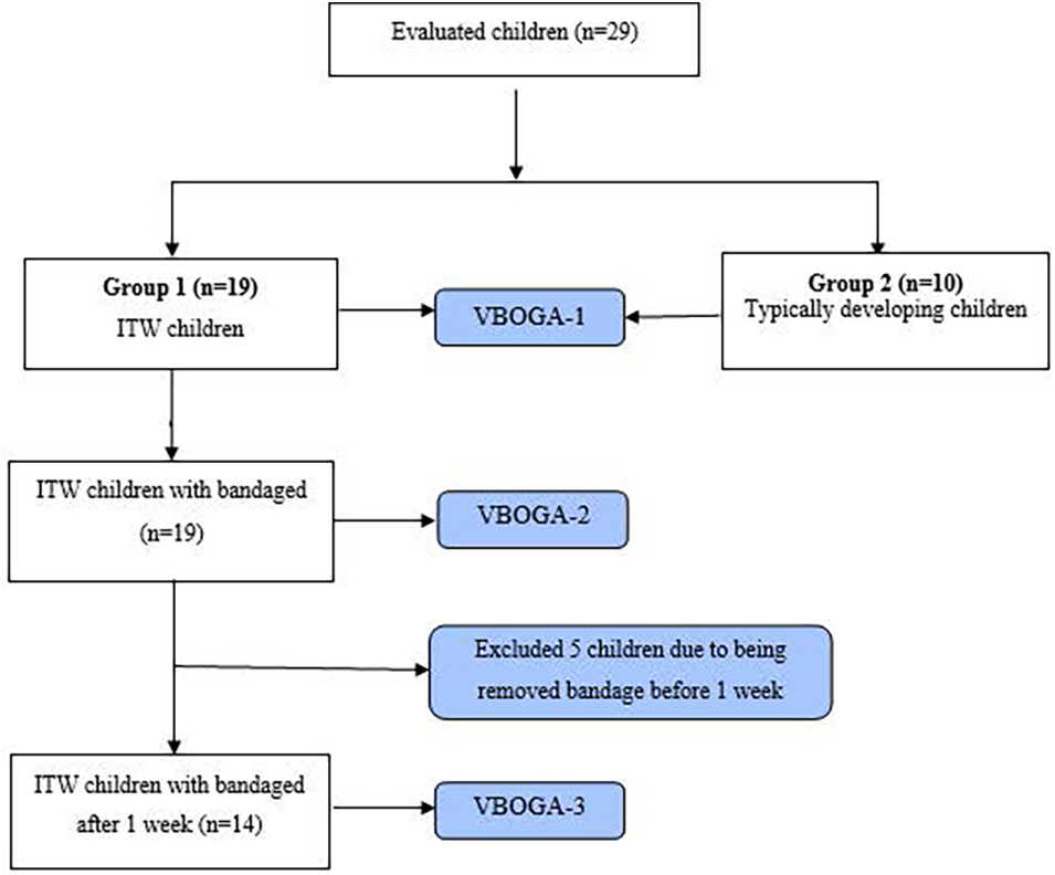
To compare heel contact between participants with ITW and those with typical gait, and to confirm that the pressure sensors worked properly, ten typically developing children were recruited for this study (
Fig. 1).
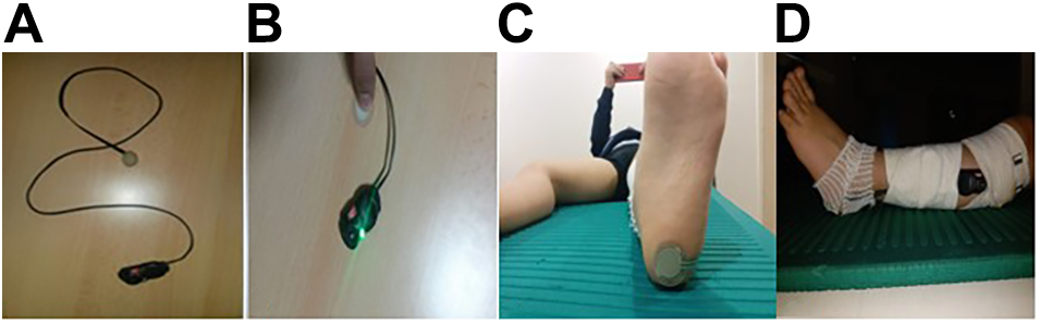
A switch-type pressure sensor (
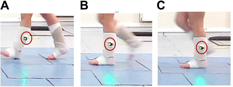
Fig. 2), which is designed to light up when the heel contacts the ground, was placed on the bilateral heels to define heel contact behavior at initial contact, loading response, and midstance. The sensor consists of a durable 25-mm-diameter membrane switch (1 mm thick and <1 g in weight, responds reliably to 200 g of load anywhere in the sensor area) with a 15-mm sensor area on a 100-mm flexible tail. The observed mean time between failure was 10,000 actuations. To find the precise location of heel contact, the bare heel was marked by ink, and the sensor was then placed on the ink mark. After the sensor was positioned, the participants were seated, and hand pressure was applied to the heel part by the same examiner to check proper sensor function. It was confirmed that every time pressure was applied, an LED connected to the sensor by a thin wire turned on. To help the camera see the LED easily, the LED was wrapped on the lateral shank by using a standard nonadhesive elastic bandage. This electronic sensor was used elsewhere to understand whether the heel was in contact with the ankle-foot orthosis (AFO) during walking, and it worked well and was consistent [
26].
A digital video camera (Canon PowerShot A2200; Canon, Tokyo, Japan) that can capture 30 frames per second and a tripod video camera stand were used to record each participant’s gait pattern. The camera was placed 3 m away, perpendicular to the middle of the 12-m walkway, to record the natural way of walking by asking participants to walk as they usually do outdoors.
Because gait speed can affect heel contact [
27,
28], cadence was controlled for each condition using a metronome set for 80/min to 100/min to ensure consistent timing of steps. Before the video recording, the participants practiced for at least 3 min to adapt to the timing of the metronome beat. Adaptation to a specific cadence is difficult for young children, so when the participant caught a cadence range of 80/min to 100/min for 3 min, the participant was assumed to have adapted to the cadence. We repeated trials until the child adapted to the metronome cadence. Parents, before the recording, also checked the walking pattern of their children, if they walk the way they usually walk outside. After each participant adapted to the proper cadence, five full gait cycles midway on the walkway were recorded on the left and the right in the sagittal plane, followed by video analysis.
Intervention
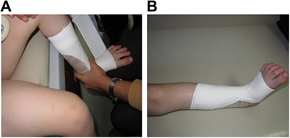
For T1, Tensoplast Sport (BSN Medical GmbH, Hamburg, Germany), a latex-free elastic adhesive bandage 2.5 m long and 5 cm wide, was used to facilitate ankle dorsiflexion during gait. Tensoban foam (BSN Medical GmbH), a foam wrap that prevents skin irritation, was applied to each participant’s shank and foot after spraying Tensospray (BSN Medical GmbH) to prevent sliding of the Tensoban foam and Tensoplast Sport couple. According to the manufacturer, there are no known complications related to these bandage materials. To minimize risk of complications, such as discomfort or skin irritation, the parents were informed about the material properties of the bandage and were instructed to remove the child’s bandage if the child expressed discomfort. With the hamstring and gastrocnemius muscles relaxed, each child was positioned in a comfortable sitting position with the lower legs and feet freely hanging. The ankle was passively set to 5° of dorsiflexion with a goniometer. Bandaging was applied by starting from the dorsal forefoot (one circular wrap) in parallel to the distal metatarsals, crossing the front-ankle region from the lateral foot to the medial half of the shank (one circular wrap again), and then wrapping was continued down likewise, crossing the front-ankle region to the distal metatarsals until using up all 2.5 m of bandage (
Fig. 3) [
29]. The same therapist (D.T.) applied all of the bandaging for all of the participants. After wrapping the bandage, the children could not achieve ankle plantarflexion even if they tried.
In T1, before the video recording, the participant’s toes and comfort were checked verbally and visually to record comfortable barefoot walking without reduced blood circulation. After T1, an appointment 1 week later was set for each family to check the bandage for T2. During the 1-week period, the family was warned to keep the bandage on the child unless some unusual complications occurred, such as itching under the bandage, pain, or edema in the toes or lower legs. No exercise program was recommended, and the bandages were never removed during the 1-week period. Children were allowed to continue their daily activities with the bandages, and the parents were instructed to let the children take a shower by covering the bandaged area with a waterproof material.
One week later, immediately after the third video recording, the bandage was removed with a solution called Leukotape Remover (BSN Medical GmbH, Hamburg, Germany), which inactivates adhesive property.
There was no intervention to the typically developing children; they were analyzed only with the VBOGA and EVGS for gait comparison with the participants with ITW.
Data Analysis
The video records of each participant for each condition were analyzed with the VBOGA by using the frame-by-frame function of the video player software QuickTime 7.75.80.95 (Apple Inc, Cupertino, California). All of the video recordings for the participants with ITW and the typically developing children were analyzed using the EVGS. The VBOGA and EVGS analyses were performed by the physical therapist (N.E.A.). The mean scores of five gait trials for each side, corresponding to the center of the walkway, were used for statistical analyses.
The VBOGA was used to analyze three gait subphases: 1, initial contact, defined as the instant the foot contacts the floor; 2, loading response, defined as the sole of the foot coming in contact with the floor; and 3, midstance, defined as the ipsilateral tibia rotating over the stationary foot (the instant the opposite foot crosses the stationary limb) by additionally using the pressure sensor (
Fig. 4).
To evaluate the effect of bandage application on heel contact in children with ITW, in T0 and T1, 570 subphases (19 children walked five times and three subphases of the stance phase in each tour were analyzed for bilateral sides) were evaluated by using a light pressure sensor. After 1 week, in T2, the analysis was repeated for 420 subphases (14 children walked five times and three subphases of the stance phase in each tour were analyzed for bilateral sides). None of the participants had heel contact at any of the subphases in T0.
All of the videos were analyzed based on whether the sensors placed on the heels lighted in three subphases of stance and were scored as 0 (light off) and 1 (light on). All of the scores for light on were summed and recorded for each child.
All of the statistical analyses were performed using a software program (IBM SPSS Statistics for Windows, Version 20.0; IBM Corp, Armonk, New York), with a significance level of 5% in all of the tests. Mann-Whitney U tests were used for comparisons of age, height, weight, averaged leg length, bilateral active and passive dorsiflexion, and EVGS differences between the participants with ITWs and the typically developing children. The Friedman test was used with post hoc pairwise comparisons using the Wilcoxon signed rank test, with a Bonferroni correction being used to account for increased type I error associated with multiple pairwise comparisons in VBOGA and EVGS for T0, T1, and T2 for children with ITW.
Nineteen participants was above the minimum sample size needed to ensure a 99% confidence level (significance level of .05 [β = 0.2]) by considering mean changes in the interested parameters (VBGO [averaged: 80%] and EVGS [averaged: 84%] scores) as the effect size (>80%) for the basis of prospective studies.[
30] With 5% error and 70% effect size, 24 children could provide power of 80%.
Results
Nineteen children with ITW and ten typically developing children were enrolled in the study. No statistically significant differences were found between groups on age, height, weight, averaged leg length, and measurements of active and passive dorsiflexion with knee extended (
P > .05) (
Table 1).
According to the TWT questionnaire, children with ITW in the present study have no neurogenic, neuromuscular, or traumatic indicators (
Table 2). All of the children with ITW achieved ankle dorsiflexion despite variable baseline range-of-motion values between participants. Except for the six children whose dorsiflexion range of motion had slightly restricted (mean ± SD range of motion: active, 6.16° ± 1.32°; passive, 11.5° ± 1.97°), for all of the patients with ITWs, the Silfverskiold test result was negative for the bilateral extremities, and there was no isolated gastrocnemius muscle tightness.
In this study, we measured the VBOGA scores and EVGSs in pre (T0), post (T1), and 1 week later with bandaging (T2) of children with ITW and compared with typically developing children. In T2, five of the 19 participants were excluded because the bandage was removed before 1 week. Therefore, 14 of the 19 children with ITW participated in T2.
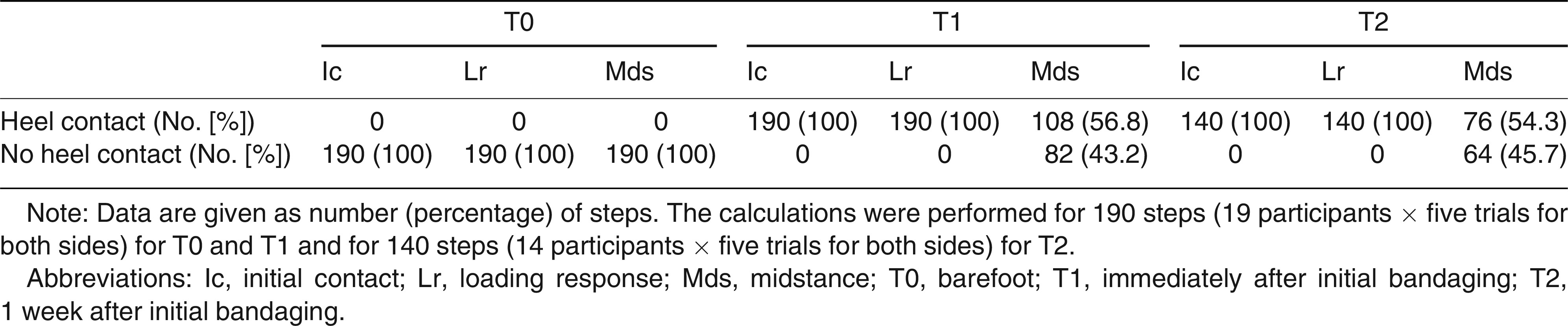
The mean ± SD total VBOGA scores in T0, T1, and T2 were as follows: 2.45 ± 5.87, 24.24 ± 9.78, and 23.81 ± 10.68, respectively. In the VBOGA, the children with ITW did not perform heel strike in all of the gait phases on their dominant and nondominant sides without bandaging (T0), all of the healthy participants activated the lights during all of the subphases in T0. The children with ITW who could not make heel contact (light off) in any subphases in stance under the T0 condition made heel contact (light on) in initial contact and loading response after the bandage application. Although in midstance, the heel was in contact for 56.8% of the 190 subphases (108 of 190 steps) (
Table 3). In T2, the heel continued to contact during initial contact and loading response for all of the phases in all of the cases. Although the percentage of heel contact in midstance was reduced from 56.8% (108 of 190 steps) in T1 to 54.3% (76 of 140 steps) in T2. The Friedman test (χ
22 = 66,797;
P < .001) post hoc Wilcoxon signed rank test with a Bonferroni correction (
P < .05/3 < 0.017) revealed significant differences between T0-T1 and T0-T2 (
P < .001). No difference was found between T1-T2 (
P = .913).
In T1, 53.7% of the steps on the dominant side and 60% of the steps on the nondominant side succeeded in heel contact. In T2, heel contact occurred in 52.9% of the steps on the dominant side and in 55.7% of the steps on the nondominant side. No significant difference in the number of heel contacts between the dominant and nondominant sides was found in T1 and T2 (P = .232 and .556, respectively).
The mean ± SD total EVGSs in T0, T1, and T2 were as follows: 7.32 ± 2.06, 1.47 ± 1.88, and 1.12 ± 0.95, respectively (according to total sagittal plane EVGSs of 0 [normal], 12 [slight deviation], or 24 [severe deviation]). The Friedman test (χ22 = 23,231; P < .001) post hoc Wilcoxon signed rank tests with a Bonferroni correction (P < .05/3 < 0.017) revealed a significant difference between T0-T1 (P < .001) and T0-T2 (P < .001), although no difference was found between T1-T2 (P = .450).
The EVGS also showed a significant difference between the children with ITW and the typically developing children in T0 (P < .001). However, no significant difference was found between the two groups in T1 (P = .054) and T2 (P = .063).
According to the parent-reported VAS, before bandaging the mean ± SD VAS score was 84.32 ± 12.11, and 1 week later with the same bandage the VAS score was 38.34 ± 17.66. There was a significant difference between the duration before bandaging and after 1 week with the bandage (P < .001).
Discussion
To our knowledge, this is the first study in the literature to evaluate the acute effects of functional bandaging in children with ITW. Dorsiflexion assistive functional bandaging is an effective tool to help achieve heel contact with the ground and improve gait quality for children with ITW.
According to the present results, immediately and 1 week after the application, functional bandaging was highly effective in providing the proper heel contact behavior in the early stance subphases (initial contact and loading response) and highly promising in the midstance phase of walking. Compared with typically developing children, participants with ITW had significantly higher EVGSs (atypical walking quality) before the bandage application (T0), but no significant difference was found between the two groups after the bandage application and after 1 week of wearing the bandage.
When detecting gastrocnemius and soleus muscle tightness and ankle hypomobility into dorsiflexion, serial casting is mostly chosen as a conservative treatment [
22,
31]. In the present study, all of the children with ITW achieved ankle dorsiflexion despite variable baseline range of motion values among participants. The bandaging probably will be less effective if the child has a fixed contracture. However, after the serial casting process, the child is given AFOs for daytime use and AFOs set in dorsiflexion for night use for the alignment. After the desired range-of-motion and alignment is achieved, the child may need to continue to wear AFOs for years after casting [
31]. As an assistive tool, functional bandaging seems to provide enough dorsiflexion moment to achieve proper heel contact at initial contact. The AFOs are difficult to fabricate due to casting, take time to make, and need to be modified as the child grows. In addition, parents need to purchase larger shoe sizes to fit the AFOs, and one shoe has to be larger than the other if the child uses the AFOs unilaterally. With elastic bandaging, which is more tolerable than orthotic devices, we achieved similar foot contact behavior and gait quality as with orthotic devices [
21,
32]. It may also be an alternative or assistive treatment tool for AFO use to support the treatment programs of children with ITW and for children who do not like to wear AFOs or larger shoes for cosmetic reasons. Because the bandage can be concealed under the socks and allows free choice of use, the child may agree to use the bandage over a longer period in daily life as an alternative to AFOs, which can enhance the benefits of an exercise program and encourage the child to participate in the rehabilitation program.
After functional bandaging, in the initial contact phase, heel strike was achieved in all of the children who could not attain a heel strike in T0. Although none of the participants had heel contact with the floor at midstance in T0, 43.2% of the children in T1 and 45.7% in T2 demonstrated premature heel-off at the midstance phase. However, because the benefits of the bandage seem limited in midstance because the participants had no heel contact at loading response and midstance in T0, the premature heel rise in midstance was a noteworthy achievement. By incorporating functional bandaging to assist dorsiflexion, an exercise program in the present study could have been more effective in increasing heel contact in midstance in children with ITW. An exercise program, such as a motor control intervention or stretching, was not included in this study because 1) the participation of young children in an exercise program is sometimes difficult, 2) there are challenges with standardizing the amount of exercise in young children, and 3) the advantages of verifying children with ITW by using the TWT and ruling out medical conditions contributing to toe-walking has not been studied.
In this pilot study, a Coban bandage, which is an elastic and nylon thread fabric with a structure containing latex, was used to gain assistive dorsiflexion ankle moment. However, it caused skin irritation and sensitivity on the applied area and, thus, was terminated immediately. On the basis of the preliminary results of the pilot study, because the other kinesiologic tapes available on the market (Kinesio tape, Pinotape, etc) were not believed to have the strength to generate enough dorsiflexion moment at the initial contact and midstance phases, the impact of Tensoplast bandages is thought to be more promising to address lack of heel contact in toe-walking. We believe that a comparison of the effects of these two functional taping applications in future studies will be useful.
In another pilot study, we attempted to control the fixed rhythm and walking speed range by using a metronome. Gait velocity differences cause various changes in gait pattern. A metronome is a tool used commonly in the literature to control the cadence and gait speed for not only heel-toe walkers but also tip-toe walkers and for different gait patterns, such as jump knee gait, crouch gait, and true equine [
33,
34]. However, the older children adapted well to the cadence, but the young children had difficulty maintaining the same cadence. In the young children, controlling their walking speed by using a metronome caused changes in their walking patterns. Therefore, instead of forcing the children to a constant cadence, a specific cadence range (80–100/min) was used. The child’s walking cadence was then monitored using a metronome to ensure that the child’s cadence was within the specified range during the analysis. In future studies, to maintain the same walking speed for objective evaluation, the use of a treadmill or technical equipment such as photosensors is recommended.
In addition, in the present study, the EVGS was used only in the sagittal plane on the dominant and nondominant sides. Nevertheless, no specific VBOGA method has been developed for children with ITW, although this was validated for children with cerebral palsy in the literature [
25]. We believe that the EVGS may be useful for evaluating the sagittal plane gait alterations in ITW, which are similar to those in mild spastic diplegia [
35,
36]. Because this is the first study correlating results of light sensors with VBOGA score and EVGS, this study, although not specifically analyzed using correlative statistics, shows that EVGS is a promising tool for evaluating gait deviations in ITW. For children with ITW, validity and reliability studies of the EVGS are strongly recommended.
The EVGSs also showed the effects of the bandage applications on proximal segments. The VBOGA scores and EVGSs revealed no statistically significant differences between T1 and T2. On the basis of the EVGS, although heel contact improved with the bandage application, 11 of 19 children showed moderate (6°–15°) forward trunk posture in T0, and eight of them continued forward trunk lean in T1 and T2. Eight of the 19 children with ITW demonstrated increased pelvic rotation (pelvic protraction of 11°–20°) at midstance in T1, and five of them continued this in T2. We observed that the children’s gait postures approached normal with the bandage application, but some proximal gait alterations remained.
The bandage application in 5° of dorsiflexion may have forced the child to bend the knee at first walking with the bandage. Accordingly, 11 of the 19 children showed increased knee flexion in stance (16°–25°) in T1, which continued in T2 in five children. Owing to the absence of previous literature for comparison, bandaging in a 5° dorsiflexion position was chosen due to the elastic property of the bandage, which may resist the generated plantarflexion moment during stance. Although observational gait analysis is often used in clinical practice, three-dimensional gait analysis will provide more accurate data and outcomes.
To distinguish the participating ITWs from neurogenic influences, neuromuscular influences, and trauma, which may affect the assistive bandage application by spasticity, motor control, and sensation problems, TWT was used. Bandage application may have different effects on gait in children with medical diagnoses causing toe-walking [
23] One study[
37]. revealed normative reference values for lower-limb range of motion in children aged 4 to 16 years; mean ± SD passive dorsiflexion range of motion with the knee extended is represented as 21.3° ± 5.4°. In the present study, six children with ITW had mildly restricted passive dorsiflexion range of motion with the knee extended (< 20°). Because we evaluated gastrocnemius muscle tightness with goniometric measurement instead of the lunge test, we indicated this in the tool as nonweightbearing goniometric measurement.
Ankle plantar flexor muscle tightness was found in six participants in the TWT. However, in the TWT, ankle flexibility is assessed by the lunge test using an inclinometer in weightbearing [
38].
In clinical practice, pressure sensor placement on the heel might be a reliable, inexpensive, and easy tool for assessing heel contact behavior during walking. The sensor is also promising for providing auditory feedback as well as visual feedback for children and parents in home- and clinic-based treatment programs.
Limitations
Due to the fear of possible local allergic reactions, five of the 19 children felt uncomfortable and requested removal of the bandage. Despite the latex-free cotton and breathable fabric used, skin reactions such as itching and irritation occurred in five children. Because of the absence of T2 evaluations for these children, the statistical comparison was made between T1 and T2 in only 14 participants. Therefore, the allergic reactions of the skin that were observed with other bandages should be monitored [
39,
40]. In addition, by dropping five participants, the confidence level of the study was reduced in the comparison between T1 and T2.
The walking cadence—which is influenced by leg length, ethnicity, and age—of the participants in this study was controlled by metronome. For future research, walking analysis may be planned to allow the children to self-direct their walking pattern to eliminate artificial bias.
Only the acute effects of the bandage application were investigated in the present study, which gives only a hint of its subacute and chronic effects. The influences of the dorsiflexion assistive bandage application on heel contact behavior and quality of gait should be investigated in the long term for children with ITW. To help us understand the duration of its functional benefits, future studies investigating the effects of the bandage application after removing the bandage for 1 or more weeks and comparing the VBOGA scores and EVGSs would be useful.
This intervention was effective for children with at least neutral active dorsiflexion, but it may not be appropriate if the child cannot passively get the heel on the ground due to the tightness.
However, ankle flexibility was measured in the nonweightbearing condition with the goniometer, a limitation of the present study.
Conclusions
This study shows that functional bandaging to assist dorsiflexion is effective for achieving heel strike at initial contact and maintaining heel contact in loading response. It is also promising for improving heel contact behavior in the midstance phase of gait. Walking quality improved after the bandage application, and the benefits continued to be observed after 1 week of bandaging.
However, although it is effective in the short term for patients with ITW who can tolerate the tape and self-correct their gait, further studies with larger sample sizes and longer follow-up are needed to understand whether functional bandaging is an assistive treatment option in the long term. In addition, the computerized gait analysis may help detect the self-selected gait speed variables more sensitively.