


Abstract
Background: Tarsal tunnel syndrome (TTS) can be divided into proximal TTS and distal TTS (DTTS). Research on methods to differentiate these two syndromes is sparse. A simple test and treatment is described as an adjunct to assist with diagnosing and providing treatment for DTTS. Methods: The suggested test and treatment is an injection of lidocaine mixed with dexamethasone administered into the abductor hallucis muscle at the site of entrapment of the distal branches of the tibial nerve. This treatment was studied with a retrospective medical record review in 44 patients with clinical suspicion of DTTS. Results: The lidocaine injection test and treatment (LITT) was positive in 84% of patients. Of patients available for follow-up evaluation (35), 11% of those with a positive LITT test (four) had complete lasting symptom relief. One-quarter of patients with initial complete symptom relief at LITT administration (four of 16) maintained this level of symptom relief at follow-up. Thirty-seven percent of patients evaluated at follow-up (13 of 35) who had a positive response to the LITT experienced partial or complete symptom relief. No association was found between level of symptom relief maintenance and the immediate level of symptom relief (Fisher exact test = 0.751; P = .797). The results showed no difference in the distribution of immediate symptom relief by sex (Fisher exact test = 1.048; P = .653). Conclusions: The LITT is a simple, safe, invasive method to help diagnose and treat DTTS, and it provides an additional method to assist with differentiating DTTS from proximal TTS. The study also provides additional evidence that DTTS has a myofascial etiology. The proposed mechanism of action of the LITT suggests a new paradigm in diagnosing muscle-related nerve entrapments that may lead to nonsurgical treatments or less invasive surgical interventions for DTTS.
The term tarsal tunnel syndrome (TTS) was introduced in 1962 by Keck [1] and has traditionally been thought to be compression of the posterior tibial nerve as it courses below the medial malleolus beneath the lacinate ligament. This aspect of entrapment of the posterior tibial nerve has been referred to as proximal TTS (PTTS) as a means of differentiating it from distal TTS (DTTS).
Heimkes et al, [2,3,4] who first suggested DTTS, performed an anatomical analysis of the structures that cause compression of one of the distal branches of the posterior tibial nerve: the medial or lateral plantar nerve. [4] (Distal TTS should not be confused with anterior TTS, which involves the deep peroneal nerve and primarily affects the dorsum of the foot.) DiGiovanni et al [2] described DTTS more broadly as the entrapment of one or more plantar nerves or the first branch of the lateral plantar nerve (also referred to as the Baxter nerve). This description of DTTS is used in this study.
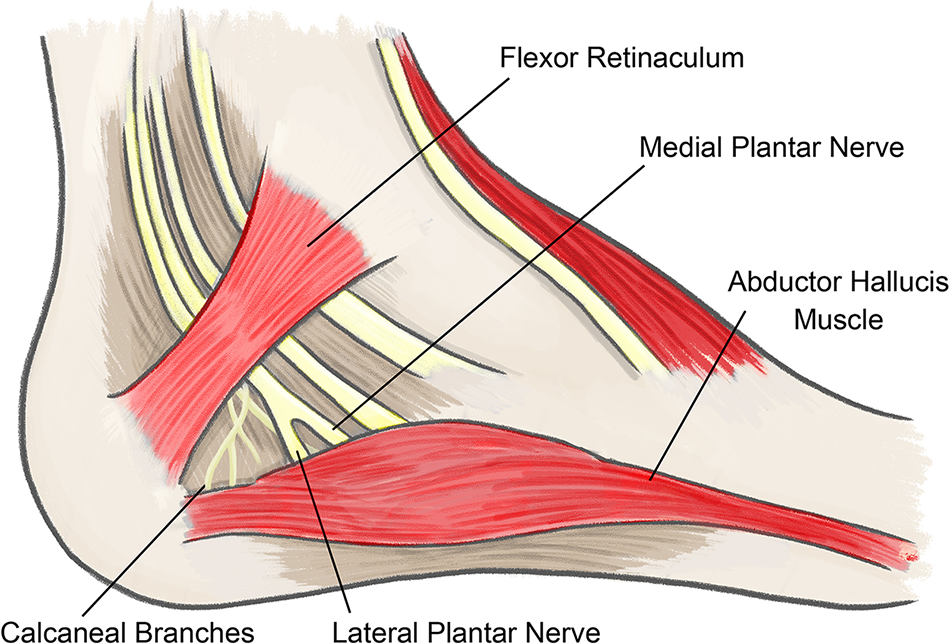
The mechanism of DTTS was described, in part, by McGill [5] in 1964, wherein the medial plantar nerve was found to be compressed by a fascial band beneath the abductor hallucis muscle. Edwards et al [6] in 1969 described entrapment of both the medial and lateral plantar nerves as they pass through the fibrous opening under the abductor hallucis muscle (Fig. 1). In later years, this mechanism has been described further, with additional detail on nerve compression due to the abductor hallucis muscle [7,8,9,10,11,12].
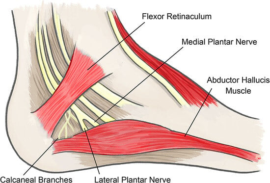
Figure 1.
Anatomy of the medial and lateral plantar nerves as they pass under the abductor hallucis muscle.
Patients with DTTS often present with symptoms similar to those with PTTS: burning pain, numbness, paresthesia, a sensation of cold, an abnormal feeling of walking on a rough surface such as sandpaper or gravel, and pain in the arch or ball of the foot [10,12–16].
Diagnosis of DTTS is based primarily on the patient’s history and the clinical examination findings, which generally include the same clinical tests as used for TTS but focused on the distal tarsal tunnel (DTT). These tests include eliciting a Tinel sign, provocative sign, diagnostic nerve blocks, and a provocative maneuver test [15,17–22]. Gould describes a provocative point of tenderness—a “soft spot”—on the abductor hallucis muscle where the medial and lateral plantar nerves and the first branch of the lateral plantar nerve enter the foot [2,23,24].
Investigative tests, such as use of the Pressure Specified Sensory Device (Axogen, Jacksonville, Florida), electromyography, nerve conduction studies, magnetic resonance imaging, and diagnostic ultrasonography, may be of value in the diagnosis of TTS in some cases. However, the literature is limited as to their use in distinguishing DTTS, although it has been shown to help [2,12,15,18,25,26,27,28,29].
There is sparse research on tests and clinical examination methods to differentiate DTTS from PTTS, and often the two cannot be distinguished from each other. As a result, the incidence of PTTS and DTTS is unknown, and surgery for TTS typically addresses both areas of the tarsal tunnel and, at times, the plantar fascia [2,7,8,11,24,30]. Watson et al [30] state:
Differentiating between variants of tarsal tunnel syndrome and plantar fasciitis can be very difficult in the most astute clinician’s hands. If a specific diagnosis cannot be made, a combination of surgical procedures may be necessary, including neurolysis and a soft-tissue release.
The gap in a sound method to differentiate PTTS from DTTS was the impetus for this research.
Extrapolating from a study that found that diagnosis of a common peroneal nerve entrapment could be assisted by injecting a small amount of lidocaine into the peroneus longus muscle at the site of the nerve’s compression and the long-standing practice of treating myofascial trigger points with injections, it was hypothesized that the same test could be used to assist in the diagnosis of other nerve compressions related to muscle impingement [31,32,33]. In the case of compression in DTTS where the distal tibial nerve branches are generally entrapped as they course under the abductor hallucis muscle [15,18,34,35,36], this study premises that the muscle itself—not only its fascia and the underlying septum—is the primary source of the nerves’ compression. As such, this study explores the possible use of an injection of lidocaine into the abductor hallucis muscle as an adjunctive clinical test to assist with the diagnosis of DTTS.
Furthermore, because injections of a local anesthetic combined with a corticosteroid have successfully alleviated pain and sensory disturbances from entrapment [14,15,18,37,38], this study also sought to consider the treatment benefits of this clinical test by adding a small amount of corticosteroid to the lidocaine injection.
Methods
Patients
This retrospective medical record review study considered 44 patients (18 men and 26 women; mean ± SD age, 52.0 ± 13.7 years; age range, 22–78 years) who presented with TTS symptoms that suggested a clinical suspicion of DTTS. The study was approved by Heartland Institutional Review Board (Belleville, Illinois). All procedures performed in studies involving human participants were in accordance with the ethical standards of the 1964 Helsinki declaration and its amendments or comparable ethical standards. For this type of study, formal consent is not required. Personal identity information was removed to maintain patient confidentiality.
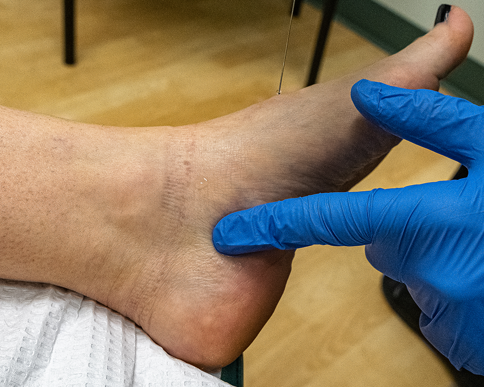
The inclusion criteria were 1) neurologic symptoms consistent with TTS in the ball of the foot or toes, either while at rest or walking (if symptoms were not present at the time of the medical visit, the LITT was not performed); 2) pain elicited on palpation of the abductor hallucis muscle at the point where the tibial nerve passes beneath the muscle, as previously described by Gould [23] as the soft spot (Fig. 2); 3) reduced sensation on the ball of the foot as determined by 2-point discrimination testing; 4) no obvious ischemia of the foot, as determined by palpation of the dorsalis pedis and posterior tibial arteries and evaluation of the capillary fill time to the great toe (the presence of adequate circulation reduced the chance of ischemic pain mimicking DTTS); 5) to avoid violating an independence in statistical analysis, no two feet were from the same patient [39]; and 6) no self-reported history of allergy or hypersensitivity to lidocaine or dexamethasone phosphate [7]. Patients gave their consent for injection.
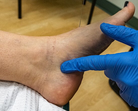
Figure 2.
Palpation of the abductor hallucis muscle at the point where the tibial nerve branches pass beneath the muscle (known as the soft spot).
The exclusion criteria were presence of a rearfoot mass, trauma, pregnancy, history of coagulopathy, human immunodeficiency virus, previous rearfoot surgery, fat pad atrophy, history of alcohol or drug abuse, current narcotic use, or history of radiculopathy, sciatica, fibromyalgia, rheumatoid arthritis, or other systemic illnesses that could affect the feet regarding the diagnosis of symptoms of PTTS or DTTS.
Lidocaine Injection Test and Treatment
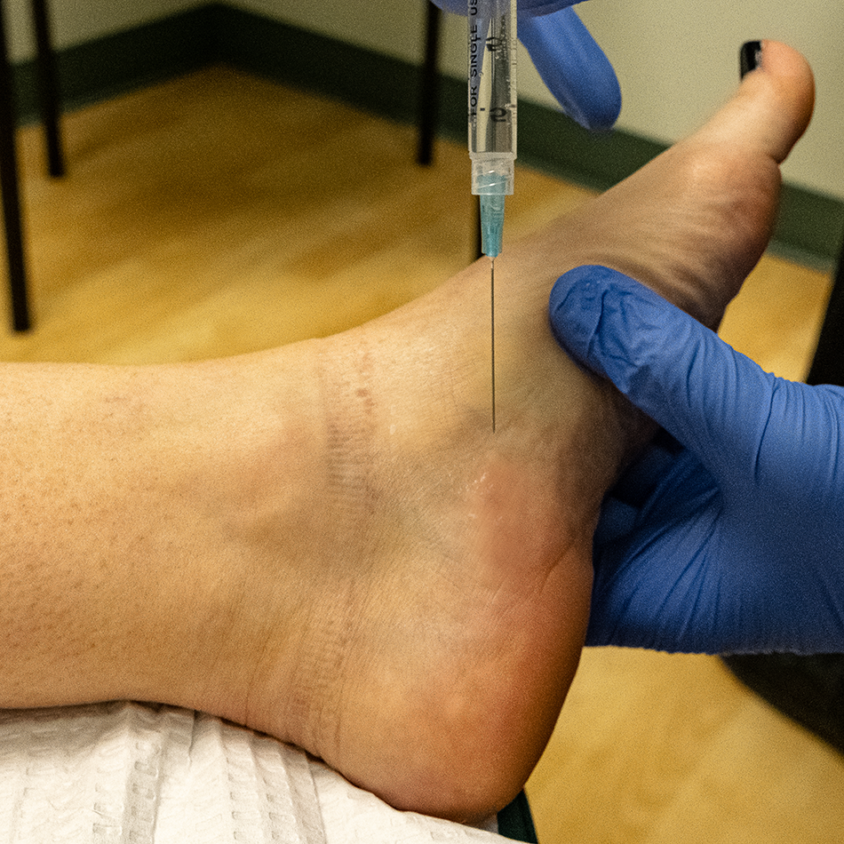
To perform the lidocaine injection test and treatment (LITT), the abductor hallucis muscle and the relevant landmarks were identified. The distal branches of the posterior tibial nerve travel under the muscle as it courses from its insertion at the medial-posterior process of the calcaneal tuberosity to its insertion into the proximal phalanx of the great toe (Fig. 1) [34]. The Gould soft spot, described previously herein, where the tibial nerve’s branches course beneath the abductor hallucis muscle, was palpated and identified (Fig. 2). The injection was performed using aspectic technique and administered using a 27-gauge needle with 3 mL of lidocaine plain mixed with 2 mg of dexamethasone phosphate. The needle is positioned just distal and superior to the soft spot, angled posteroinferiorly and as parallel to muscle as possible (typically about 20°). By gently pressing down on the inside of the front of the foot, the abductor muscle bulges upward slightly, making the muscle easier to localize (Fig. 3). The injection is administered slowly as the needle moves across the soft spot toward the bottom edge of the muscle. To avoid injury to the underlying nerves, it is important to inject along the most superficial aspect of the muscle, bathing the muscle with the lidocaine and corticosteroid, which causes the skin to bulge upward slightly as the injection is administered. Injecting distal-superior to the soft spot and ending proximal to it ensures that the area of the muscle that passes over the tibial nerve branches will be covered with lidocaine and corticosteroid.
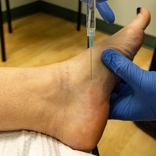
Figure 3.
Using aseptic technique, a 27-guage needle is angled almost parallel to the muscle as 3 mL of lidocaine 1% plain mixed with 2 mg of dexamethasone phosphate is slowly injected along the abductor hallucis muscle. The injection should be performed only superficially so as to not injure the underlying nerves.
A few minutes after the injection, the patient was instructed to walk briefly in the office’s hallway and report whether their symptoms had improved significantly (any amount beyond minimal). The positive effect of the test, or lack thereof, was quickly recognizable by patients. If symptoms improved, the LITT was deemed positive. Patients with a positive LITT were asked to grade the improvement as either completely resolved (complete) or partly improved (partial). (Patients who had primarily numbness, not pain, had difficulty providing feedback after the LITT using a 10-point pain scale; therefore, a simplified feedback method was implemented.) Some patients who reported a positive LITT stated that the strength and movement of their toes had dramatically improved. If the patient’s symptoms improved only minimally or not at all, the LITT was deemed negative. Patients who were unsure whether their symptoms improved were also considered negative.
Patients were instructed to return to the office, and this initial follow-up visit generally occurred within 1 to 4 weeks. Patients were questioned about any residual adverse effects from the LITT, and those whose LITT had been positive were asked whether their symptoms had returned (no relief) or worsened or whether the improvement had continued at the same level (maintained) or at a reduced level (partly maintained). Patients with adverse effects or any level of symptom improvement were followed for a minimum of 3 months, or until the adverse effects resolved or improvement ceased.
Test Results
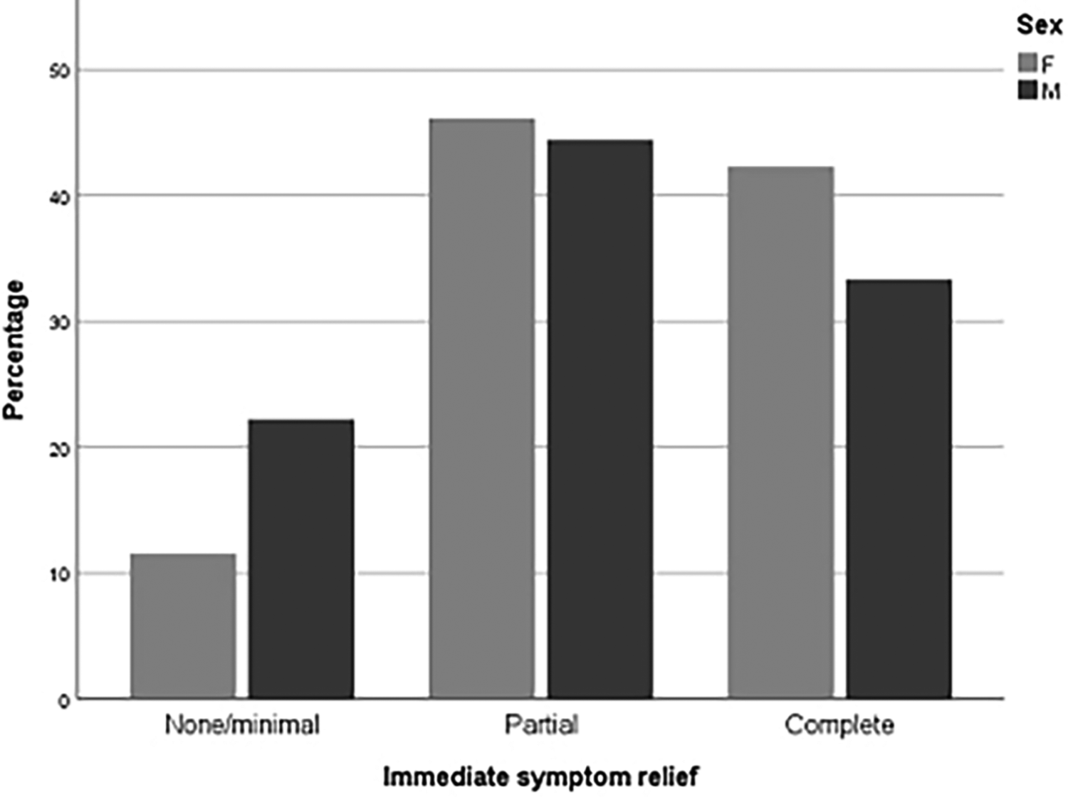
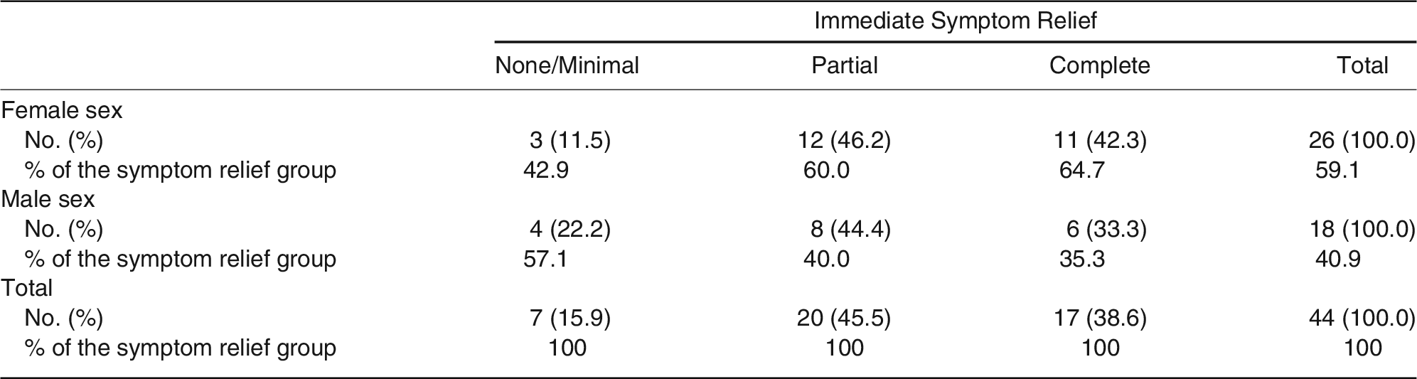
Of the 44 study patients, 37 exhibited a positive LITT. The symptom relief reported by the participants a few minutes after the injection was stated as complete by 17 (39%), partial by 20 (45%), and none or minimal by seven (16%). As a result, a positive outcome of the LITT was reported by 84% of those who were injected with the lidocaine-dexamethasone phosphate mixture. This is shown in Table 1 and Figure 4, where these results are also broken down by sex. Given that this is a preliminary exploratory study, the results are presented using descriptive statistics. Comparisons of outcomes between groups used the χ2 test or the Fisher exact test where cell frequencies were small. There was no difference in the distribution of immediate symptom relief by sex (Fisher exact test = 1.048; P = .653).

Table 1.
Immediate Symptom Relief Outcome by Sex
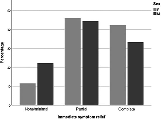
Figure 4.
Clustered bar chart showing immediate symptom relief reported by sex.
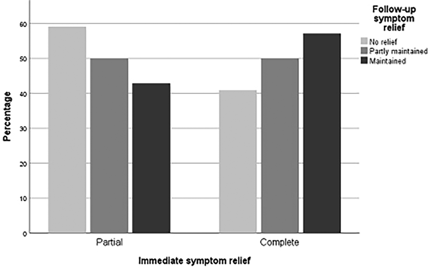
Patients reported level of symptom relief at the follow-up consultation (2–4 weeks after the LITT). It was described as no relief, partly maintained, or maintained. These results are shown in Table 2 and Fig. 5. Follow-up data were missing for two of the 37 patients who had an initial positive LITT because they did not return for follow-up.
Table 2.
Level of Symptom Relief at Follow-up (2–4 Weeks After the LITT) for Patients with Positive Immediate Outcomes
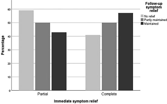
Figure 5.
Clustered bar chart showing the degree of symptom relief maintenance at the follow-up visit (2–4 weeks after lidocaine injection test and treatment) for patients with partial or complete immediate symptom relief.
Of the 16 patients who experienced initial complete relief, this level was maintained in four (25%). Of patients available for follow-up evaluation (35), 11% of those with a positive LITT (four) had complete, lasting symptom relief at follow-up. Thirteen of 35 patients who had a positive initial response to LITT experienced partial or complete symptom relief at follow-up. This study found no association between level of symptom relief maintenance and immediate level of symptom relief (Fisher exact test = 0.751; P = .797).
The main symptom of numbness was present in 12 patients, with eight experiencing a significant improvement in sensation almost immediately after the LITT and two of them maintaining their improvement at follow-up.
During the physical examination, palpation of the Gould soft spot resulted in a Tinel-type sensation or sudden worsening of symptoms in 17 patients. Thirty-five patients had a Tinel sign elicited over the tarsal tunnel: 14 over the proximal tarsal tunnel (PTT) and 12 over the DTT (individuals with a Tinel sign elicited at the DTT should not be confused with those who had a Tinel-type sensation from palpation on the soft spot), with the remaining nine not clearly localized to the DTT or the PTT.
The only adverse effect or complication observed was a transient discomfort or soreness in the area of the injection experienced by three patients. This was described as mild in severity, and there was no evidence of hematoma, infection, or injury to the underlying nerves.
Discussion
Within the diagnosis of TTS, the clinician has few methods of differentiating DTTS from PTTS. The present study assessed the LITT to diagnose and treat 44 patients with DTTS, finding that 84% of patients had a positive LITT, with 11% of the 35 patients seen at follow-up experiencing lasting relief. These results suggest that the LITT is useful, safe, and inexpensive method to assist with the diagnosis and treatment of DTTS. (Patients who complain of numbness only may opt to not have the issue investigated or treated, although reduced sensation could predispose their foot or feet to increased risk of injury and ulceration.)
The reliability of the test may be impaired if the physician inadvertently injects lidocaine deep to the abductor hallucis muscle, anesthetizing the underlying nerves and possibly producing a false-positive result. In this regard, it was observed that patients who had a Tinel sign over the PTT continued to experience the Tinel sign at the PTT after the LITT, evidencing that the LITT did not anesthetize the nerves at the DTT. Injecting deep to the abductor hallucis muscle could also, in theory, injure the nerves, and, as such, depending on the physician’s experience and the patient’s anatomy, diagnostic ultrasound may be used to assist with the injection. Although the DTT’s nerves were not anesthetized in any of the study participants, because lidocaine has a short duration of action, if this untoward effect occurs, the patient can be reassured that the effects are temporary.
In addition to its short duration of action, lidocaine was chosen for the LITT due to its quick onset. Other anesthetics may be used for the LITT, although these should not be mixed with epinephrine because this could cause the muscle to contract, worsening the patient’s symptoms [40].
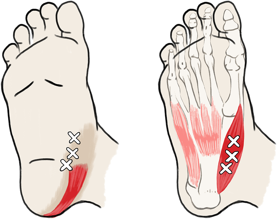
The abductor hallucis muscle has been recognized as a taut muscle band that has three myofascial trigger points (Fig. 6), which are foci of taut palpable bands of muscles [32,33,36,41,42]. Injections of local anesthetics (with and without corticosteroids) into such trigger points have been used to diagnose and alleviate pain [32,33,36,41,42,43,44].
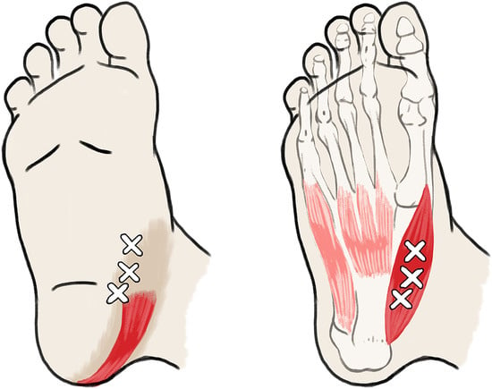
Figure 6.
Myofascial trigger points of the abductor hallucis muscle.
Wada et al [36] examined the innervation of the abductor hallucis muscle by the medial plantar nerve, finding that the nerve’s entry points correspond to the muscle’s trigger points. The authors opined that the high nerve concentration into this area of the abductor hallucis muscle is related to the pathophysiology of the muscle’s myofascial pain. Histologic analysis of the nerves beneath the abductor hallucis muscle show signs of compression neuropathy in the form of demyelination. [9,44]
Based on this previous research, it is hypothesized that the LITT’s mechanism of action lies in the use of lidocaine to cause the abductor hallucis muscle to undergo flaccid paralysis or to “relax.” [13,45]. This flaccid state allows compression of the muscle and its fascia on the underlying distal tibial nerve branches to lessen, alleviating their entrapment. Early research on the use of botulinum toxin for heel pain syndrome related to nerve compression associated with the abductor hallucis and quadratus plantae muscles is encouraging [46].
Physicians should be aware that DTTS can be complicated by other issues that may mimic the symptoms of DTTS or that may occur concomitantly, including PTTS, which when present with DTTS is often referred to simply as TTS [7,8,11]. Other differential diagnoses include other nerve entrapments, such as calcaneal nerve entrapment, soleal sling syndrome, or radiculopathy; neuropathy, as in diabetes; or other issues, including vascular disease or venous congestion, tumor, or fracture [2,17,47,48,49,50,51,52.]
Limitations of this study include the small sample size, the length of follow-up, the variable period of the patients’ return visit, and the exclusion of adolescents and children. As such, lengthier and more extensive studies exploring the LITT should be performed, perhaps incorporating a standardized period for patients to return to the clinic. Due to the limitations of this study, physicians using the LITT in the diagnosis and treatment of DTTS should interpret the findings with caution. These results should not be interpreted as discounting the importance of the physician’s history, physical examination, and diagnostic testing (when required) and the value of other treatments.
Because this research suggests that the abductor hallucis muscle is the primary compressive force, future research may be aimed at addressing the compression of this muscle. Measures could possibly include physical therapy techniques or altering the muscle’s biomechanics through foot orthoses or the use of pharmacologic agents, as well as investigating the outcome of a narrowed surgical approach to TTS that decompresses only the PTT or the distal tarsal tunnel.
Conclusions
A distinct clinical entity, DTTS can occur as part of the broader diagnosis of TTS. This exploratory study shows that the LITT provides a simple, rapid, and safe adjunctive tool that may assist in diagnosing and treating DTTS. It is hoped that the proposed mechanism of the LITT—focused on muscle as a primary cause of the nerve entrapment—leads to new, nonsurgical treatments for DTTS and other muscle-involved nerve compressions and sparks further discussion and research in this area.
Funding
None reported.
Acknowledgments
Jackie Campbell for assistance with the statistical analysis.
Conflicts of Interest
None reported.
References
- Keck, C. The Tarsal tunnel syndrome. J Bone Joint Surg Am 1962, 44, 180. [Google Scholar]
- DiGiovanni, BF; Abuzzahab, FS; Gould, JS. Plantar fascia release with proximal and distal tarsal tunnel release: a surgical approach to chronic, disabling plantar fasciitis with associated nerve pain. Tech Foot Ankle Surg 2003, 2, 254. [Google Scholar]
- Fernández-Gibello, A; Moroni, S; Camuñas, G; et al. Ultrasound-guided decompression surgery of the tarsal tunnel: a novel technique for the proximal tarsal tunnel syndrome—part II. Surg Radiol Anat 2019, 41, 43. [Google Scholar]
- Heimkes, B; Posel, P; Stotz, S; et al. The proximal and distal tarsal tunnel syndromes: an anatomical study. Int Orthop 1987, 11, 193. [Google Scholar]
- McGill, DA. Tarsal tunnel syndrome. Proc R Soc Med 1964, 57, 1125. [Google Scholar]
- Edwards, WG; Lincoln, CR; Bassett, FH, III; et al. The tarsal tunnel syndrome: diagnosis and treatment. JAMA 1969, 207, 716. [Google Scholar]
- Dellon, AL. The four medial ankle tunnels: a critical review of perceptions of tarsal tunnel syndrome and neuropathy. Neurosurg Clin N Am 2008, 19, 629. [Google Scholar]
- Franson, J; Baravarian, B. Tarsal tunnel syndrome: a compression neuropathy involving four distinct tunnels. Clin Podiatr Med Surg 2006, 23, 597. [Google Scholar]
- Ghosh, SK; Raheja, S; Tuli, A. Potential sites of compression of tibial nerve branches in foot: a cadaveric and imaging study. Clin Anat 2013, 26, 768. [Google Scholar]
- Inthasan, C; Mahakkanukrauh, P. Tarsal tunnel syndrome: anatomical facts and clinical implications. J Anat Soc India 2019, 68, 236. [Google Scholar]
- Mullick, T; Dellon, AL. Results of decompression of four medial ankle tunnels in the treatment of tarsal tunnel syndrome. J Reconstr Microsurg 2008, 24, 119. [Google Scholar]
- Oh; Shin, J. Neuropathies of the foot. Clin Neurophysiol 2007, 118, 954. [Google Scholar]
- Appiah-Ankam, J; Hunter, JM. Pharmacology of neuromuscular blocking drugs. Contin Educ Anaesth Crit Care Pain 2004, 4, 2. [Google Scholar]
- Baxter, DE; Thigpen, CM. Heel pain: operative results. Foot Ankle 1984, 5, 16. [Google Scholar]
- Brown, MN; Pearce, BS; Vanetti, TK; et al. Medial Planter Nerve Entrapment. In Peripheral Nerve Entrapments: Clinical Diagnosis and Management; Trescot, AM, Ed.; Springer International Publishing: Cham, Switzerland, 2016; p. 844. [Google Scholar]
- Schon, LC; Baxter, DE. Neuropathies of the foot and ankle in athletes. Clin Sports Med 1990, 9, 489. [Google Scholar]
- Antoniadis, G; Scheglmann, K. Posterior tarsal tunnel syndrome: diagnosis and treatment. Dtsch Ärztebl Int 2008, 105, 776. [Google Scholar]
- Brown, MN; Pearce, BS; Vanetti, TK; et al. Lateral Planter Nerve Entrapment. In Peripheral Nerve Entrapments: Clinical Diagnosis and Management; Trescot, AM, Ed.; Springer International Publishing: Cham, Switzerland; 2016; p. 832. [Google Scholar]
- Hendrix, CL; Jolly, GP; Garbalosa, JC; et al. Entrapment neuropathy: the etiology of intractable chronic heel pain syndrome. J Foot Ankle Surg 1998, 37, 273. [Google Scholar]
- Lee, CH; Dellon, AL. Prognostic ability of Tinel sign in determining outcome for decompression surgery in diabetic and nondiabetic neuropathy. Ann Plast Surg 2004, 53, 523. [Google Scholar]
- Rinkel, WD; Cabezas, MC; van Neck, JW; et al. Validity of the Tinel sign and prevalence of tibial nerve entrapment at the tarsal tunnel in both diabetic and nondiabetic subjects: a cross-sectional study. Plast Reconstr Surg 2018, 142, 1258. [Google Scholar]
- Trepman, E; Kadel, NJ; Chisholm, K; et al. Effect of foot and ankle position on tarsal tunnel compartment pressure. Foot Ankle Int 1999, 20, 721. [Google Scholar]
- Gould, JS. Tarsal tunnel syndrome. Foot Ankle Clin 2011, 16, 275. [Google Scholar]
- Watson, TS; Anderson, RB; Davis, WH; et al. Distal tarsal tunnel release with partial plantar fasciotomy for chronic heel pain: an outcome analysis. Foot Ankle Int 2020, 23, 530. [Google Scholar]
- Hochman, MG; Zilberfarb, JL. Nerves in a pinch: imaging of nerve compression syndromes. Radiol Clin N Am 2004, 42, 221. [Google Scholar]
- Oishi, C; Ichikawa, Y; Chiba, A. The usefulness of electrophysiological examinations in the diagnosis of tarsal tunnel syndrome. Clin Neurophysiol 2018, 129, e34. [Google Scholar]
- Patel, AT; Gaines, K; Malamut, R; et al. Usefulness of electrodiagnostic techniques in the evaluation of suspected tarsal tunnel syndrome: an evidence‐based review. Muscle Nerve 2005, 32, 236. [Google Scholar]
- Sodani, A; Dube, M; Jain, R. Value of motor nerve conduction studies in the diagnosis of idiopathic tarsal tunnel syndrome: a single-center prospective observational study from India. Ann Indian Acad Neurol 2018, 21, 35. [Google Scholar]
- Takakura, Y; Kumai, T; Takaoka, T; et al. Tarsal tunnel syndrome caused by coalition associated with a ganglion. J Bone Joint Surg Br 1998, 80, 130. [Google Scholar]
- Hong, JT; Lee, SW; Son, BC; et al. Surgical experiences of the tarsal tunnel syndrome. J Korean Neurosurg Soc 2004, 36, 443. [Google Scholar]
- Nirenberg, MS. A simple test to assist with the diagnosis of common fibular nerve entrapment and predict outcomes of surgical decompression. Acta Neurochir 2020, 162, 1. [Google Scholar]
- Travell, JG; Simons, LS. Myofascial Pain and Dysfunction: The Trigger Point Manual; Lippincott Williams & Wilkins: Philadelphia; 1993. [Google Scholar]
- Scott, NA; Bing, G; Barton, PM; et al. Trigger point injections for chronic non-malignant musculoskeletal pain: a systematic review. Pain Med 2009, 10, 54. [Google Scholar]
- Kelikian, AS; Sarrafian, SK. Sarrafian's Anatomy of the Foot and Ankle: Descriptive, Topographic, Functional; Lippincott Williams & Wilkins: Philadelphia; 2011. [Google Scholar]
- Macchi, V; Tiengo, C; Porzionato, A. Correlation between the course of the medial plantar artery and the morphology of the abductor hallucis muscle. Clin Anat 2005, 18, 580. [Google Scholar]
- Wada, JT; Akamatsu, F; Hojaij, F; et al. An anatomical basis for the myofascial trigger points of the abductor hallucis muscle. Biomed Res Int 2020, 2020, 1. [Google Scholar]
- Beskin, JL. Nerve entrapment syndromes of the foot and ankle. J Am Acad Orthop Surg 1997, 5, 261. [Google Scholar]
- Kurashige, T. Hypertrophy of the abductor hallucis muscle: a case report and review of the literature. SAGE Open Med Case Rep 2017, 5, 2050313X17727638. [Google Scholar]
- Menz, HB. Two feet, or one person? problems associated with statistical analysis of paired data in foot and ankle medicine. The Foot 2004, 14, 2. [Google Scholar]
- Williams, JH; Barnes, WS. The positive inotropic effect of epinephrine on skeletal muscle: a brief review. Muscle Nerve 1989, 12, 968. [Google Scholar]
- Nguyen; Bang, M. Trigger point therapy and plantar heel pain: a case report. Foot 2010, 20, 158. [Google Scholar]
- Giamberardino, MA; Affaitati, G; Fabrizio, A; et al. Myofascial pain syndromes and their evaluation. Best Pract Res Clin Rheumatol 2011, 25, 185. [Google Scholar]
- Qureshi, NA; Alsubaie, HA; Ali, GI. Myofascial pain syndrome: a concise update on clinical, diagnostic and integrative and alternative therapeutic perspectives. Int Neuropsychiatr Dis J 2019, 13, 1. [Google Scholar]
- Mackinnon, SE; Dellon, AL; Daneshvar, A. Histopathology of the tarsal tunnel syndrome: examination of a human tibial nerve. Contemp Orthop 1984, 9, 43. [Google Scholar]
- Naguib, M; Lien, CA; Meistelman, C. Pharmacology of Neuromuscular Blocking Drugs. In Miller’s Anesthesia, 8th ed.; Miller, RD, Ed.; Elsevier Saunders: Philadelphia; 2015; p. 958. [Google Scholar]
- Radovic, P. Treatment of “plantar fasciitis”/plantar heel pain syndrome with botulinum toxin: a novel injection paradigm pilot study. Foot 2020, 45, 01711. [Google Scholar]
- Bowley, MP; Doughty, CT. Entrapment neuropathies of the lower extremity. Med Clin North Am 2019, 103, 371. [Google Scholar]
- Kushner, S; Reid, DC. Medial tarsal tunnel syndrome: a review. J Orthop Sports Phys Ther 1984, 6, 39. [Google Scholar]
- Labib, SA; Gould, JS; Rodriguez-del-Rio, FA; et al. Heel pain triad (HPT): the combination of plantar fasciitis, posterior tibial tendon dysfunction and tarsal tunnel syndrome. Foot Ankle Int 2002, 23, 212. [Google Scholar]
- Rinkel, WD; Cabezas, MC; Birnie, E; et al. The natural history of tarsal tunnel syndrome in diabetic subjects. J Plast Reconstr Aesthet Surg 2020, 73, 1482. [Google Scholar]
- Rosenbaum, AJ; DiPreta, JA; Misener, D. Plantar heel pain. Med Clin North Am 2014, 98, 339. [Google Scholar]
- Williams, EH; Rosson, GD; Hagan, RR; et al. Soleal sling syndrome (proximal tibial nerve compression): results of surgical decompression. Plast Reconstr Surg 2012, 129, 454. [Google Scholar]

© 2023 American Podiatric Medical Association